. 2023 Apr;299(4):103041.
doi: 10.1016/j.jbc.2023.103041. Epub 2023 Feb 18.

UHMK1 is a novel splicing regulatory kinase

Affiliations

UHMK1 is a novel splicing regulatory kinase

Vanessa C Arfelli et al. J Biol Chem. 2023 Apr.

Abstract

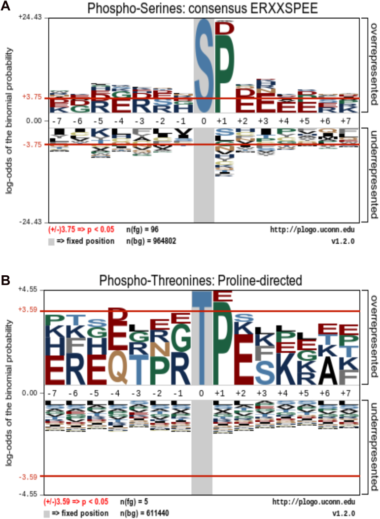
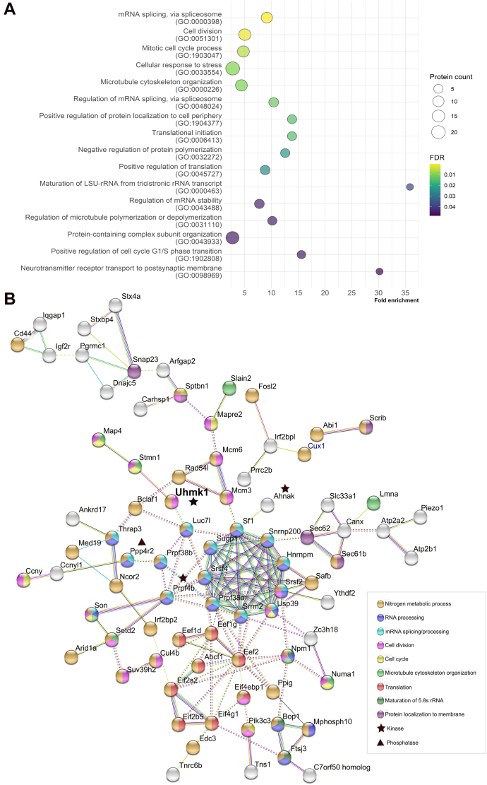
The U2AF Homology Motif Kinase 1 (UHMK1) is the only kinase that contains the U2AF homology motif, a common protein interaction domain among splicing factors. Through this motif, UHMK1 interacts with the splicing factors SF1 and SF3B1, known to participate in the 3' splice site recognition during the early steps of spliceosome assembly. Although UHMK1 phosphorylates these splicing factors in vitro, the involvement of UHMK1 in RNA processing has not previously been demonstrated. Here, we identify novel putative substrates of this kinase and evaluate UHMK1 contribution to overall gene expression and splicing, by integrating global phosphoproteomics, RNA-seq, and bioinformatics approaches. Upon UHMK1 modulation, 163 unique phosphosites were differentially phosphorylated in 117 proteins, of which 106 are novel potential substrates of this kinase. Gene Ontology analysis showed enrichment of terms previously associated with UHMK1 function, such as mRNA splicing, cell cycle, cell division, and microtubule organization. The majority of the annotated RNA-related proteins are components of the spliceosome but are also involved in several steps of gene expression. Comprehensive analysis of splicing showed that UHMK1 affected over 270 alternative splicing events. Moreover, splicing reporter assay further supported UHMK1 function on splicing. Overall, RNA-seq data demonstrated that UHMK1 knockdown had a minor impact on transcript expression and pointed to UHMK1 function in epithelial-mesenchymal transition. Functional assays demonstrated that UHMK1 modulation affects proliferation, colony formation, and migration. Taken together, our data implicate UHMK1 as a splicing regulatory kinase, connecting protein regulation through phosphorylation and gene expression in key cellular processes.

Keywords: KIS; RNA processing; RNA splicing; RNA-seq; SF1; SUGP1; UHM motif; gene expression; phosphoproteomics; protein kinase.


Conflict of interest statement

Conflict of interest The authors declare that they have no conflicts of interest with the contents of this article.

Figures

Figure 1
Figure 1
Global impact of UHMK1 on phosphoproteome.A, experimental workflow used in the phosphoproteome experiment. UHMK1 modulation in NIH3T3 cells was achieved expressing the shUHMK1#2 (sh#2) and shUHMK1#3 (sh#3) sequences, for knockdown, and UHMK1WT (WT) or the kinase-dead mutant UHMK1K54R (K54R) for overexpression. A scrambled shRNA sequence (shCTRL) and the empty vector (EV) MIY were used as control. Scheme created with ©BioRender.com. B, Western blot confirming the efficient modulation of UHMK1 expression in NIH3T3 cells used in the phosphoproteome experiment. The numbers 1 to 3 represent the biological replicates from each condition. Membranes were blotted with anti-UHMK1, anti-GFP, and anti-Actin (loading control). Total protein: 100 μg (knockdown) and 50 μg (overexpression). CF, the volcano plots show the global impact of UHMK1 knockdown (C and D) and overexpression (E and F) on phosphoproteome. The red and blue circles represent the significantly upregulated and downregulated phosphopeptides in each comparison, respectively. Phosphopeptides with p-value <0.01 and log2 fold change >|2| have their gene names assigned. G, overview of the absolute number of phosphopeptides and corresponding number of proteins differentially phosphorylated in the UHMK1 phosphoproteome. H, percentage of phosphorylated Serine, Threonine, and Tyrosine residues in the UHMK1 phosphoproteome. UHMK1, U2AF Homology Motif Kinase 1.
Figure 2
Figure 2
Distribution and phosphorylation pattern of the differentially phosphorylated proteins.A, Venn Diagram comparing the number of unique DPPs among the experimental conditions. KD = UHMK1-KD (knockdown); WT = UHMK1WT overexpression; K54R= UHMK1K54R overexpression. BD, percentage of phosphosites upregulated and downregulated in the DPPs found exclusively in UHMK1-KD (B), UHMK1WT (C), and UHMK1K54R (D). EH, phosphorylation pattern of the phosphopeptides (and respective phosphosites) from the DPPs (indicated by their gene names) shared by UHMK1-KD (sh#2 and sh#3), UHMK1WT and UHMK1K54R cells (E), UHMK1WT and UHMK1K54R cells (F), UHMK1-KD and UHMK1K54R (G), and the only DPP (Med19) shared by UHMK1-KD and UHMK1WT (H). DPP, differentially phosphorylated protein; UHMK1, U2AF Homology Motif Kinase 1.
Figure 3
Figure 3
UHMK1 phosphorylation consensus sequence retrieved from pLogo analysis. Amino acid heights are scaled according to their statistical significance, as well as the stacking order (the most significant residues are positioned closest to the x axis). The red horizontal lines represent a threshold for Bonferroni-corrected statistical significance values. Positive values higher than the threshold correspond to statistically significant (p < 0.05) overrepresented amino acids. Negative values lower than the threshold correspond to statistically significant underrepresented amino acids. A, UHMK1 preferentially phosphorylates proline-directed serine within the consensus sequence ERXXSPEE. Input sequences = 96. Log-odds of the binomial probability: E (−4) = 3.89; R (−3) = 3.81; P (+1) = 20.32; E (+2) = 4.36; E (+3) = 5.07. B, proline-directed threonine is also preferentially phosphorylated by UHMK1. Input sequences = 5. P (+1) = 4.03. UHMK1, U2AF Homology Motif Kinase 1.
Figure 4
Figure 4
UHMK1 putative substrates are implicated in a variety of cellular processes and form a complex network.A, Biological Process (BP) terms retrieved from Gene Ontology (GO) analysis. Only the most specific terms within the hierarchy presented in PANTHER are shown (complete results in Table S3, specific terms highlighted in gray). B, interaction network of UHMK1 and the 117 putative substrates (DPPs). Only the interacting proteins (represented by their gene names) are shown in the figure: the pink edges represent the experimentally determined interactions; blue edges represent interactions registered in curated databases; green edges represent interactions extracted from text mining; black edges represent coexpression. Each node represents one protein. Node colors indicate the most representative BP terms from GO analysis. Intercluster interactions are represented by dashed edges. Protein-protein interaction (PPI) enrichment p-value: 4.55e-15. The kinases and the phosphatase in this network are marked with a black star and a triangle, respectively. UHMK1 is highlighted in bold. DPP, differentially phosphorylated protein; UHMK1, U2AF Homology Motif Kinase 1.
Figure 5
Figure 5
Besides SF1 and SUGP1, 10 RNA/splicing-related DPPs contain ULM motifs. Clustal Omega alignment of proteins containing well characterized ULM motifs (highlighted in gray) and the ten novel RNA/splicing-related putative UHMK1 substrates. Uniprot identification code for the mouse proteins are shown (left). The conserved tryptophan (W) within the ULM domain is highlighted in yellow, and its position within the protein is depicted on the right. The preferred amino acids in the positions +1 and +2 are highlighted in purple, and the preferred amino acids at −1 and previous positions are highlighted in green. TNRC6B and SF1 (red) were the only RNA-related proteins from our study considered as bona fide ULM-containing proteins in ScanProsite analysis (not shown). DPP, differentially phosphorylated protein; UHMK1, U2AF Homology Motif Kinase 1; ULM, UHM ligand motif.
Figure 6
Figure 6
UHMK1 impacts mRNA splicing.AF, splicing analysis from UHMK1 RNA-seq data, comparing UHMK1 knockdown (UHMK1-KD) with shCTRL cells and UHMK1WT overexpressing cells with the empty vector (EV) control cells. A and B, significant alternative splicing events (ASEs) in UHMK1-KD and UHMK1WT overexpressing cells, respectively; C and D, predicted impact on coding sequence by ASE; E and F, percent spliced in (PSI) levels for the top 20 most significant Exon inclusion/exclusion ASEs. G, splicing reporter assay. The ratio between β-galactosidase and luciferase is an indirect measure of splicing occurring between both genes. Average values of four independent experiments. Constant amount of the pTN24 reporter plasmid is present in all conditions. One way ANOVA followed by Bonferroni correction was used to compare UHMK1WT and UHMK1K54R to the empty vector control MIY (not significant). Increase in splicing in SF1 over pcDNA (p = 0.0244) and difference between SF1+UHMK1WT and SF1+UHMK1K54R (p = 0.0294) was confirmed by Student t test. Controls of gene expression levels of UHMK1 and SF1 are provided in Fig. S5. UHMK1, U2AF Homology Motif Kinase 1.
Figure 7
Figure 7
UHMK1 impacts gene expression.A and B, Principal Component Analysis (PCA) plots showing the overall effect of UHMK1 knockdown mediated by shUHMK1#1 (sh#1), shUHMK1#2 (sh#2), and shUHMK1#3 (sh#3) compared to shCTRL and UHMK1WT overexpression compared to empty vector (EV) control, respectively. C, volcano plot showing the differentially expressed genes (DEGs) in UHMK1-KD cells (shUHMK1#1, shUHMK1#2, and shUHMK1#3 combined as one single artificial condition) compared to shCTRL cells. p-value <0.05 was used as cutoff. Genes with log2 fold change >|0.5| are labeled. D, heatmap of expression of the 32 DEGs represented as z-score of normalized counts, in each replicate of UHMK1 knockdown (KD) and shCTRL (CTRL) cells. E, gene sets enriched in UHMK1-KD in GSEA analysis. F, heatmap showing the expression of the Epithelial Mesenchymal Transition (EMT)-enriched gene set in UHMK1 knockdown cells. Gene expression is represented as z-score of normalized counts. G, enrichment plot for EMT gene signature. GSEA, Gene Set Enrichment Analysis; UHMK1, U2AF Homology Motif Kinase 1.
Figure 8
Figure 8
UHMK1 affects proliferation, colony formation, and migration of NIH3T3 cells.A, proliferation was evaluated by the percentage of cells that incorporated BrdU. Charts represent the ratio of BrdU incorporation in UHMK1 knockdown mediated by shUHMK1#1 (sh#1), shUHMK1#2 (sh#2), and shUHMK1#3 (sh#3) relative to the scrambled control (shCTRL) cells and UHMK1WT overexpressing cells relative to empty vector (EV) cells. Data from three independent experiments (∗p < 0.05, One-way ANOVA followed by Bonferroni’s multiple comparison test). B, clonogenic assay. The bar plots represent the percentage of colonies in UHMK1 knockdown and UHMK1WT overexpressing cells relative to the shCTRL or empty vector (EV), respectively. Results from three independent experiments, carried out in triplicate (∗p = 0.0286, Mann-Whitney test). The representative images of crystal violet-stained colonies are shown. One colony was defined by the minimum of 50 cells. C, viability assay. For UHMK1 knockdown cells, the chart represents the average of four experiments performed in sextuplicate. For UHMK1WT overexpressing cells, the chart represents the average of five experiments, two performed in triplicate and three performed in sextuplicate. D, apoptosis evaluation by Annexin V assay. Mean from three independent experiments. Early apoptosis is defined by the cells that are positive only for Annexin V; late apoptosis comprises the cells that are positive for both Annexin V and propidium iodide (PI); Necrotic cells are positive only for PI and Viable cells are negative for both markers. E, migration assay. Representative images of two independent experiments (I and II) with UHMK1 knockdown and UHMK1WT overexpressing cells. The experiments were performed in duplicates. Three images from each replicate were taken: in the center, bottom, and top of the membrane, relative to position of the circumference in the plate. The images in this figure are representative of the center. The control representing spontaneous migration towards lower chamber containing 1% FBS and the images acquired in the top and bottom of the wells are provided in Figs. S7–S9. Images were acquired with Microscope Leica DMi8, 10× magnification. FBS, fetal bovine serum; UHMK1, U2AF Homology MotifKinase 1.
Figure 9
Figure 9
Schematic view of the role of the 28 RNA-related UHMK1 substrates (DPPs) in different layers of gene expression. We searched the role of the 28 RNA-related DPPs in the literature, UniProt (90) and Spliceosome (91) databases. The RNA-related DPPs are represented by their gene names and highlighted in red. Proteins that act in more than one function related to RNA metabolism are marked with an asterisk (∗). The main functions are highlighted in a box: Chromatin modification, Transcription, mRNA splicing, Splicing regulation, mRNA stability, and rRNA processing. Approximately, half of the novel RNA-related UHMK1 substrates (15 proteins) are components of the spliceosome. Six DPPs have a role in mRNA stability, through mechanisms involving the poly(A) tail (TNRC6B (92) and NPM1 (93)), the 5′ Cap (EDC3 (94)), binding to N6-methyladenosine mRNAs (YTHDF2 (95)), nonsense-mediated decay (NMD)-independent decay (THRAP3 (96)), and binding to 3′ UTR (CRHSP-24 - Carhsp1) (97)). Four of the RNA-related DPPs act in transcription, either by acting on RNA pol II regulation (SAFB (98) and NPM1 (99)) or as transcription factors (FRA-2 - Fosl2) (100) and Eef1d (101)). SETD2, a methyltransferase responsible for H3K36me3 mark, act in chromatin modification (102). Moreover, three DPPs act specifically in the cleavage of the tricistronic rRNA transcript: MPP10 (Mphosph10) (component of the U3 snoRNP) and FTSJ3 (2′-O-methyltransferase) are involved in the processing of the 18S rRNA (103, 104), while BOP1 (component of the PeBoW complex) is necessary for the processing of 5.8S and 28S rRNAs (105). NPM1 also acts in the nucleolus as a histone chaperone, affecting chromatin status of rDNA (99). Created with ©BioRender.com. DPP, differentially phosphorylated protein; PRFs, preribosomal factors; RPs, ribosomal proteins; UHMK1, U2AF Homology Motif Kinase 1.

References

    1. Ardito F., Giuliani M., Perrone D., Troiano G., Lo Muzio L. The crucial role of protein phosphorylation in cell signaling and its use as targeted therapy. Int. J. Mol. Med. 2017;40:271–280. - PMC - PubMed
    1. Maucuer A., Le Caer J.P., Manceau V., Sobel A. Specific Ser-Pro phosphorylation by the RNA-recognition motif containing kinase KIS. Eur. J. Biochem. 2000;267:4456–4464. - PubMed
    1. Kielkopf C.L., Lücke S., Green M.R. U2AF homology motifs: protein recognition in the RRM world. Genes Dev. 2004;18:1513–1526. - PMC - PubMed
    1. Manceau V., Swenson M., Le Caer J.P., Sobel A., Kielkopf C.L., Maucuer A. Major phosphorylation of SF1 on adjacent Ser-Pro motifs enhances interaction with U2AF65. FEBS J. 2006;273:577–587. - PMC - PubMed
    1. Manceau V., Kielkopf C.L., Sobel A., Maucuer A. Different requirements of the kinase and UHM domains of KIS for its nuclear localization and binding to splicing factors. J. Mol. Biol. 2008;381:748–762. - PMC - PubMed

MeSH terms