. 2023 Aug 7;119(9):1869-1882.
doi: 10.1093/cvr/cvad022.

A Nrf2-OSGIN1&2-HSP70 axis mediates cigarette smoke-induced endothelial detachment: implications for plaque erosion

Affiliations

A Nrf2-OSGIN1&2-HSP70 axis mediates cigarette smoke-induced endothelial detachment: implications for plaque erosion

Sandro Satta et al. Cardiovasc Res. .

Abstract

Aims: Endothelial erosion of plaques is responsible for ∼30% of acute coronary syndromes (ACS). Smoking is a risk factor for plaque erosion, which most frequently occurs on the upstream surface of plaques where the endothelium experiences elevated shear stress. We sought to recreate these conditions in vitro to identify potential pathological mechanisms that might be of relevance to plaque erosion.

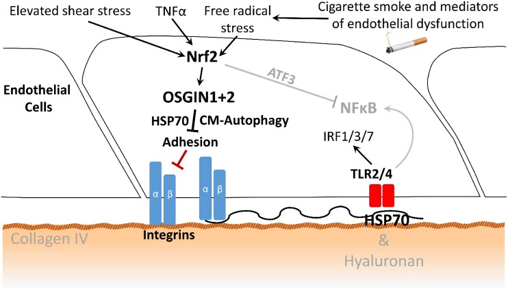
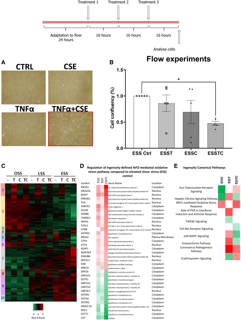
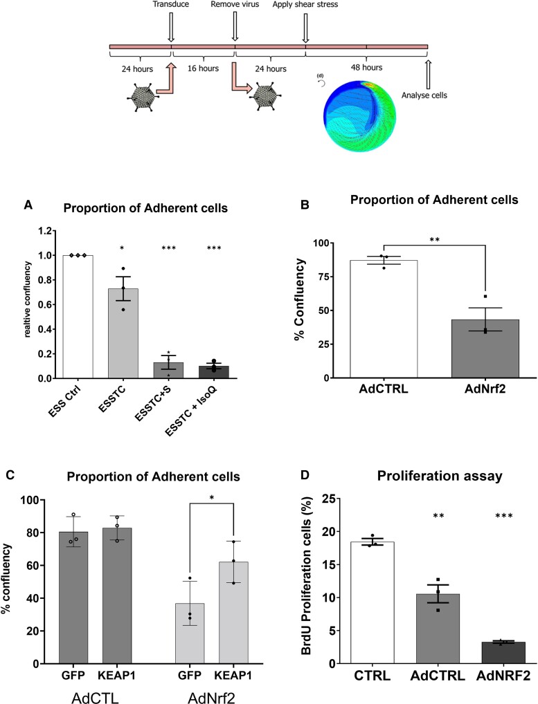
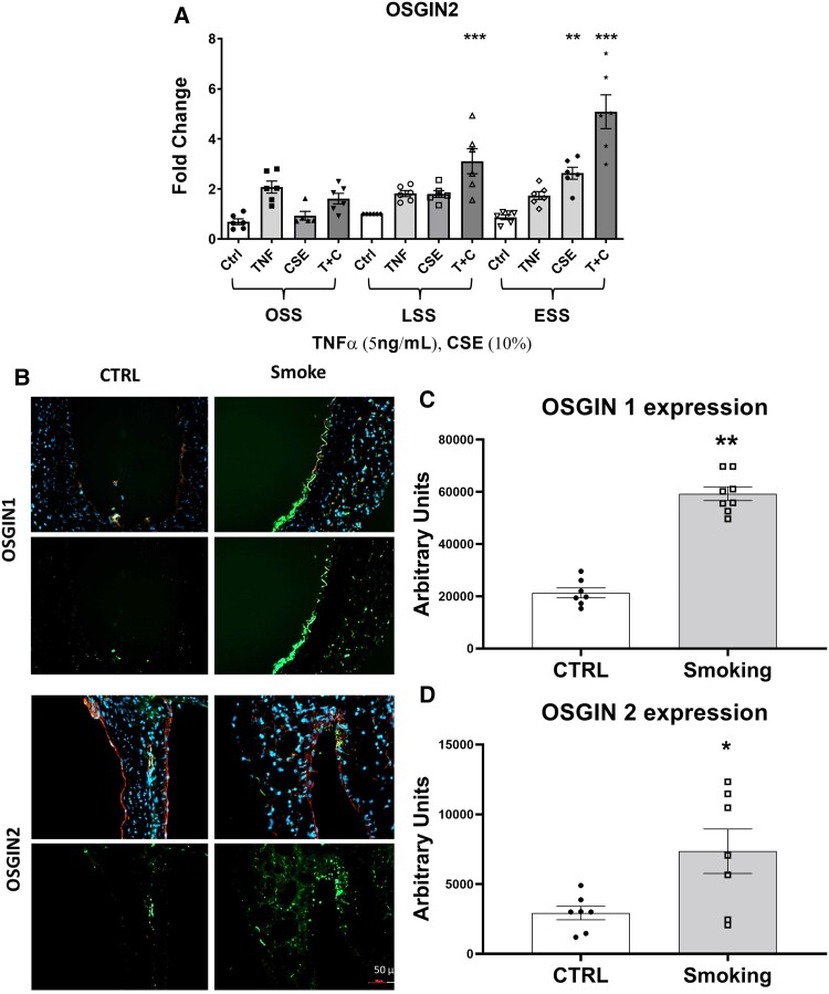
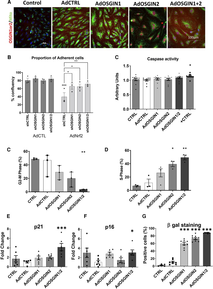
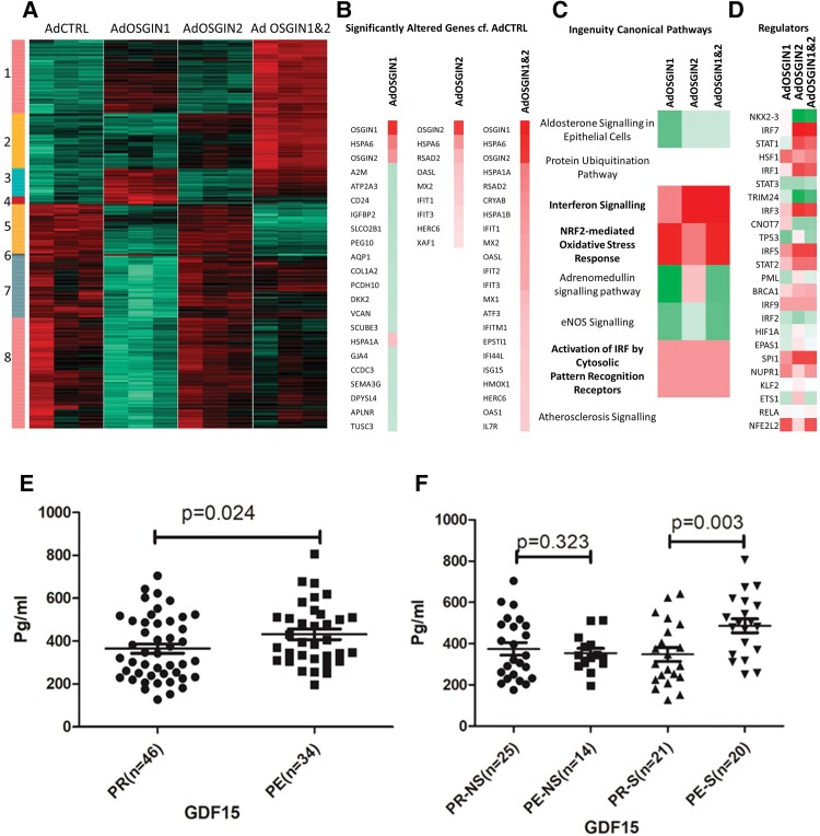
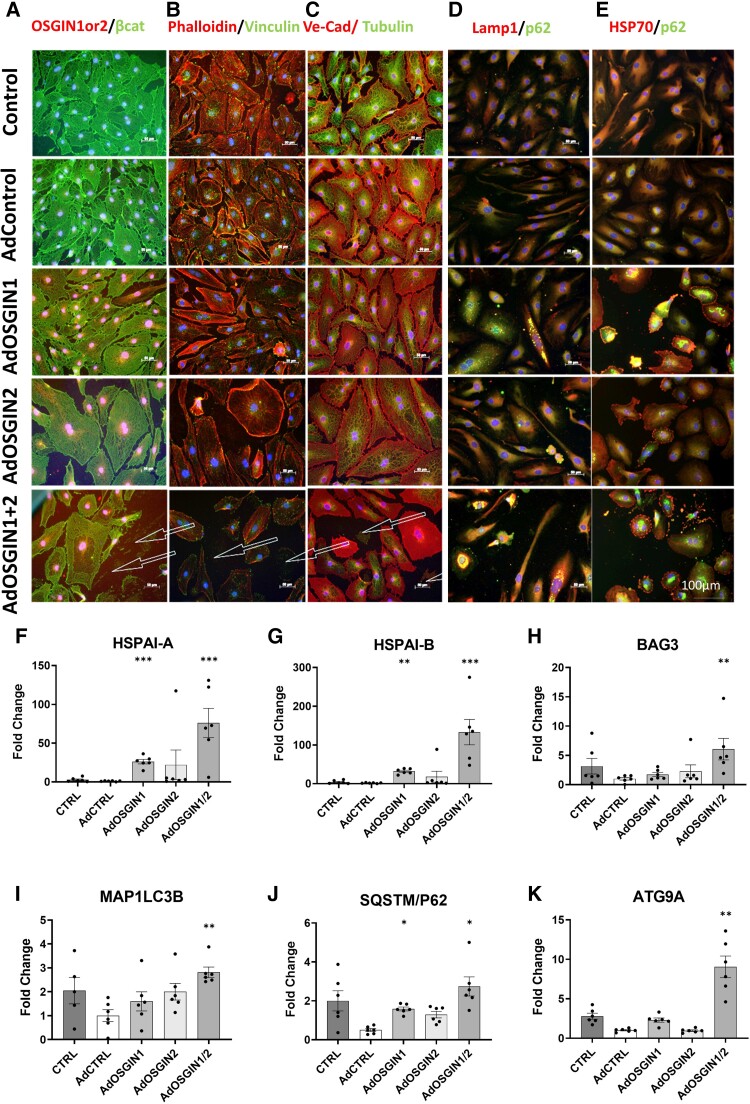
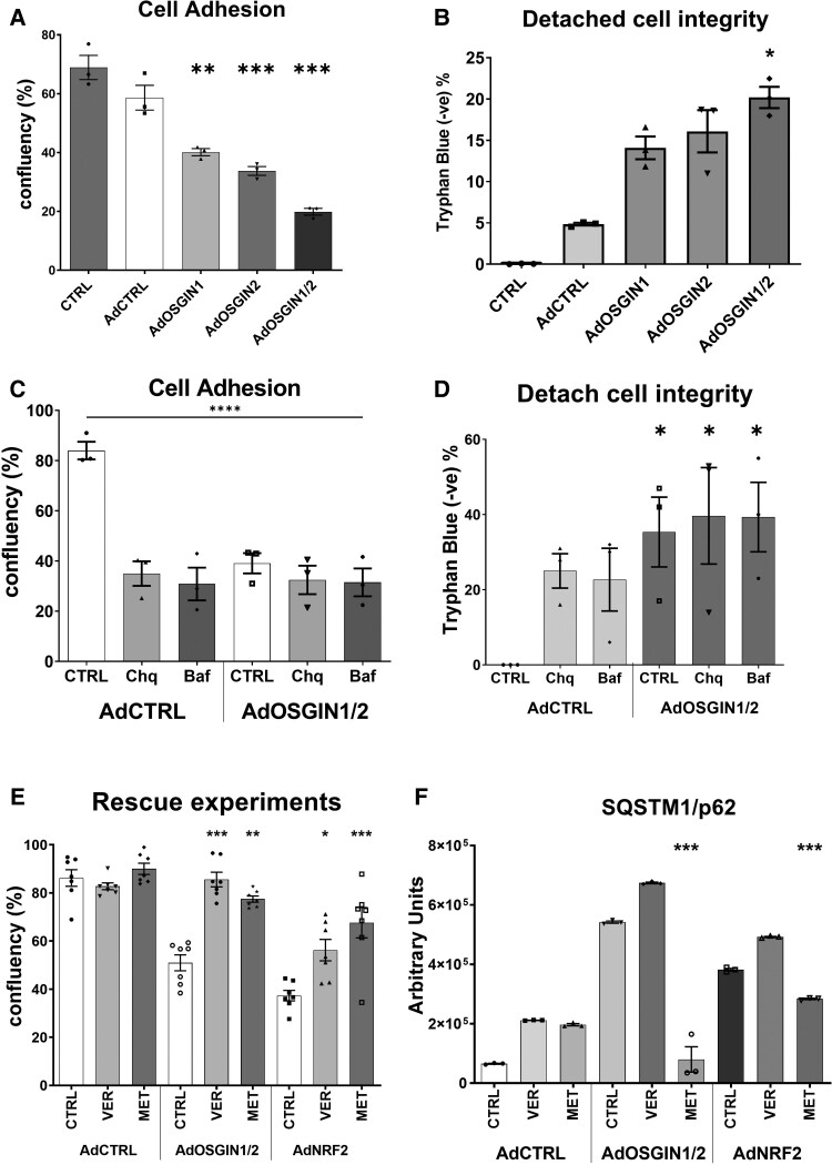
Methods and results: Culturing human coronary artery endothelial cells (HCAECs) under elevated flow (shear stress of 7.5 Pa) and chronically exposing them to cigarette smoke extract (CSE) and tumour necrosis factor-alpha (TNFα) recapitulated a defect in HCAEC adhesion, which corresponded with augmented Nrf2-regulated gene expression. Pharmacological activation or adenoviral overexpression of Nrf2 triggered endothelial detachment, identifying Nrf2 as a mediator of endothelial detachment. Growth/Differentiation Factor-15 (GDF15) expression was elevated in this model, with protein expression elevated in the plasma of patients experiencing plaque erosion compared with plaque rupture. The expression of two Nrf2-regulated genes, OSGIN1 and OSGIN2, was increased by CSE and TNFα under elevated flow and was also elevated in the aortas of mice exposed to cigarette smoke in vivo. Knockdown of OSGIN1&2 inhibited Nrf2-induced cell detachment. Overexpression of OSGIN1&2 induced endothelial detachment and resulted in cell cycle arrest, induction of senescence, loss of focal adhesions and actin stress fibres, and disturbed proteostasis mediated in part by HSP70, restoration of which reduced HCAEC detachment. In ACS patients who smoked, blood concentrations of HSP70 were elevated in plaque erosion compared with plaque rupture.

Conclusion: We identified a novel Nrf2-OSGIN1&2-HSP70 axis that regulates endothelial adhesion, elevated GDF15 and HSP70 as biomarkers for plaque erosion in patients who smoke, and two therapeutic targets that offer the potential for reducing the risk of plaque erosion.

Keywords: Autophagy; Endothelial erosion; Nrf2; adhesion.


Conflict of interest statement

Conflict of interest: None of the authors have received any financial, personal, or professional support, other than the funding disclosed above that has a bearing on the data presented here. Dr Libby is an unpaid consultant to or involved in clinical trials for Amgen, AstraZeneca, Baim Institute, Beren Therapeutics, Esperion Therapeutics, Genentech, Kancera, Kowa Pharmaceuticals, Medimmune, Merck, Norvo Nordisk, Novartis, Pfizer, and Sanofi-Regeneron. Dr Libby is a member of the scientific advisory board for Amgen, Caristo Diagnostics, Cartesian Therapeutics, CSL Behring, DalCor Pharmaceuticals, Dewpoint Therapeutics, Kancera, Kowa Pharmaceuticals, Olatec Therapeutics, Medimmune, Novartis, PlaqueTec, TenSixteen Bio, and XBiotech, Inc. Dr Libby’s laboratory has received research funding in the last 2 years from Novartis. Dr Libby is on the Board of Directors of XBiotech, Inc. Dr Libby has a financial interest in XBiotech, a company developing therapeutic human antibodies. Dr Libby has a financial interest in TenSixteen Bio, a company targeting somatic mosaicism and clonal haematopoiesis of indeterminate potential (CHIP) to discover and develop novel therapeutics to treat age-related diseases. Dr Libby’s interests were reviewed and managed by Brigham and Women’s Hospital and Mass General Brigham in accordance with their conflict-of-interest policies.

Figures

Graphical Abstract
Graphical Abstract
Elevated flow, cigarette smoke extract, and TNFα can trigger endothelial detachment, replicating features of plaque erosion in patients. Nrf2 activation or overexpression of OSGIN1&2 induces detachment of human coronary artery endothelial cells, likely through dysregulation of chaperone-mediated autophagy, that can be rescued by inhibition of HSP70 and 5′ AMP-activated protein kinase activation.
Figure 1
Figure 1
In vitro modelling of the conditions found in endothelial erosion. (A) Photomicrographs of HCAECs, cultured under elevated flow for 72 h (7.5 Pa), with the experimental design of the in vitro model illustrated above. (B) Quantification of detachment in HCAECS cultured under elevated flow with the addition of TNFα (T-5 ng/mL) and CSE (C-10%) or both (TC), with the combined treatment resulting in a significant loss of cell adhesion (*P < 0.05, n = 6 donors). (C) Heatmap with identified clusters of gene expression changes identified in the transcriptomic analysis of the combined effects of oscillatory (OSS), laminar (LSS), and elevated (ESS) shear stress on HCAECs treated with control, 3 doses of 5 ng/mL TNFα (T), 3 doses of 10% CSE (C), or the combination of TNFα and CSE (TC), n = 3 donors. (D) Regulation of Ingenuity-defined Nrf2-mediated oxidative stress pathway compared with ESS control. (E) Top predicted canonical pathways by Ingenuity analysis, red indicates a predicted activation and green a decrease, with white suggesting dysregulation.
Figure 2
Figure 2
Evidence for a role for Nrf2 in endothelial detachment. (A) Quantification of cell number in elevated laminar shear stress control (ESS), with the addition of TNFα and CSE [mean ± SD, one-way ANOVA, elevated flow + TNFα + CSE (ESSTC), 30% reduction vs. ESS, P < 0.05, n = 3], and Nrf2 activator sulforaphane (ESSTC-S, 2.5 μM, 5-fold reduced adhesion vs. ESSTC, P < 0.05, n = 3) or isoliquiritigenin (ESS-IsoQ, 10 μM, 9-fold reduced adhesion vs. ESSTC, P < 0.05, n = 3). (B) Adenoviral overexpression of Nrf2 (200 pfu/cell and 200 pfu/cell AdCTRL combined to match later experiments) promotes 50% of cell detachment compared with AdCTRL (400 pfu/cell) (****P < 0.0001, n = 3) using the experimental design illustrated above. (C) Transduction with lentiviral control (GFP) or lentiKEAP1 prior to adenoviral overexpression of Nrf2 (as C) resulted in a significant reduction of Nrf2-dependent detachment (*P < 0.05, n = 3). (D) BrdU proliferation assay in HCAECs treated with adenoviral overexpression of wild-type Nrf2, % BrdU positive cells, mean and SEM obtained from n = 6, *P < 0.05 and **P < 0.01, compared with control.
Figure 3
Figure 3
Regulation of OSGIN1&2 by cigarette smoke. (A) OSGIN2 mRNA expression in HCAECs cultured under OSS, LSS, or ESS, with TNFα (5 ng/mL) or CSE (10%) or both (*P = 0.05, **P < 0.01, ***P < 0.001 vs. LSS CTRL, mean, and SEM, n = 6, two-way ANOVA). (B) Immunohistochemical staining on 8 µm sections of aortas from male mice exposed to cigarette smoke for 3 months with quantification of staining; green: OSGIN1 or OSGIN2; red: CD31; and blue: DAPI nuclear stain. (C and D) Quantification of immunofluorescence staining (**P < 0.01, n = 8, OSGIN1 vs. 7 CTRL; *P < 0.05, n = 7, OSGIN2 vs. 7 CTRL, mean, and SEM).
Figure 4
Figure 4
OSGIN1&2 overexpression effects endothelial proliferation and senescence. (A) Immunocytochemical staining for OSGIN1 and OSGIN2 (in red) shows their nuclear localization. Mitochondrial staining (in green) confirmed no localization of OSGIN1 and OSGIN2 (scale bar 100 µm). (B) Quantification of adhesion upon transduction with lentiviral shCtrl or lentiviral vectors overexpressing short hairpin targeted against OSGIN1 (shOSGIN1), OSGIN2 (shOSGIN2), or both vectors (shOSGIN1/2) with subsequent transduction with AdCtrl or AdNrf2 as described in Figure 2C). (C) Caspase 3/7 activity of HCAECs transfected with adenovirus overexpressing OSGIN1, OSGIN 2, and both together, compared with AdCtrl transfections and positive control (0.2 mM H2O2). The caspase 3/7 activity was measured using Caspase-Glo 3/7 assay. Data are presented as mean ± SEM (n = 6). **P < 0.01. (D and E) Flow cytometry analysis after adenoviral-mediated overexpression of OSGIN1 and OSGIN2 showed inhibition of cell cycle in HCAECs, with cells accumulating in S-phase, without proceeding to division (mean ± SD, one-way ANOVA, *P < 0.05, and ***P < 0.001, n = 3). (F and G) Changes in mRNA expression of p-21Waf-1 and p-16INK4a with OSGIN1, and/or 2 overexpression (***P < 0.001; *P < 0.05 vs. AdCTRL, mean ± SEM, n = 6). (H) Increased staining for senescence-associated β-galactosidase and up-regulation of p-21Waf-1 and p-16INK4a demonstrates the induction of the senescence pathway and lysosomal accumulation by OSGIN1&2 (***P < 0.001, median and inter-quartiles, n = 3; Supplementary material online, Figure S16).
Figure 5
Figure 5
Changes in gene expression in HCAECs overexpressing OSGIN1, 2, or 1&2 and analysis of GDF15 expression in ACS patients. (A) Heatmap of gene expression significantly changed between AdCTRL or AdOSGIN1, 2, or 1&2 (n = 3); green indicated decreased, black no change, and red increased gene expression. Clustering analysis revealed eight clusters. (B) The genes with the most significant/largest fold change by AdOSGIN1, 2, or 1&2 compared with AdCTRL. Red indicates an increase in expression and green a decrease. (C) Predicted upstream transcriptional regulators; red indicates a predicted activation, white no change, and green a decrease. (D) Top predicted canonical pathways; red indicates a predicted activation and green a decrease, with white suggesting dysregulation. (E) Quantification of circulating GDF15 levels in serum from patients with OCT-defined plaque rupture (PR) and OCT-defined plaque erosion (PE). (F) Subgroup analysis of circulating GDF15 levels in smokers (S) and non-smokers (NS), with OCT-defined PR and OCT-defined PE.
Figure 6
Figure 6
OSGIN1&2 effects on cell structure and autophagy-related gene expression. (A–C) Immunocytochemical analysis of HCAECs with adenoviral-mediated overexpression of OSGIN 1 + 2 (scale bar 100 µm). β-Catenin (a marker of intercellular junctional stability), vinculin (focal adhesions) and phalloidin (Actin), Tubulin and VE-Cadherin (intercellular junctions in red), visualized by immunofluorescence microscopy. OSGIN1 + 2 overexpression profoundly affects cell structure, and reduces cytoskeletal integrity and focal adhesions with cells detaching even in static culture denoted by arrows. (D and E) Immunofluorescence of SQSTM/p62 (in green) and LAMP1 or HSP70 (in red) demonstrates an accumulation of SQSTM/p62 and LAMP1 positive vesicles, indicative of a block in autophagic flux. Detachment was observed under static conditions (arrows). (F–K) Changes in mRNA expression of key regulators of the chaperone-mediated autophagy pathway by OSGIN1 + 2 overexpression: (F) HSPA1A; (G) HSPA1B; (H) BAG3; (I) MAP1LC3B; (J) SQSTM1/p62; (K) ATG9A (mean ± SD, one-way ANOVA, **P < 0.01 and ***P < 0.001; n = 6).
Figure 7
Figure 7
Quantification of endothelial cell detachment using orbital shaker model. (A and B) Adenoviral overexpression of OSGIN1, 2, and 1&2 triggers cell detachment (mean ± SD, one-way ANOVA, **P < 0.01, ***P < 0.001 vs. AdCTRL; n = 3), with detached cells displaying a significant maintenance of cell membrane integrity (*P < 0.05 vs. AdCTRL; n = 4). (C and D) Chloroquine (150 µM), bafilomycin (50 nM), or OSGIN1&2 overexpression induced comparable, non-synergistic detachment (mean ± SD, two-way ANOVA, **P < 0.01, ***P < 0.001 vs. AdCTRL, n = 4), with a similar maintenance of cell membrane integrity (*P < 0.05 vs. AdCTRL, n = 4). (E) Co-treatment with Ver155008 (15 µM) or Metformin (100 µM) reduced OSGIN1&2 or Nrf2-mediated cell detachment (mean ± SEM, two-way ANOVA, *P < 0.05, **P < 0.01, ***P < 0.001, n = 7). (F) Metformin, but not VER-155008 treatment reversed SQSTM1/p62 protein accumulation following AdOSGIN1 + 2 and AdNRF2 overexpression (mean ± SEM, two-way ANOVA, ***P < 0.001, n = 3).

Comment in

References

    1. Virmani R, Burke AP, Farb A, Kolodgie FD. Pathology of the vulnerable plaque. J Am Coll Cardiol 2006;47:C13–C18. - PubMed
    1. White SJ, Newby AC, Johnson TW. Endothelial erosion of plaques as a substrate for coronary thrombosis. Thromb Haemost 2016;115:509–519. - PubMed
    1. Libby P, Pasterkamp G, Crea F, Jang I-K. Reassessing the mechanisms of acute coronary syndromes. Circ Res 2019;124:150–160. - PMC - PubMed
    1. Dai J, Xing L, Jia H, Zhu Y, Zhang S, Hu S, Lin L, Ma L, Liu H, Xu M, Ren X, Yu H, Li L, Zou Y, Zhang S, Mintz GS, Hou J, Yu B. In vivo predictors of plaque erosion in patients with ST-segment elevation myocardial infarction: a clinical, angiographical, and intravascular optical coherence tomography study. Eur Heart J 2018;39:2077–2085. - PubMed
    1. Yamamoto E, Yonetsu T, Kakuta T, Soeda T, Saito Y, Yan BP, Kurihara O, Takano M, Niccoli G, Higuma T, Kimura S, Minami Y, Ako J, Adriaenssens T, Boeder NF, Nef HM, Fracassi F, Sugiyama T, Lee H, Crea F, Kimura T, Fujimoto JG, Fuster V, Jang IK. Clinical and laboratory predictors for plaque erosion in patients with acute coronary syndromes. J Am Heart Assoc 2019;8:e012322. - PMC - PubMed

Publication types

Substances