. 2023 Feb 20;13(1):63.
doi: 10.1038/s41398-023-02357-x.

Chemogenetic rectification of the inhibitory tone onto hippocampal neurons reverts autistic-like traits and normalizes local expression of estrogen receptors in the Ambra1+/- mouse model of female autism

Affiliations

Chemogenetic rectification of the inhibitory tone onto hippocampal neurons reverts autistic-like traits and normalizes local expression of estrogen receptors in the Ambra1+/- mouse model of female autism

Annabella Pignataro et al. Transl Psychiatry. .

Abstract

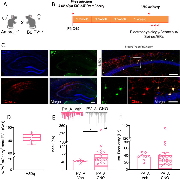
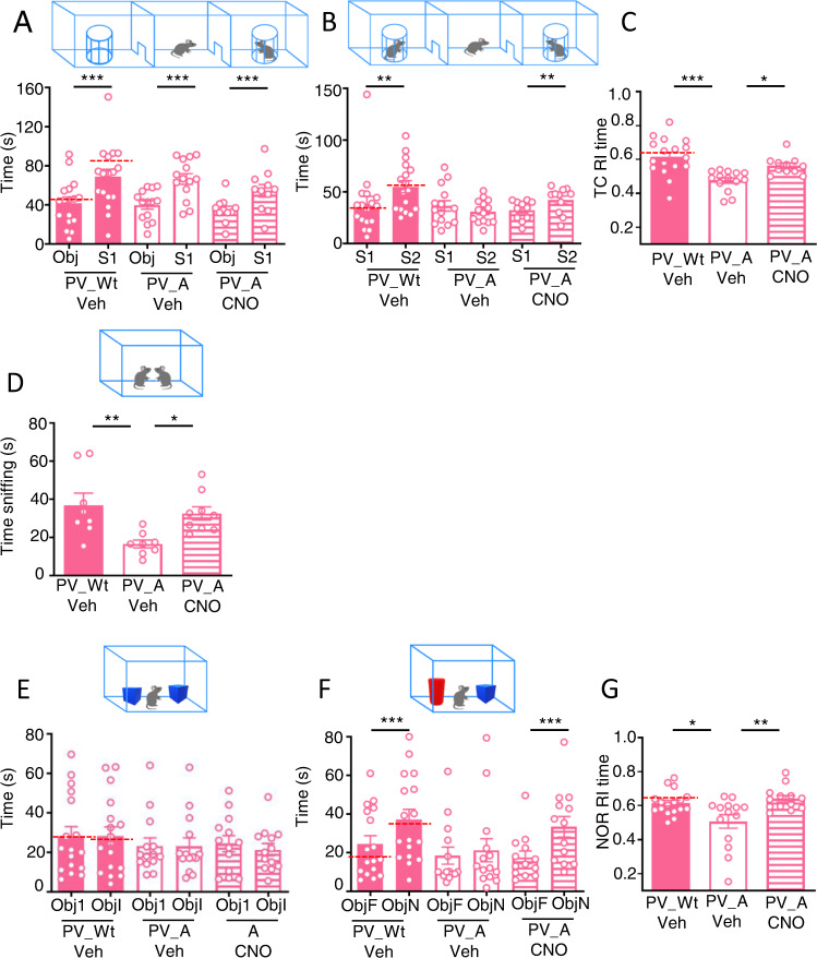
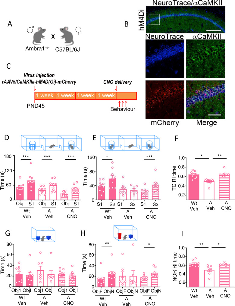
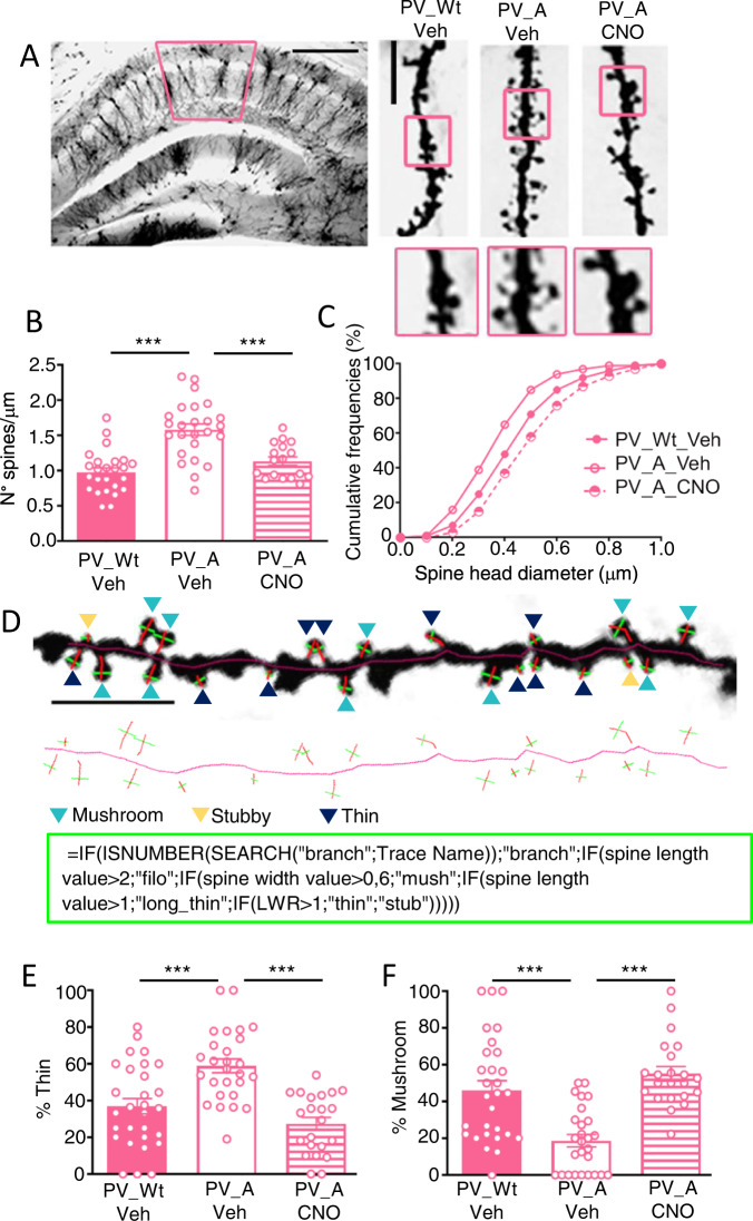
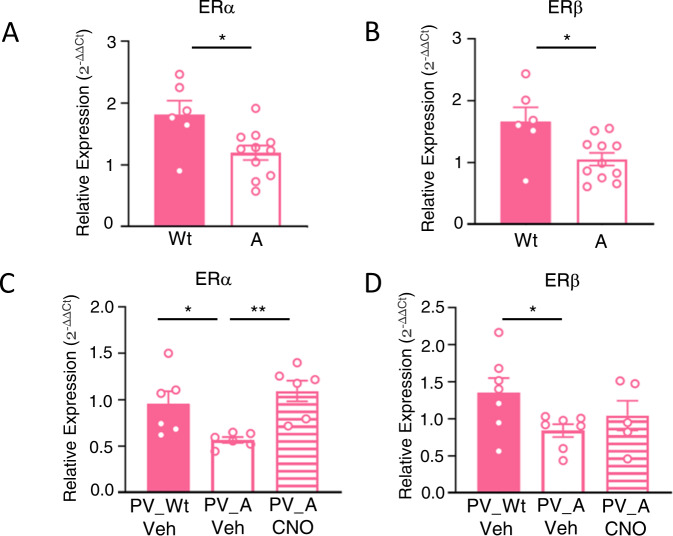
Female, but not male, mice with haploinsufficiency for the proautophagic Ambra1 gene show an autistic-like phenotype associated with hippocampal circuits dysfunctions which include loss of parvalbuminergic interneurons (PV-IN), decrease in the inhibition/excitation ratio, and abundance of immature dendritic spines on CA1 pyramidal neurons. Given the paucity of data relating to female autism, we exploit the Ambra1+/- female model to investigate whether rectifying the inhibitory input onto hippocampal principal neurons (PN) rescues their ASD-like phenotype at both the systems and circuits level. Moreover, being the autistic phenotype exclusively observed in the female mice, we control the effect of the mutation and treatment on hippocampal expression of estrogen receptors (ER). Here we show that excitatory DREADDs injected in PV_Cre Ambra1+/- females augment the inhibitory input onto CA1 principal neurons (PN), rescue their social and attentional impairments, and normalize dendritic spine abnormalities and ER expression in the hippocampus. By providing the first evidence that hippocampal excitability jointly controls autistic-like traits and ER in a model of female autism, our findings identify an autophagy deficiency-related mechanism of hippocampal neural and hormonal dysregulation which opens novel perspectives for treatments specifically designed for autistic females.


Conflict of interest statement

The authors declare no competing interests.

Figures

Fig. 1
Fig. 1. Chemogenetic activation of PV-IN enhances the GABAergic tone onto CA1 pyramidal neurons.
A Breeding. B Experimental design: AAV-hSyn-DIO-hM3Dq-mCherry (hM3Dq) was bilaterally injected in CA1. Tests were performed 4 weeks later. Vehicle (Veh) or CNO were injected i.p. 40 min before behavioral testing and electrophysiological recordings, and 70 min before rtPCR experiments, or diluted at the same concentration in the drinking water 24 h before mice were sacrificed for dendritic spine analysis. C Left panels: low magnification image depicting the selective stereotaxic infusion of AAV-hSyn-DIO-hM3Dq-mCherry in the CA1. Scale bar: 200 μm. Right panels: representative images of CA1 NeuroTrace/PV/mCherry labelling showing overlapping signals (yellow) of mCherry (red) and PV + interneurons (green). Scale bars: top, 250 μm; below, 50 μm. D Percentage estimation of PV-IN infected with the hM3Dq AAV (PV+mCherry+/total PV+). In box-and-whisker plots the centre lines denote median values, edges are upper and lower quartiles, whiskers show minimum and maximum values and points are individual experiments (N = 7 for hM3Dq). EF sIPSCs in CA1 neurons from hM3Dq PV_Ambra1+/− females injected with Veh (left) or CNO (right). Scale bar: 1 s, 20 pA. CNO increased the inhibitory tone onto CA1 neurons. E Peak amplitude (p = 0.013, Mann-Whitney test). F Instantaneous frequency. N = 8, 17 neurons, from 3 Veh- and 5 from CNO females. Data are expressed as mean ± s.e.m. *p < 0.05.
Fig. 2
Fig. 2. Chemogenetic activation of inhibitory PV-IN in Ambra1+/− females normalizes autistic-like behaviors.
A Histogram report mean time spent exploring the object (Obj) and the conspecific stranger (S1) during the sociability phase of the TC test. All mice explore more the conspecific stranger than the object (PV_Wt/Veh: Obj vs S1: t(16)=7.116, p = 0.0001; PV_A/Veh: Obj vs S1: t(13)=7.714, p = 0.0001; PV_A/CNO: Obj vs S1: t(10)=5.507, p = 0.0003; Dotted-line indicate mean values detected in PV_Wt/CNO control mice infused with vehicle instead of DREADDs. B Histogram report mean-time spent exploring the familiar female (S1) and the novel female (S2) during the social novelty phase of the TC test. Only PV_A/Veh females show impairment in recognition of the social novelty as they spent equal amounts of time sniffing S1 and S2. (PV_Wt/Veh: S1 vs S2: W = 119, p = 0.0032; PV_A/Veh: S1 vs S2: t(13)=1.740, p = 0.1; PV_A/CNO: S1 vs S2: W = 55, p = 0.002). Dotted-line indicate mean values detected in PV_Wt/CNO control mice infused with vehicle instead of DREADDs. C Histogram reporting the mean recognition index (RI) calculated during the social novelty phase of TC. Social novelty impairments in A/Veh females are rescued in A/CNO females. TC social novelty phase: PV_Wt/Veh (N = 17), PV_A/Veh, (N = 14), PV_A/CNO (N = 11), F(2,39)=11.26 p = 0.0001; PV_Wt/Veh vs PV_A/Veh, p = 0.00086; PV_A/Veh vs PV_A/CNO, p = 0.037. Dotted-line indicate mean values detected in PV_Wt/CNO control mice infused with vehicle instead of DREADDs. D Histogram reports mean time spent exploring an unknown conspecific during the social interaction in pair test (SIP). Social interaction impairments of PV_A/Veh females in SIP test are rescued in PV_A/CNO females (SIP: PV_Wt/Veh (N = 8), PV_A/Veh (N = 8), PV_A/CNO (N = 9), F(2,22)=6.186, p = 0.007; PV_Wt/Veh vs PV_A/Veh, p = 0.009; PV_A/Veh vs PV_A/CNO, p = 0.04). E Histogram report mean time spent exploring the two identical objects (Object 1 (Obj1) and Identical Object (Obj I)) during the object exploration phase of the NOR test by PV_Wt/Veh females and PV_A females injected with Veh or CNO. Data indicate that regardless of genotype and treatment all mice similarly explore the identical objects (PV_Wt/Veh: Obj1 vs ObjI: W = 7, p = 0.88; PV_A/Veh: Obj1 vs ObjI: W = -38, p = 0.24; PV_A/CNO: Obj1 vs ObjI: t(12)=1.102, p = 0.29). Dotted-line indicate mean values detected in PV_Wt/CNO control mice infused with vehicle instead of DREADDs. F Histogram report mean time spent exploring the familiar object (ObjF) and the novel object (Obj N) during the test phase of the NOR by PV_Wt/Veh females and PV_A females injected with Veh or CNO. Data indicate that recognition impairments of the novel object in A/Veh females are rescued in A/CNO females. (ObjF vs ObjN: PV_Wt/Veh: t(16)=7.06, p = 0.0001; PV_A/Veh: W = -29, p = 0.38; PV_A/CNO: W = -91, p = 0.0002). Dotted-line indicate mean values detected in PV_Wt/CNO control mice infused with vehicle instead of DREADDs. G Histogram reporting the mean recognition index (RI) calculated during the test phase of NOR test. Novel object recognition impairments in PV_A/Veh females are rescued in PV_A/CNO females. (PV_Wt/Veh (N = 17), PV_A/Veh (N = 14), PV_A/CNO (N = 13), F(2,41)=6.59, p = 0.003; PV_Wt/Veh vs PV_A/Veh, p = 0.023; PV_A/Veh vs PV_CNO, p = 0.004). Dotted-line indicate mean values detected in PV_Wt/CNO control mice infused with vehicle instead of DREADDs. Data are expressed as mean ± s.e.m. *p < 0.05, **p < 0.01, ***p < 0.001.
Fig. 3
Fig. 3. Chemogenetic inhibition of CA1 principal neuron activity in Ambra1+/− females rescues autistic-like behaviors.
A Breeding. B Representative images of NeuroTrace/CamKII/mCherry labelling showing overlapping signals (yellow) of mCherry (red) and CamKII- neurons (green). hM4Di receptors are selectively expressed in CA1 principal neurons. Scale bars: top, 200 μm; below, 50 μm. C Experimental design: the inhibitory rAAV5/CaMKIIa-hM4D(Gi)-mCherry vector was bilaterally injected in CA1 of Wt and Ambra1+/− females. Behavioral tests were performed 4 weeks later. Vehicle (Veh) or CNO were injected i.p. 40 min before testing. D Histogram reporting mean time spent exploring the object (Obj) and the conspecific stranger (S1) during the sociability phase of the TC test. All mice explore more the conspecific stranger than the object (Wt/Veh: Obj vs S1: t(8)=5.43, p = 0.0006; A/Veh: Obj vs S1: t(7)=7.389, p = 0.0002; A/CNO: Obj vs S1: t(7)=5.531, p = 0.0009). E Histogram reports mean time spent exploring the familiar female (S1) and the novel female (S2) during the social novelty phase of the TC test. Only A/Veh females show impairment in recognition of the social novelty as they spent equal amount of time sniffing S1 and S2. (Wt/Veh: S1 vs S2: t(8)=2.517, p = 0.036; A/Veh: S1 vs S2: t(7)=0.562, p = 0.59; A/CNO: S1 vs S2: t(7)=5.251, p = 0.001). F Histogram reporting the mean recognition index (RI) calculated during the social novelty phase of TC. Social novelty impairments in A/Veh females are rescued in A/CNO females. (TC social novelty phase: Wt/Veh (N = 9), A/Veh (N = 8) and A/CNO (N = 8); Kruskal-Wallis H = 12.01, p = 0.002; Dunn’s multiple comparison Wt/Veh vs A/Veh, p = 0.029; A/Veh vs A/CNO, p = 0.005). G Histogram reports mean time spent exploring the two identical objects during the object exploration phase of the NOR test by Wt and A females injected with Veh or CNO. Data indicate that regardless of genotype and treatment all mice similarly explore the identical objects (Obj1 vs ObjI: Wt/Veh: W = -9, p = 0.63; A/Veh: W = -17, p = 0.17; A/CNO: t(6)=0.98, p = 0.36). H Histogram reports mean time spent exploring the familiar object (ObjF) and the novel object (ObjN) during the test phase of the NOR by Wt/Veh females and A females injected with Veh or CNO. Data indicate that recognition impairments of the novel object in A/Veh females are rescued in A/CNO females (ObjF vs ObjN: Wt/Veh: W = 45, p = 0.0039; A/Veh: W = 12, p = 0.37; A/CNO: W = -26, p = 0.031). I Histogram reporting the mean recognition index (RI) calculated during the test phase of NOR test. Novel object recognition impairments in A/Veh females are rescued in A/CNO females (Wt/Veh (N = 9), A/Veh (N = 7), and A/CNO (N = 7); F(2,20)=7.473, p = 0.0038; Wt/Veh vs A/Veh p = 0.004 A/Veh vs A/CNO, p = 0.021). Data are expressed as mean ± s.e.m. *p < 0.05, **p < 0.01, ***p < 0.001.
Fig. 4
Fig. 4. Chemogenetic activation of inhibitory PV-IN in Ambra1+/− females normalizes autistic-like dendritic spines.
A Representative Golgi-stained dorsal hippocampal section. Left panel: 5x magnification, scale bar: 250 μm. Right panels: 100x magnification, scale bar: 5 μm. B Histogram reporting the mean spine density (mean ± s.e.m.): number of spines is similar in PV_Wt/Veh and PV_A/CNO females [PV_Wt/Veh (N = 6, 25 neurons), PV_A/Veh (N = 5, 25 neurons), PV_A/CNO (N = 4, 17 neurons), F(2,64)=21.63, p = 0.0001; PV_A/Veh vs PV_A/CNO, p = 0.0001; PV_Wt/Veh vs PV_A/CNO, p = 0.55]. C Spine head diameters length are expressed as cumulative frequencies (~1000 spines per group). Spine head diameters in PV_A/CNO females exceed those in PV_Wt/Veh females. (Kolmogorov Smirnoff (KS) test: PV_Wt/Veh vs PV_A/Veh, KS: D = 0.243, p < 0.001; PV_A/Veh vs PV_A/CNO, D = 0.431 p < 0.001; PV_Wt/Veh vs PV_A/CNO, D = 0.259, p < 0.001). D Upper panel: enlarged 100x magnification (scale bar: 5 μm) of a representative dendritic segment where dendritic spines types of CA1 pyramidal neurons are pointed out (mushroom spines by light blue arrows, stubby by yellow arrows and thin by dark blue arrows). Immediately below, the skeleton of the same dendritic segment with the visualization of the neck length and head diameter of each spine. Bottom square: the critical formula used for objective classification of spine-type (see methods for details). E, F Histograms reporting the percentage of thin (E) and mushroom (F) spines: percentage of thin (E) and mushroom (F) spines is similar in PV_Wt/Veh and PV_A/CNO females [PV_Wt/Veh (N = 4, neurons: 8; segments: 29), PV_A/Veh (N = 4, neurons: 8; segments: 27), PV_A/CNO (N = 4, neurons: 8; segments: 22). Thin spines: F(2,75)=16.68, p = 0.00002; PV_Wt/Veh vs PV_A/Veh, p = 0.0002; PV_A/Veh vs PV_A/CNO, p = 0.0001 = ; PV_Wt/Veh vs PV_A/CNO, p = 1; Mushroom spines: main effect Kruskal-Wallis H = 28, p = 0.0001; Dunn’s multiple comparison PV_Wt/Veh vs PV_A/Veh, p = 0.001; PV_A/Veh vs PV_A/CNO, p = 0.001]. Data are expressed as mean ± s.e.m. *p < 0.05, **p < 0.01, ***p < 0.001.
Fig. 5
Fig. 5. Chemogenetic activation of inhibitory PV-IN in Ambra1+/− females normalizes hippocampal mRNA ERs.
A, B Histogram reports the relative expression (2-ΔΔCt) of estrogen receptors α (ERs α) and β (ERs β) in the hippocampus of Wt and Ambra1+/− females. Baseline levels of ER α and β are significantly decreased in Ambra1+/− females compared to Wt females. (Wt (N = 6), A (N = 11), for ERα: t(15)=2.76, p = 0.0145; for ERβ: t(15)=2.746, p = 0.015). C, D Histogram reports the relative expression (2-ΔΔCt) of estrogen receptors α (ER α) and β (ER β) in the hippocampus of PV_Wt/Veh females and PV_A females injected with Veh or CNO. ER α and β levels are decreased in PV_Ambra1+/− compared to PV_Wt females. C ER α deficit is significantly rescued in PV_A/CNO females, whereas no differences in (D) ER β level are found in PV_A/CNO females, as compared to their vehicle counterpart. [For ER α: PV_Wt/Veh (N = 6), PV_A/Veh (N = 6) and PV_A/CNO (N = 6); F(2,15)=6.96, p = 0.007; PV_Wt/Veh vs PV_A/Veh p = 0.05, PV_A/Veh vs PV_A/CNO p = 0.007. For ER β: PV_Wt/Veh (N = 7), PV_A/Veh (N = 7) and PV_A/CNO (N = 5); F(2,16)=2.761, p = 0.09; PV_Wt/Veh vs PV_A/Veh: t(12)=2.410, p = 0.032; PV_A/Veh vs PV_A/CNO: t(10)=1.04, p = 0.32)]. *p < 0.05, **p < 0.01.

Similar articles

Cited by

References

    1. Hodges H, Fealko C, Soares N. Autism spectrum disorder: definition, epidemiology, causes, and clinical evaluation. Transl Pediatr. 2020;9:S55–S65. - PMC - PubMed
    1. De Rubeis S, He X, Goldberg AP, Poultney CS, Samocha K, Cicek AE, et al. Synaptic, transcriptional and chromatin genes disrupted in autism. Nature. 2014;515:209–15. - PMC - PubMed
    1. Blatt GJ. The neuropathology of autism. Sci (Cairo) 2012;2012:703675. - PMC - PubMed
    1. Ajram LA, Pereira AC, Durieux AMS, Velthius HE, Petrinovic MM, McAlonan GM. The contribution of [1H] magnetic resonance spectroscopy to the study of excitation-inhibition in autism. Prog Neuropsychopharmacol Biol Psychiatry. 2019;89:236–44. - PubMed
    1. Lee E, Lee J, Kim E. Excitation/Inhibition imbalance in animal models of Autism Spectrum Disorders. Biol Psychiatry. 2017;81:838–47. - PubMed

Publication types

Substances

LinkOut - more resources