. 2023 Sep;143(9):1724-1734.e15.
doi: 10.1016/j.jid.2023.01.024. Epub 2023 Feb 17.

Prediction and Demonstration of Retinoic Acid Receptor Agonist Ch55 as an Antifibrotic Agent in the Dermis

Affiliations

Prediction and Demonstration of Retinoic Acid Receptor Agonist Ch55 as an Antifibrotic Agent in the Dermis

David M Dolivo et al. J Invest Dermatol. 2023 Sep.

Abstract

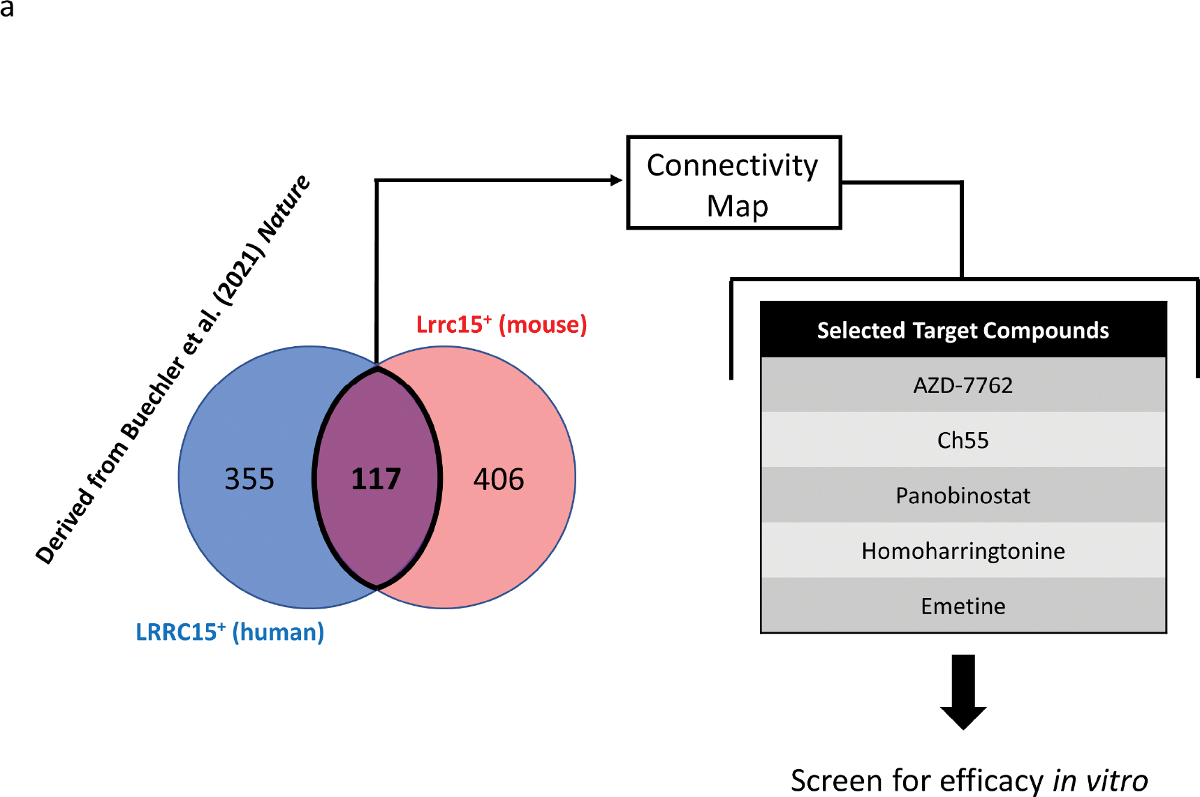
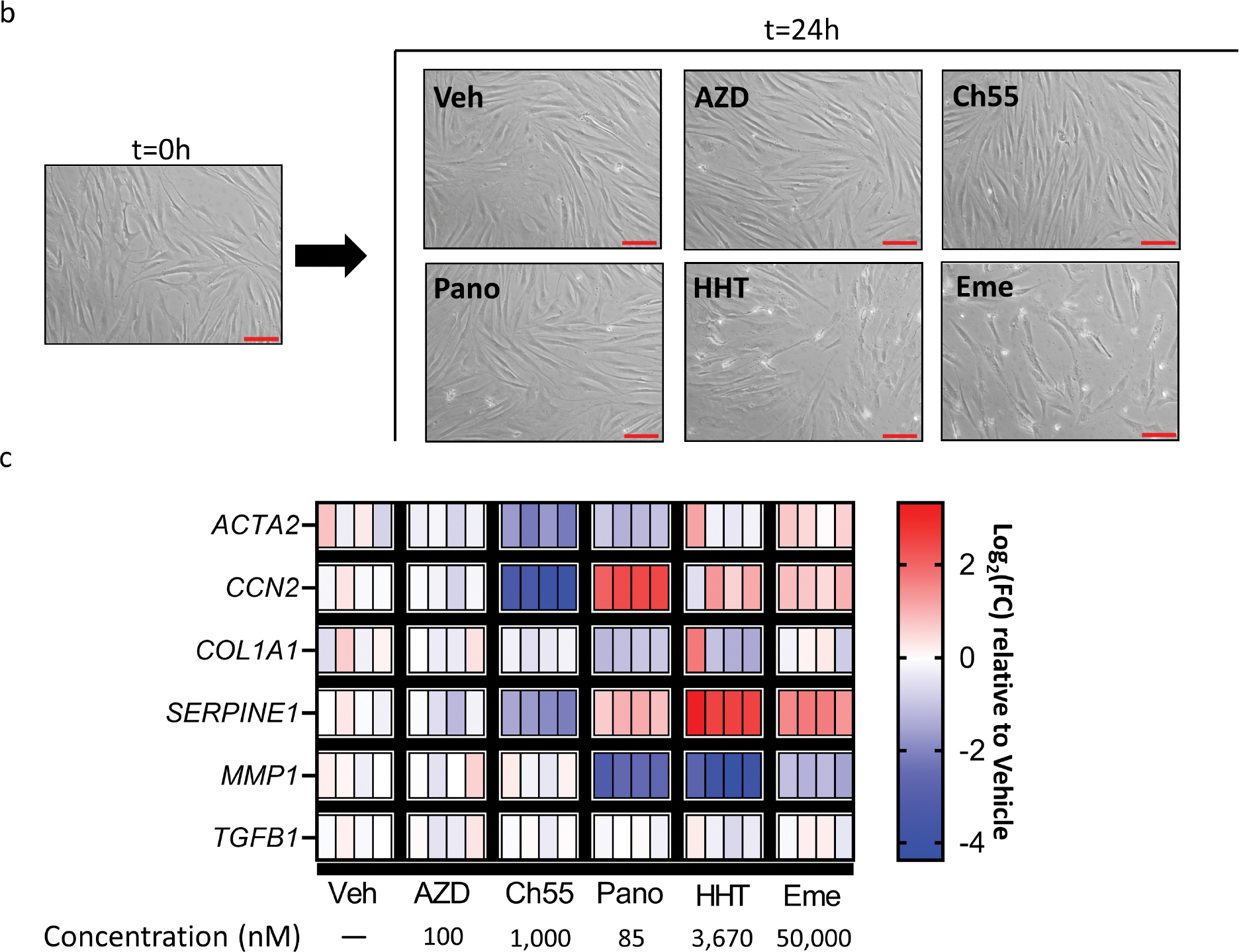
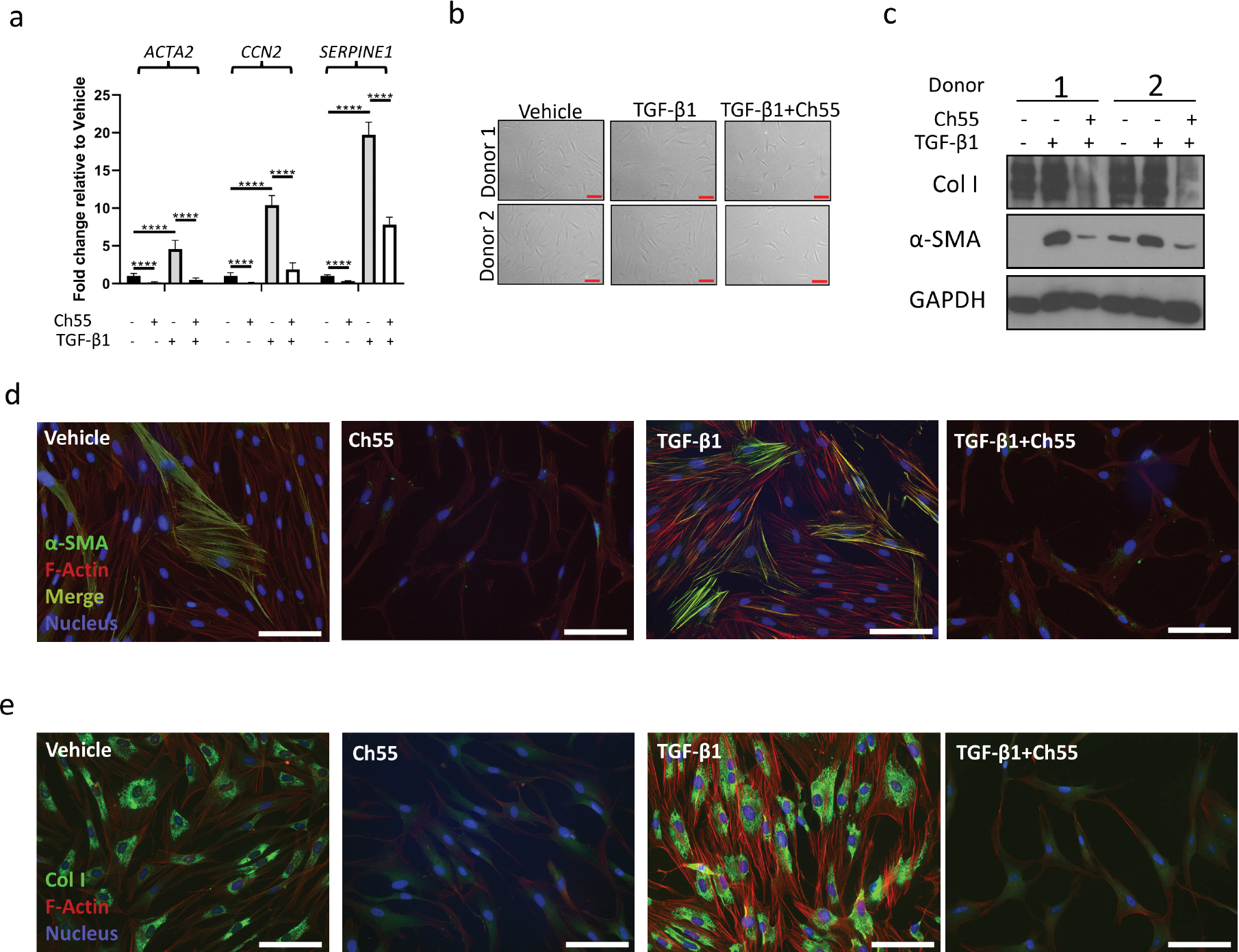
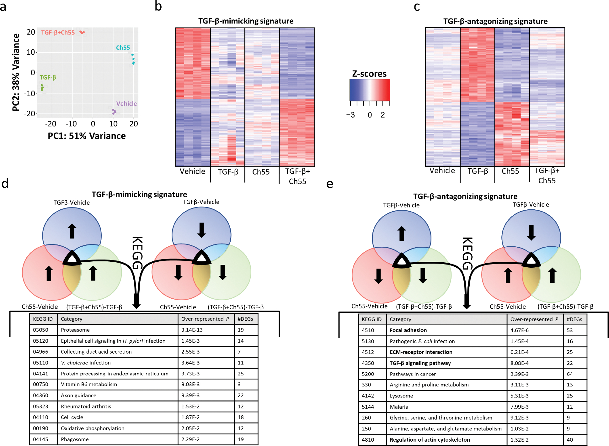
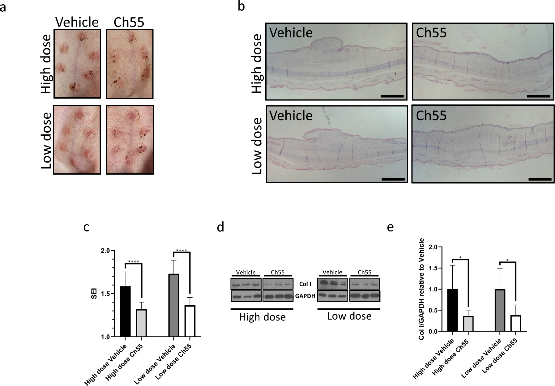
The prevalence of fibrotic diseases and the lack of pharmacologic modalities to effectively treat them impart particular importance to the discovery of novel antifibrotic therapies. The repurposing of drugs with existing mechanisms of action and/or clinical data is a promising approach for the treatment of fibrotic diseases. One paradigm that pervades all fibrotic diseases is the pathological myofibroblast, a collagen-secreting, contractile mesenchymal cell that is responsible for the deposition of fibrotic tissue. In this study, we use a gene expression paradigm characteristic of activated myofibroblasts in combination with the Connectivity Map to select compounds that are predicted to reverse the pathological gene expression signature associated with the myofibroblast and thus contain the potential for use as antifibrotic compounds. We tested a small list of these compounds in a first-pass screen, applying them to fibroblasts, and identified the retinoic acid receptor agonist Ch55 as a potential hit. Further investigation exhibited and elucidated the antifibrotic effects of Ch55 in vitro as well as showing antiscarring activity upon intradermal application in a preclinical rabbit ear hypertrophic scar model. We hope that similar predictions to uncover antiscarring compounds may yield further preclinical and ultimately clinical success.


Conflict of interest statement

CONFLICTS OF INTEREST

All authors declare no conflicts of interest.

Figures

Figure 1.
Figure 1.. Pilot screen for anti-fibrotic effects in primary fibroblasts
(A) The gene expression signature of interest was extracted by taking the intersection of human LRRC15+ fibroblast-enriched marker genes (472 genes) and mouse Lrrc15+ fibroblast-enriched marker genes (523 genes), which yielded a common signature of 117 genes. This signature was used to query the CMap in order to predict small molecules to reverse the transcriptional paradigm. Five target compounds were then manually selected for screening in fibroblasts in vitro. (B,C) Primary human foreskin fibroblasts were grown in culture and exposed to vehicle control or indicated drugs at a single concentration for 24 hours. (B) Representative brightfield microscopy before (left) and after (right) treatment. Scale bar=100μm. (C) Heatmap representing log2fold change values of each replicate relative to mean expression of vehicle control replicates for indicated genes. GAPDH was used as an internal control. Veh=vehicle. AZD=AZD-7762. Ch55=Ch55. Pano=panobinostat. HHT=homoharringtonine. Eme=emetine. n=4 replicates per condition.
Figure 2.
Figure 2.. Effects of Ch55 antagonistic to TGF-β1 stimulation in human fibroblasts
Primary human foreskin fibroblasts were grown in culture and exposed to vehicle control (Vehicle), vehicle+10ng/mL rhTGF-β1 (TGF-β1), 1,000nM Ch55 (Ch55), or 10ng/mL rhTGF-β1+1,000nM Ch55 (TGF-β1+Ch55) in vitro. (A) Transcript quantification of ACTA2, CCN2, and SERPINE1 by qRT-PCR in cells harvested after 24 hours of treatment, expressed relative to vehicle-treated cells. GAPDH was used as an internal control. n=6 replicates per group. Statistical analysis was performed by one-way ANOVA followed by Tukey’s post-hoc test, with selected statistical comparisons visualized in the figure. (B) Representative brightfield microscopy of fibroblasts from two donors immediately before harvest at 48 hours. Scale bar=100μm. (C) Western blot analysis of samples in (B), analyzing expression of α-SMA and type I collagen. GAPDH was used as an internal control. (D,E) Representative immunostaining of (D) α-SMA or (E) Collagen I in cells treated and fixed at 48 hour harvest. F-actin was counterstained with Alexafluor-568-conjugated phalloidin, and nuclei were counterstained with DAPI. Scale bar=100μm.
Figure 3.
Figure 3.. RNA-seq analysis of Ch55 effects in primary human foreskin fibroblasts
Primary human foreskin fibroblasts were grown in culture and exposed to vehicle control (Vehicle), vehicle+10ng/mL rhTGF-β1 (TGF-β1), 1,000nM Ch55 (Ch55), or 10ng/mL rhTGF-β1+1,000nM Ch55 (TGF-β1+Ch55) in vitro. RNA was harvested and expression profiling performed by RNA-seq. (A) PCA representation of variations in transcriptional profiles between and among treatment groups. (B,C) Heatmaps depicting normalized gene expression, represented as Z-scores, of Ch55-induced signatures (B) mimicking and (C) antagonizing TGF-β-induced effects. (D,E) Results of KEGG database query of signatures depicted in (B) and (C), respectively.
Figure 4.
Figure 4.. Anti-fibrotic effects of Ch55 in a rabbit ear hypertrophic scar model in vivo
Rabbit ear excisional wounds were performed and treated with either Ch55 (high or low dose, 10μg/wound or 2μg/wound, respectively) or corresponding vehicle control via intradermal injections to closed wounds over the course of scar development, before terminating experiments on POD28. (A) Representative photographs of rabbit ear scars at time of harvest. (B) Visualization of representative hypertrophic scar cross-sections by H&E staining. Scale bar=1mm. (C) Quantification of scar elevation index (SEI) for scar tissues at harvest. n=11–12 samples/condition. (D) Representative Western blot detecting expression of type I collagen in protein isolated from scar dermis at harvest for high dose and low dose Ch55 treatment groups and their respective controls. GAPDH was used as a loading control. (E) Densitometric quantification of type I collagen relative to GAPDH, as detected by Western blot. Density values for each sample are normalized to the mean of the respective vehicle controls for that dose. n=5–6 samples per group.

Comment in

Similar articles

References

    1. Abbasi S, Sinha S, Labit E, Rosin NL, Yoon G, Rahmani W, et al. Distinct regulatory programs control the latent regenerative potential of dermal fibroblasts during wound healing. Cell stem cell 2020;27(3):396–412. e6. - PubMed
    1. Abergel RP, Meeker CA, Oikarinen H, Oikarinen AI, Uitto J. Retinoid modulation of connective tissue metabolism in keloid fibroblast cultures. Arch Dermatol 1985;121(5):632–5. - PubMed
    1. Al Tanoury Z, Piskunov A, Andriamoratsiresy D, Gaouar S, Lutzing R, Ye T, et al. Genes involved in cell adhesion and signaling: a new repertoire of retinoic acid receptor target genes in mouse embryonic fibroblasts. Journal of Cell Science 2014;127(3):521–33. - PubMed
    1. Ambinder AJ, Norsworthy K, Hernandez D, Palau L, Paun B, Duffield A, et al. A Phase 1 Study of IRX195183, a RARα-Selective CYP26 Resistant Retinoid, in Patients With Relapsed or Refractory AML. Frontiers in Oncology 2020;10:587062. - PMC - PubMed
    1. Amiri N, Golin AP, Jalili RB, Ghahary A. Roles of cutaneous cell-cell communication in wound healing outcome: An emphasis on keratinocyte-fibroblast crosstalk. Experimental Dermatology 2022;31(4):475–84. - PubMed

Publication types

LinkOut - more resources