. 2023 May 3;31(5):1468-1479.
doi: 10.1016/j.ymthe.2023.02.016. Epub 2023 Feb 18.

Delivery of mitochondria confers cardioprotection through mitochondria replenishment and metabolic compliance

Affiliations

Delivery of mitochondria confers cardioprotection through mitochondria replenishment and metabolic compliance

Alian Zhang et al. Mol Ther. .

Abstract

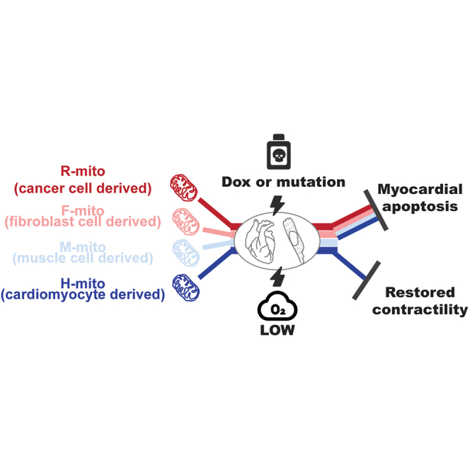
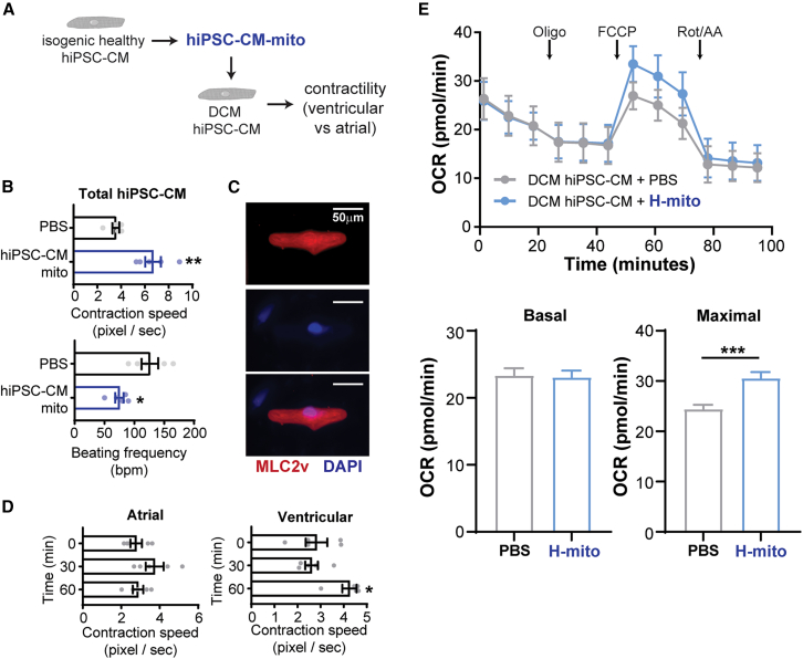
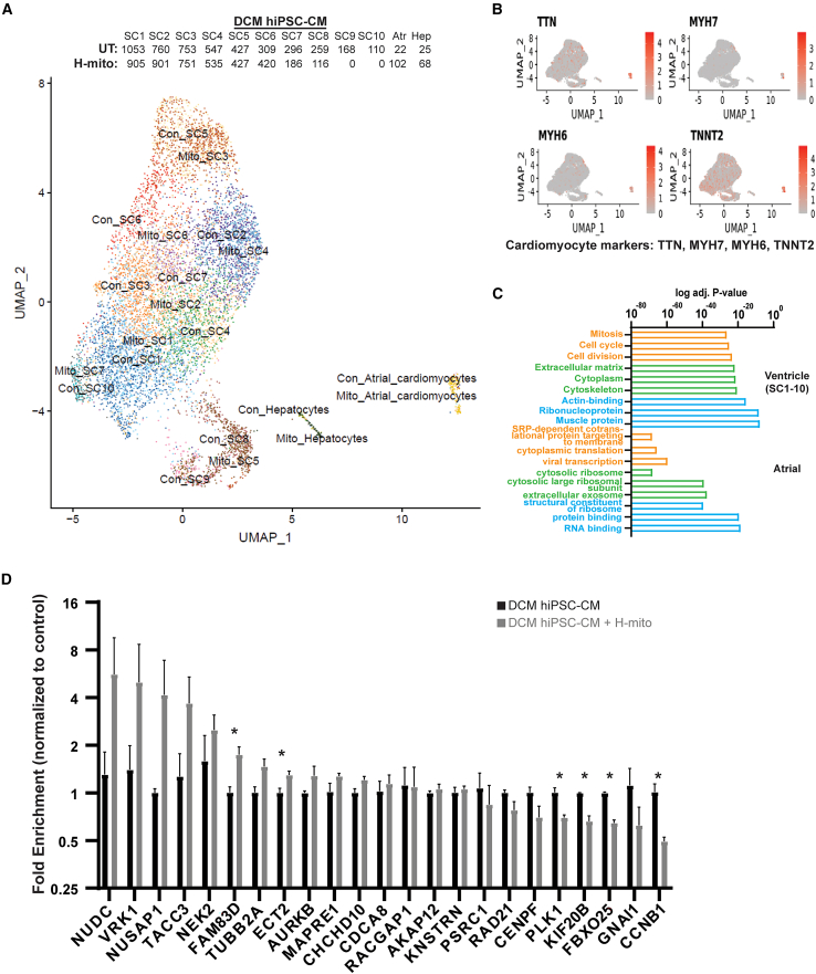
Mitochondrial dysfunction is a hallmark of heart failure. Mitochondrial transplantation has been demonstrated to be able to restore heart function, but its mechanism of action remains unresolved. Using an in-house optimized mitochondrial isolation method, we tested efficacy of mitochondria transplantation in two different heart failure models. First, using a doxorubicin-induced heart failure model, we demonstrate that mitochondrial transplantation before doxorubicin challenge protects cardiac function in vivo and prevents myocardial apoptosis, but contraction improvement relies on the metabolic compatibility between transplanted mitochondria and treated cardiomyocytes. Second, using a mutation-driven dilated cardiomyopathic human induced pluripotent stem cell-derived cardiomyocyte model, we demonstrate that mitochondrial transplantation preferentially boosts contraction in the ventricular myocytes. Last, using single-cell RNA-seq, we show that mitochondria transplantation boosts contractility in dystrophic cardiomyocytes with few transcriptomic alterations. Together, we provide evidence that mitochondria transplantation confers myocardial protection and may serve as a potential therapeutic option for heart failure.

Keywords: dilated cardiomyopathy; doxorubicin; iPSC; mitochondria delivery.


Conflict of interest statement

Declaration of interests None.

Figures

None
Graphical abstract
Figure 1
Figure 1
Isolation and quantification of mitochondria using flow cytometry (A) Immunoelectron microscopy and immunofluorescence images of isolated mitochondria. (B) Mitochondrial amount (Mitotracker green), mitochondrial membrane potential (TMRM) and mitochondrial superoxide levels (MitoSox) in cells and isolated mitochondria were evaluated using flow cytometry (n = 3 independent cultures; 30,000–100,000 events per run). Data are shown as mean ± standard error of the mean. Significance was determined by one-way ANOVA for the remaining panels. ∗p < 0.05, ∗∗p < 0.01, ∗∗∗p < 0.001, ∗∗∗∗p < 0.0001.
Figure 2
Figure 2
Mitochondrial transplantation protects cardiomyocytes from DOX-induced mitochondrial dysfunction (A) Human R-mito was injected into wild-type murine hearts and amount of transplanted R-mito mitochondria were measured using human specific mtDNA primers normalized to mouse copy gene in isolated cardiomyocytes (N = 3 animals each). Data are shown as mean ± standard error of the mean. After 4 weeks of weekly DOX injection and 2 weeks of recovery, animals were subjected to Langendorff isolation. (B–D) Purified cardiomyocytes were stained for intracellular ROS (CellROX, N = 6 animals, total N = 50 cells), mitochondrial oxidative status (MitoSox, N = 6 animals, total n = 50 cells), and mitochondrial membrane potential (TMRM, N = 6 animals, total n = 100 cells). Data are shown as violin plots. Scale bar, 20 μm. Significance was determined by one-way ANOVA for the remaining panels. ∗p < 0.05, ∗∗p < 0.01, ∗∗∗p < 0.001, ∗∗∗∗p < 0.0001.
Figure 3
Figure 3
Mitochondrial transplantation ameliorates DOX-induced myocardial dysfunction (A) Experimental setup testing if mitochondrial pre-transplantation can prevent DOX-induced cardiotoxicity. (B) Quantification of myocardial apoptosis and necrosis upon DOX challenge (N = 3–5 neonatal hearts were pooled per sample prep; technical n = 3; 30,000–100,000 events per run). (C, D) Analysis of cardiomyocyte contraction (n = 20 cells per group). (E–G) Analysis of mitochondrial respiration (N = 3–5 neonatal hearts were pooled per sample prep; technical n = 3–8). Data are shown as mean ± standard error of the mean. Significance was determined by one-way ANOVA for the remaining panels. ∗p < 0.05, ∗∗p < 0.01, ∗∗∗∗p < 0.0001.
Figure 4
Figure 4
Mitochondrial transplantation ameliorates contractile function in DCM hiPSC-CMs (A) Evaluating the efficacy of mitochondria transplantation for DCM using hiPSC-CM model. (B) Isogenic healthy hiPSC-CMs were used for mitochondria isolation. Mitochondrial transplantation into DCM hiPSC-CMs improves contractile speed and lowers beating frequency (n = 5). (C) Identification of ventricular hiPSC-CMs by immunofluorescence staining. (D) Mitochondria transplantation confers cardiac improvement in ventricular, not atrial, DCM cardiomyocytes (n = 5). (E) Analysis of DCM hiPSC-CM mitochondrial respiration (n = 5–6). Data are shown as mean ± standard error of the mean. Significance was determined by two-tailed, unpaired Student’s t-test for (B) or one-way ANOVA for (D). ∗p < 0.05, ∗∗p < 0.01, ∗∗∗p < 0.001.
Figure 5
Figure 5
Mitochondrial transplantation induces transcriptomic changes in DCM hiPSC-CMs (A) UMAP analysis of single cell RNA-seq transplanted with or without H-mitochondria. Number of cells per subpopulation are shown. (B) Identification of cardiomyocytes using myocardial markers. (C) Top three significantly over-represented GO terms for ventricular and atrial DCM hiPSC-CMs differentially expressed genes (DEGs) are shown. (D) Potential mitochondria transplantation regulated targets were evaluated by RT-qPCR in mitochondria transplanted DCM hiPSC-CMs. Data are shown as mean ± standard error of the mean. Significance was determined by two-tailed, unpaired Student’s t-test. ∗p < 0.05.

Comment in

References

    1. Conrad N., Judge A., Tran J., Mohseni H., Hedgecott D., Crespillo A.P., Allison M., Hemingway H., Cleland J.G., McMurray J.J.V., et al. Temporal trends and patterns in heart failure incidence: a population-based study of 4 million individuals. Lancet. 2018;391:572–580. doi: 10.1016/s0140-6736(17)32520-5. - DOI - PMC - PubMed
    1. GBD 2017 Disease and Injury Incidence and Prevalence Collaborators. James S.L., Abate D., Abate K.H., Abay S.M., Abbafati C., Abbasi N., Abbastabar H., Abd-Allah F., Abdela J., Abdelalim A., et al. Global, regional, and national incidence, prevalence, and years lived with disability for 354 diseases and injuries for 195 countries and territories, 1990–2017: a systematic analysis for the Global Burden of Disease Study 2017. Lancet. 2018;392:1789–1858. doi: 10.1016/s0140-6736(18)32279-7. - DOI - PMC - PubMed
    1. Virani S.S., Alonso A., Benjamin E.J., Bittencourt M.S., Callaway C.W., Carson A.P., Chamberlain A.M., Chang A.R., Cheng S., Delling F.N., et al. Heart disease and stroke statistics—2020 update: a report from the American heart association. Circulation. 2020;141:e139–e596. doi: 10.1161/cir.0000000000000757. - DOI - PubMed
    1. van Riet E.E., Hoes A.W., Wagenaar K.P., Limburg A., Landman M.A., Rutten F.H. Epidemiology of heart failure: the prevalence of heart failure and ventricular dysfunction in older adults over time. A systematic review. Eur. J. Heart Fail. 2016;18:242–252. doi: 10.1002/ejhf.483. - DOI - PubMed
    1. Ceia F., Fonseca C., Mota T., Morais H., Matias F., de Sousa A., Oliveira A., EPICA Investigators Prevalence of chronic heart failure in Southwestern Europe: the EPICA study. Eur. J. Heart Fail. 2002;4:531–539. doi: 10.1016/s1388-9842(02)00034-x. - DOI - PubMed

Publication types

MeSH terms