. 2023 Apr;299(4):103025.
doi: 10.1016/j.jbc.2023.103025. Epub 2023 Feb 15.

The aryl sulfonamide indisulam inhibits gastric cancer cell migration by promoting the ubiquitination and degradation of the transcription factor ZEB1

Affiliations

The aryl sulfonamide indisulam inhibits gastric cancer cell migration by promoting the ubiquitination and degradation of the transcription factor ZEB1

Jiaqi Lu et al. J Biol Chem. 2023 Apr.

Abstract

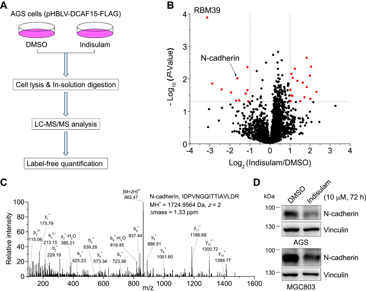
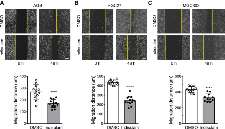
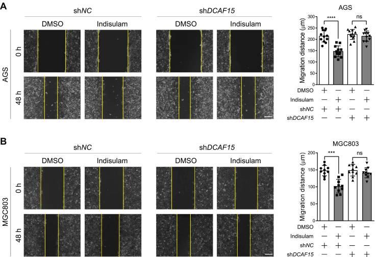
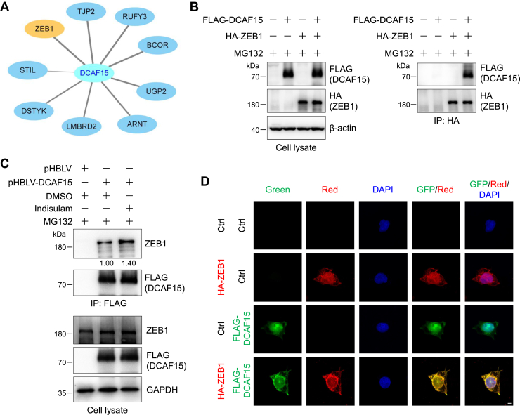
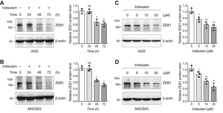
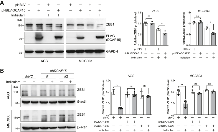
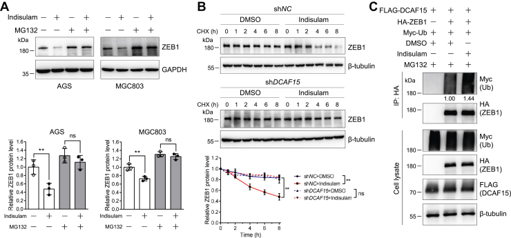
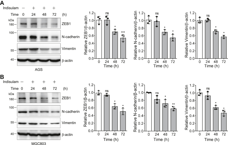
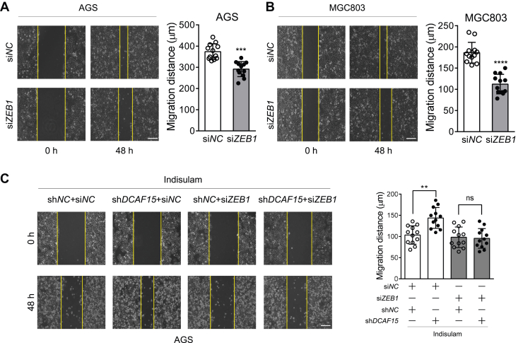
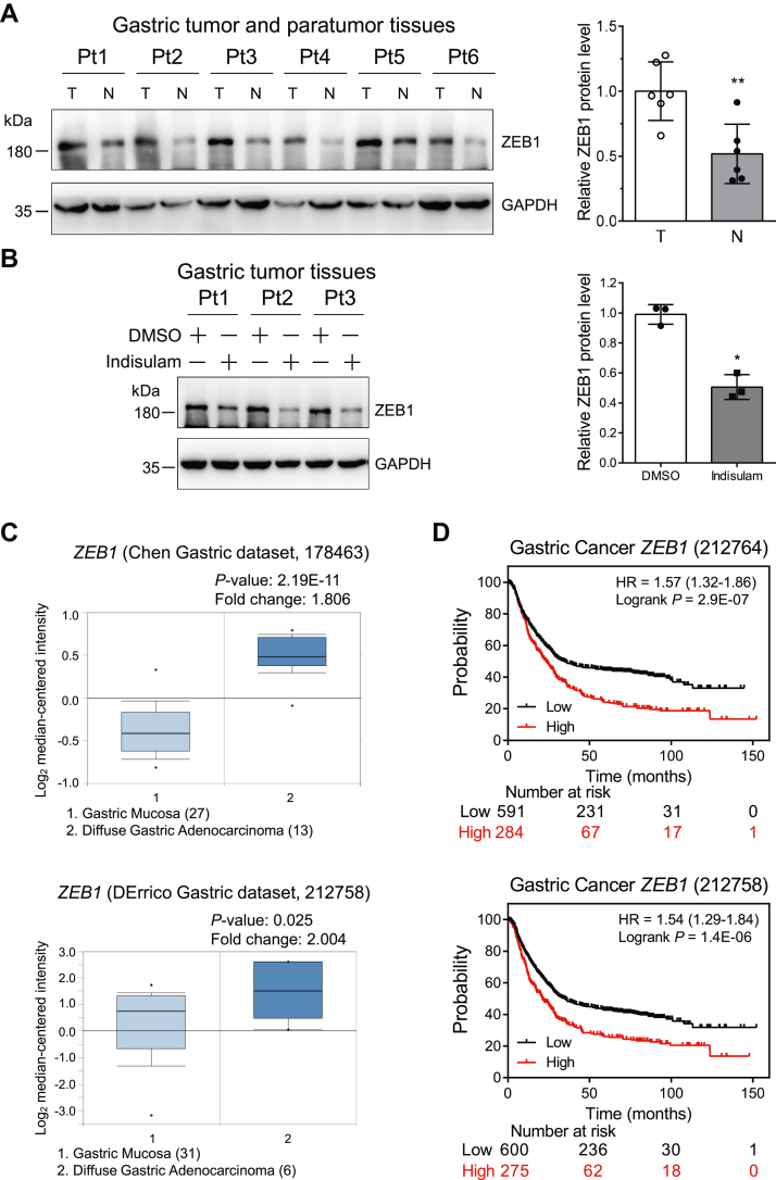
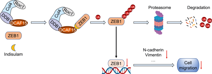
Gastric cancer is one of the cancers with high morbidity and mortality worldwide. The aryl sulfonamide indisulam inhibits the proliferation of several types of cancer cells through its function as a molecular glue to promote the ubiquitination and degradation of RNA-binding motif protein 39 (RBM39). However, it is unknown whether and how indisulam regulates the migration of cancer cells. In this work, using label-free quantitative proteomics, we discover that indisulam significantly attenuates N-cadherin, a marker for epithelial to mesenchymal transition and migration of cancer cells. Our bioinformatics analysis and biochemical experiments reveal that indisulam promotes the interaction between the zinc finger E-box-binding homeobox 1 (ZEB1), a transcription factor of N-cadherin, and DCAF15, a substrate receptor of CRL4 E3 ubiquitin ligase, and enhances ZEB1 ubiquitination and proteasomal degradation. In addition, our cell line-based experiments demonstrate that indisulam inhibits the migration of gastric cancer cells in a ZEB1-dependent manner. Analyses of patient samples and datasets in public databases reveal that tumor tissues from patients with gastric cancer express high ZEB1 mRNA and this high expression reduces patient survival rate. Finally, we show that treatment of gastric tumor samples with indisulam significantly reduces ZEB1 protein levels. Therefore, this work discloses a new mechanism by which indisulam inhibits the migration of gastric cancer cells, indicating that indisulam exhibits different biological functions through distinct signaling molecules.

Keywords: DCAF15; N-cadherin; ZEB1; degradation; indisulam; label-free quantification; migration; quantitative proteomics; ubiquitination.


Conflict of interest statement

Conflict of interest The authors declare that they have no conflicts of interest with the contents of this article.

Figures

Figure 1
Figure 1
Quantitative proteomics and biochemical approaches discover and verify that indisulam downregulates N-cadherin in gastric cancer cells.A, flowchart for the label-free quantitative proteomics used in this work. B, volcano plot for proteins identified by proteomics analysis of cell lysates obtained from the DMSO- or indisulam-treated (10 μM, 6 h) AGS cells expressing DCAF15. −Log10 (p-value) and Log2 (Indisulam/DMSO) were obtained from Proteome Discoverer database search and Perseus analysis. Proteins (red circles) with −Log10 (p-value) >1.30 (i.e., p-value < 0.05, horizontal dotted line) and Log2 (Indisulam/DMSO) >1.0 or <−1.0 (vertical dotted lines) were considered as differentially regulated by indisulam. C, tandem mass spectrometry spectrum of a representative tryptic peptide derived from N-cadherin. The amino acid sequence, charge state (z), MH+, and Δmass were provided. D, Western blotting analysis of cell lysates obtained from AGS and MGC803 cells treated with DMSO or indisulam (10 μM) for 72 h. DMSO, dimethyl sulfoxide.
Figure 2
Figure 2
Indisulam inhibits the migration of gastric cancer cells. Scratch assay was utilized to evaluate the relative migration of gastric cancer cells AGS (A), HGC27 (B), and MGC803 (C). Representative images were obtained from cells treated with DMSO or indisulam (10 μM) for 0 and 48 h. The vertical yellow lines indicated the edges of the scratch. Bar graphs showed the mean and standard deviations (mean ± SD, n = 13, 12, and 11, respectively, from three biological replicates). Student’s t test, ∗∗∗∗p < 0.0001. The scale bar represents 200 μm. DMSO, dimethyl sulfoxide.
Figure 3
Figure 3
DCAF15 knockdown eliminates the inhibitory effect of indisulam on the migration of gastric cancer cells.A and B, scratch assay was performed for shNC and shDCAF15-expressing AGS and MGC803 cells in the presence of dimethyl sulfoxide (DMSO) or indisulam (10 μM). Representative images were taken at 0 h and 48 h after treatment. Vertical yellow lines indicated the edge of the scratch. Mean ± SD (n = 12 or 10 from three biological replicates), Student’s t test, ∗∗∗p < 0.001, ∗∗∗∗p < 0.0001, ns: not significant. The scale bar represents 200 μm.
Figure 4
Figure 4
Indisulam enhances the interaction between ZEB1 and DCAF15.A, DCAF15-interacting proteins were obtained from BioGRID (https://thebiogrid.org/). The cutoff threshold for the modified CompPASS score (https://bioplex.hms.harvard.edu/comppass/) is >0.75. The score for ZEB1 is 1.00. B, ZEB1 interacts with DCAF15. HEK293T cells were transfected with FLAG-DCAF15 and/or hemagglutinin (HA)-ZEB1 plasmids and then split into two plates for 48 h. Cells were pretreated with MG132 (10 μM) for 2 h and lysed. ZEB1 was immunoprecipitated with anti-HA magnetic beads. Cell lysates and immunoprecipitates were subjected to immunoblotting analysis. C, indisulam enhances the DCAF15-ZEB1 interaction. FLAG-DCAF15 plasmid or control vector was transfected into HEK293T cells for 48 h. Cells were pretreated with MG132 (10 μM) for 2 h and then treated with dimethyl sulfoxide (DMSO) or indisulam (10 μM) for 14 h. Cells were lysed, and DCAF15 and its interacting proteins were immunoprecipitated with anti-FLAG affinity gel. Cell lysates and immunoprecipitates were subjected to immunoblotting analysis. The relative intensity of the immunoprecipitated ZEB1 was shown below the image. D, ZEB1 colocalizes with DCAF15 in the nucleus. HEK293 cells were transiently transfected with the control or indicated plasmids for 24 h, fixed, and incubated with the primary and fluorescent secondary antibodies. Immunofluorescence was detected under a confocal microscope. The scale bar represents 5 μm.
Figure 5
Figure 5
Indisulam downregulates ZEB1 in a time- and dose-dependent manner.A and B, AGS and MGC803 cells were treated with indisulam (10 μM) for different times and the resulting cell lysates were subjected to immunoblotting analysis. Mean ± SD (n = 3), Student’s t test, ∗p < 0.05, ns: not significant. C and D, AGS and MGC803 cells were treated with dimethyl sulfoxide or different concentrations of indisulam for 72 h. The cell lysates were subjected to immunoblotting analysis. Mean ± SD (n = 3), Student’s t test, ∗p < 0.05, ∗∗p < 0.01.
Figure 6
Figure 6
DCAF15 enhances the indisulam-induced degradation of ZEB1 in gastric cancer cells.A, overexpression of DCAF15 accelerates the downregulation of ZEB1 induced by indisulam. The stable pHBLV (control) and pHBLV-DCAF15-expressing AGS and MGC803 cells were treated with dimethyl sulfoxide or indisulam (10 μM) for 48 h. The resulting cell lysates were subjected to immunoblotting analysis. Mean ± SD (n = 3), Student’s t test, ∗p < 0.05, ns: not significant. B, DCAF15 knockdown abolishes the indisulam-induced downregulation of ZEB1. The stable shNC and shDCAF15-expressing AGS and MGC803 cells were treated with dimethyl sulfoxide or indisulam (10 μM) for 48 h. The resulting cell lysates were subjected to immunoblotting analysis. Mean ± SD (n = 3), Student’s t test, ∗p < 0.05, ∗∗p < 0.01, ns: not significant.
Figure 7
Figure 7
Indisulam induces ZEB1 degradation through the ubiquitin proteasome system.A, proteasome inhibitor MG132 abolishes the indisulam-induced ZEB1 degradation. AGS and MGC803 cells were pretreated with DMSO or indisulam (10 μM) for 48 h and then treated with DMSO or MG132 (10 μM) for 16 h in the presence of DMSO or indisulam. The resulting cell lysates were subjected to immunoblotting analysis. Mean ± SD (n = 3), Student’s t test, ∗∗p < 0.01, ns: not significant. B, DCAF15 knockdown eliminates the indisulam-induced degradation of ZEB1. AGS cells stably expressing shNC and shDCAF15 were treated with DMSO or indisulam (10 μM) and cycloheximide (200 μg/ml) for the indicated time. Mean ± SD (n = 3), two-way ANOVA, ∗∗p < 0.01, ns: not significant. C, indisulam promotes ZEB1 ubiquitination. HEK293T cells were transfected with the indicated plasmids for 48 h, pretreated with MG132 (10 μM) for 2 h, and treated again with DMSO or indisulam (10 μM) for 12 h in the presence of MG132. ZEB1 was immunoprecipitated with anti-HA magnetic beads. The cell lysates and immunoprecipitates were subjected to immunoblotting analysis. The relative intensity of ubiquitinated ZEB1 was shown below the image. The experiments were performed twice and similar results were obtained. DMSO, dimethyl sulfoxide; HA, hemagglutinin.
Figure 8
Figure 8
Indisulam attenuates the epithelial to mesenchymal transition markers in gastric cancer cells.A and B, the protein level of EMT markers was immunoblotted for cell lysates obtained from gastric cancer cells AGS (A) and MGC803 (B) treated with indisulam (10 μM) for different time. Mean ± SD (n = 3), Student’s t test, ∗p < 0.05, ∗∗p < 0.01, ∗∗∗p < 0.001, ns: not significant.
Figure 9
Figure 9
ZEB1 mediates the indisulam-inhibited migration of gastric cancer cells.A and B, ZEB1 knockdown reduces the migration of AGS and MGC803 cells. Cells were transfected with the control and ZEB1-specific siRNA with lipofectamine RNAiMAX. At 24 h after transfection, the scratch assay was performed. Mean ± SD (n = 12 from three biological replicates), Student’s t test, ∗∗∗p < 0.001, ∗∗∗∗p < 0.0001. The scale bar represents 200 μm. C, ZEB1 knockdown attenuates the DCAF15-mediated inhibitory effect of indisulam on the migration of AGS cells. The shNC and shDCAF15-expressing stable AGS cells were transfected with the control and ZEB1-specific siRNA with lipofectamine RNAiMAX. At 24 h after transfection, the scratch assay was performed in the presence of indisulam (10 μM). Mean ± SD (n = 12 from three biological replicates), Student’s t test, ∗∗p < 0.01, ns: not significant. The scale bar represents 200 μm.
Figure 10
Figure 10
Indisulam reduces ZEB1 in gastric cancer tissues, and high expression of ZEB1 decreases the overall survival of patients with gastric cancer.A, immunoblotting of ZEB1 in normal (paratumor) and tumor tissues from six patients with gastric cancer. Pt: patient, T: Tumor, N: Normal, Mean ± SD (n = 6), Student’s t test, ∗∗p < 0.01. B, gastric cancer tissues were treated with dimethyl sulfoxide (DMSO) and indisulam, and ZEB1 in tissue lysates was immunoblotted. Mean ± SD (n = 3), Student’s t test, ∗p < 0.05. C, the relative mRNA level of ZEB1 in gastric mucosa and diffuse gastric adenocarcinoma was obtained from Oncomine (https://www.oncomine.org). D, the correlation between the ZEB1 mRNA and the overall survival of patients with gastric cancer was obtained from Kaplan–Meier plotter (https://kmplot.com/analysis/).
Figure 11
Figure 11
Proposed model for the inhibitory effect of indisulam on the migration of gastric cancer cells.

References

    1. Sung H., Ferlay J., Siegel R.L., Laversanne M., Soerjomataram I., Jemal A., et al. Global cancer statistics 2020: GLOBOCAN estimates of incidence and mortality worldwide for 36 cancers in 185 countries. CA Cancer J. Clin. 2021;71:209–249. - PubMed
    1. Smyth E.C., Verheij M., Allum W., Cunningham D., Cervantes A., Arnold D., et al. Gastric cancer: ESMO clinical practice guidelines for diagnosis, treatment and follow-up. Ann. Oncol. 2016;27:v38–v49. - PubMed
    1. Van Cutsem E., Sagaert X., Topal B., Haustermans K., Prenen H. Gastric cancer. Lancet. 2016;388:2654–2664. - PubMed
    1. Canel M., Serrels A., Frame M.C., Brunton V.G. E-cadherin-integrin crosstalk in cancer invasion and metastasis. J. Cell Sci. 2013;126:393–401. - PubMed
    1. Zheng X., Carstens J.L., Kim J., Scheible M., Kaye J., Sugimoto H., et al. Epithelial-to-mesenchymal transition is dispensable for metastasis but induces chemoresistance in pancreatic cancer. Nature. 2015;527:525–530. - PMC - PubMed

Publication types

MeSH terms