Review
. 2023 Apr 18;67(3):399-414.
doi: 10.1042/EBC20220158.

Structure and function of microbial α-l-fucosidases: a mini review

Affiliations
Review

Structure and function of microbial α-l-fucosidases: a mini review

Haiyang Wu et al. Essays Biochem. .

Abstract

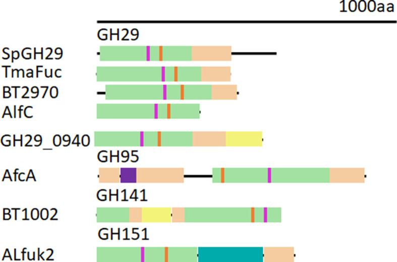
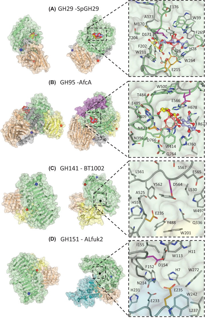
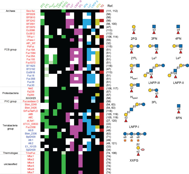
Fucose is a monosaccharide commonly found in mammalian, insect, microbial and plant glycans. The removal of terminal α-l-fucosyl residues from oligosaccharides and glycoconjugates is catalysed by α-l-fucosidases. To date, glycoside hydrolases (GHs) with exo-fucosidase activity on α-l-fucosylated substrates (EC 3.2.1.51, EC 3.2.1.-) have been reported in the GH29, GH95, GH139, GH141 and GH151 families of the Carbohydrate Active Enzymes (CAZy) database. Microbes generally encode several fucosidases in their genomes, often from more than one GH family, reflecting the high diversity of naturally occuring fucosylated structures they encounter. Functionally characterised microbial α-l-fucosidases have been shown to act on a range of substrates with α-1,2, α-1,3, α-1,4 or α-1,6 fucosylated linkages depending on the GH family and microorganism. Fucosidases show a modular organisation with catalytic domains of GH29 and GH151 displaying a (β/α)8-barrel fold while GH95 and GH141 show a (α/α)6 barrel and parallel β-helix fold, respectively. A number of crystal structures have been solved in complex with ligands, providing structural basis for their substrate specificity. Fucosidases can also be used in transglycosylation reactions to synthesise oligosaccharides. This mini review provides an overview of the enzymatic and structural properties of microbial α-l-fucosidases and some insights into their biological function and biotechnological applications.

Keywords: carbohydrate-active enzymes; fucose; fucosidases; glycoside hydrolases; gut bacteria.


Conflict of interest statement

The authors declare that there are no competing interests associated with the manuscript.

Figures

Figure 1
Figure 1. Schematic modular representation of microbial α-l-fucosidases from different GH families
Catalytic modules are shown in green and β-sandwich domains that may have carbohydrate binding properties in light brown. If a second β-sandwich domain is present, such as in GH29_0940, it is coloured yellow. AfcA has an additional helical barrel domain, colored purple. For clarity, the AfcA N-terminal domain of unknown function and the C-terminal bacterial Ig-like domain are not shown. These extend the total length of AfcA to 1959 amino acids. ALfuk2 also has a Rossman fold domain, colored teal.
Figure 2
Figure 2. Crystal structures of microbial α-l-fucosidases from different GH families with close up of active sites
Catalytic modules are shown in green and β-sandwich domains that may have carbohydrate binding properties in light brown and yellow. Catalytic nucleophile residues are coloured magenta and catalytic acid/base residues are coloured in orange. Where possible WT apo crystal structures (grey) have been aligned to their corresponding inactive mutant crystal structures (green) to highlight residue movements upon binding to a substrate like ligand. The N- and C-termini are indicated with blue and red spheres, respectively. Surface representation views are related by a 90° rotation around the y axis. If a substrate complex is not available, the location of the active site is indicated with a black sphere. (A) GH29 fucosidase (SpGH29, apo PDB = 6ORG; D171N; E215Q mutant in complex with LeX PDB = 6ORF). The catalytic domain comprises residues 11-317 and the C terminal β-sandwich module comprises residues 318-451. The bound ligand is shown with Fuc (light red), Gal (yellow) and GlcNAc (light blue). (B) GH95 fucosidase (AfcA, apo PDB = 2EAB; E566A mutant in complex with substrate PDB: 2EAD). The catalytic domain comprises residues 80-133 and 387-778, the N-terminal domain (in light brown) residues 9-79 and 134-293, and the C-terminal β-sandwich module (in yellow) residues 779-896. There is a helical barrel protruding from the N-terminal domain, residues 80-133. The substrate is shown with Gal (yellow), Fuc (light red) and Glc (light blue). C) GH141 fucosidase (BT1002, apo PDB = 5MQP). The catalytic domain comprises residues 1-108 and 296-619, the ancillary β-sandwich domain, residues 109-295 (in yellow for residues 151-251 and in wheat for residues 109-251 and 252-295, according to visual separation into sub domains). D) GH151 fucosidase (ALfuk2, apo PDB = 6TVK). The catalytic domain covers residues 1-336, the C-terminal domain (in wheat), residues 560-660 and the Rossman fold domain (in teal), residues 341-558.
Figure 3
Figure 3. Substrate specificity of microbial GH29 α-l-fucosidases
The α1,2 substrates are colored in green, α1,3 in pink, α1,4 in sky blue, and α1,6 in olive. Light versions of the above colors indicate trace activity. Black boxes correspond to no enzymatic activity and empty boxes indicate lack of data. GH29A and GH29B α-l-fucosidases are coloured in red and blue, respectively; FCB, Fibrobacteres-Chlorobi-Bacteroidetes super phylum; PVC, Planctomycetes-Verrucomicrobia-Chlamydiae bacterial superphylum; TG, transglycosylation capability. Glycan structures presentation according to Symbol Nomenclature for Glycans (SNFG) [109,110].

References

    1. Flowers H.M. (1981) Chemistry and biochemistry of D- and L-fucose. Adv. Carbohydr. Chem. Biochem. 39, 279–345 10.1016/S0065-2318(08)60208-5 - DOI - PubMed
    1. Sutherland I.W. (1971) Enzymic hydrolysis of colanic acid. Eur. J. Biochem. 23, 582–587 10.1111/j.1432-1033.1971.tb01657.x - DOI - PubMed
    1. Maverakis E., Kim K., Shimoda M., Gershwin M.E., Patel F., Wilken R.et al. . (2015) Glycans in the immune system and the altered glycan theory of autoimmunity: a critical review. J. Autoimmun. 57, 1–13 10.1016/j.jaut.2014.12.002 - DOI - PMC - PubMed
    1. Becker D.J. and Lowe J.B. (2003) Fucose: Biosynthesis and biological function in mammals. Glycobiology 13, 41R–53R 10.1093/glycob/cwg054 - DOI - PubMed
    1. Schneider M., Al-Shareffi E. and Haltiwanger R.S. (2017) Biological functions of fucose in mammals. Glycobiology 27, 601–618 10.1093/glycob/cwx034 - DOI - PMC - PubMed

Publication types

LinkOut - more resources