

Evaluation of a deep learning-based reconstruction method for denoising and image enhancement of shoulder MRI in patients with shoulder pain
- PMID: 36806569
- PMCID: PMC10289918
- DOI: 10.1007/s00330-023-09472-9
Evaluation of a deep learning-based reconstruction method for denoising and image enhancement of shoulder MRI in patients with shoulder pain
Abstract
Objectives: To evaluate the diagnostic performance of an automated reconstruction algorithm combining MR imaging acquired using compressed SENSE (CS) with deep learning (DL) in order to reconstruct denoised high-quality images from undersampled MR images in patients with shoulder pain.
Methods: Prospectively, thirty-eight patients (14 women, mean age 40.0 ± 15.2 years) with shoulder pain underwent morphological MRI using a pseudo-random, density-weighted k-space scheme with an acceleration factor of 2.5 using CS only. An automated DL-based algorithm (CS DL) was used to create reconstructions of the same k-space data as used for CS reconstructions. Images were analyzed by two radiologists and assessed for pathologies, image quality, and visibility of anatomical landmarks using a 4-point Likert scale.
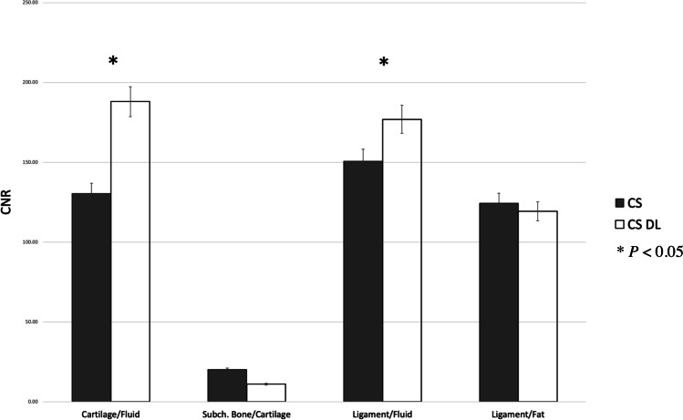
Results: Overall agreement for the detection of pathologies between the CS DL reconstructions and CS images was substantial to almost perfect (κ 0.95 (95% confidence interval 0.82-1.00)). Image quality and the visibility of the rotator cuff, articular cartilage, and axillary recess were overall rated significantly higher for CS DL images compared to CS (p < 0.03). Contrast-to-noise ratios were significantly higher for cartilage/fluid (CS DL 198 ± 24.3, CS 130 ± 32.2, p = 0.02) and ligament/fluid (CS DL 184 ± 17.3, CS 141 ± 23.5, p = 0.03) and SNR values were significantly higher for ligaments and muscle of the CS DL reconstructions (p < 0.04).
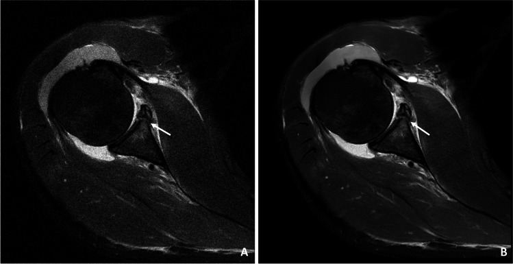
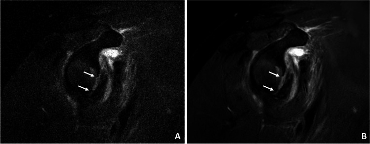
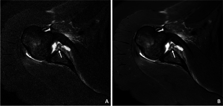
Conclusion: Evaluation of shoulder pathologies was feasible using a DL-based algorithm for MRI reconstruction and denoising. In clinical routine, CS DL may be beneficial in particular for reducing image noise and may be useful for the detection and better discrimination of discrete pathologies. Assessment of shoulder pathologies was feasible with improved image quality as well as higher SNR using a compressed sensing deep learning-based framework for image reconstructions and denoising.
Key points: • Automated deep learning-based reconstructions showed a significant increase in signal-to-noise ratio and contrast-to-noise ratio (p < 0.04) with only a slight increase of reconstruction time of 40 s compared to CS. • All pathologies were accurately detected with no loss of diagnostic information or prolongation of the scan time. • Significant improvements of the image quality as well as the visibility of the rotator cuff, articular cartilage, and axillary recess were detected.
Keywords: Compressed SENSE; Deep learning algorithm; Magnetic resonance imaging; Shoulder injury.
© 2023. The Author(s).
Conflict of interest statement
K.W. is employed by Philips GmbH Market DACH but was not involved in data acquisition or analysis. The rest of the authors of this manuscript declare no relationships with any companies, whose products or services may be related to the subject matter of the article.
Figures

References
MeSH terms
LinkOut - more resources
Full Text Sources
