

Differences in skin test reactions to official and defined antigens in guinea pigs exposed to non-tuberculous and tuberculous bacteria
- PMID: 36806813
- PMCID: PMC9941491
- DOI: 10.1038/s41598-023-30147-4
Differences in skin test reactions to official and defined antigens in guinea pigs exposed to non-tuberculous and tuberculous bacteria
Abstract
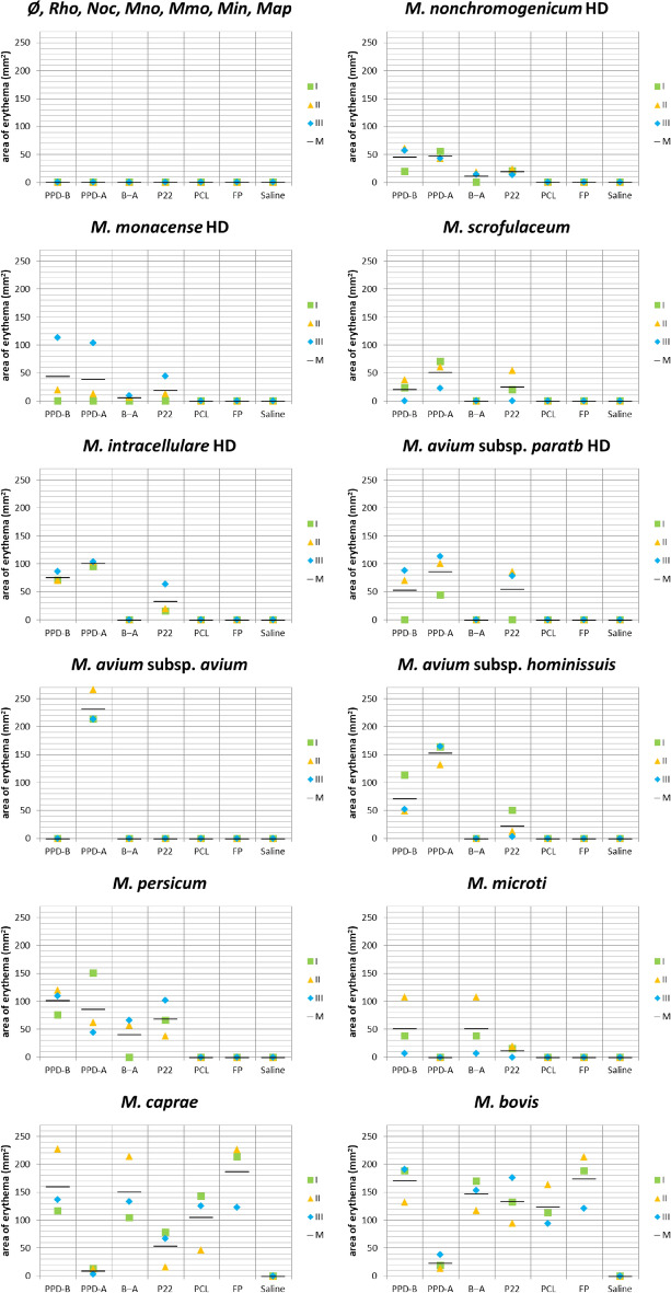
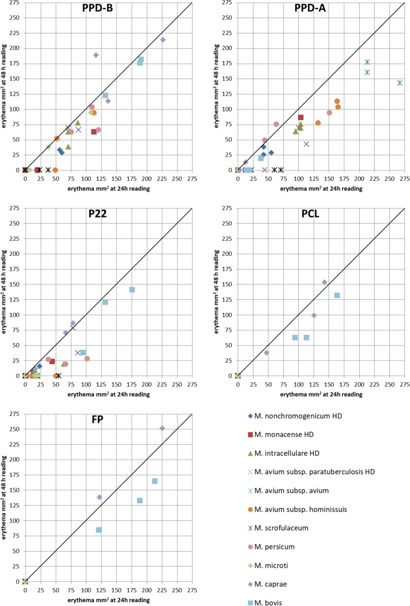
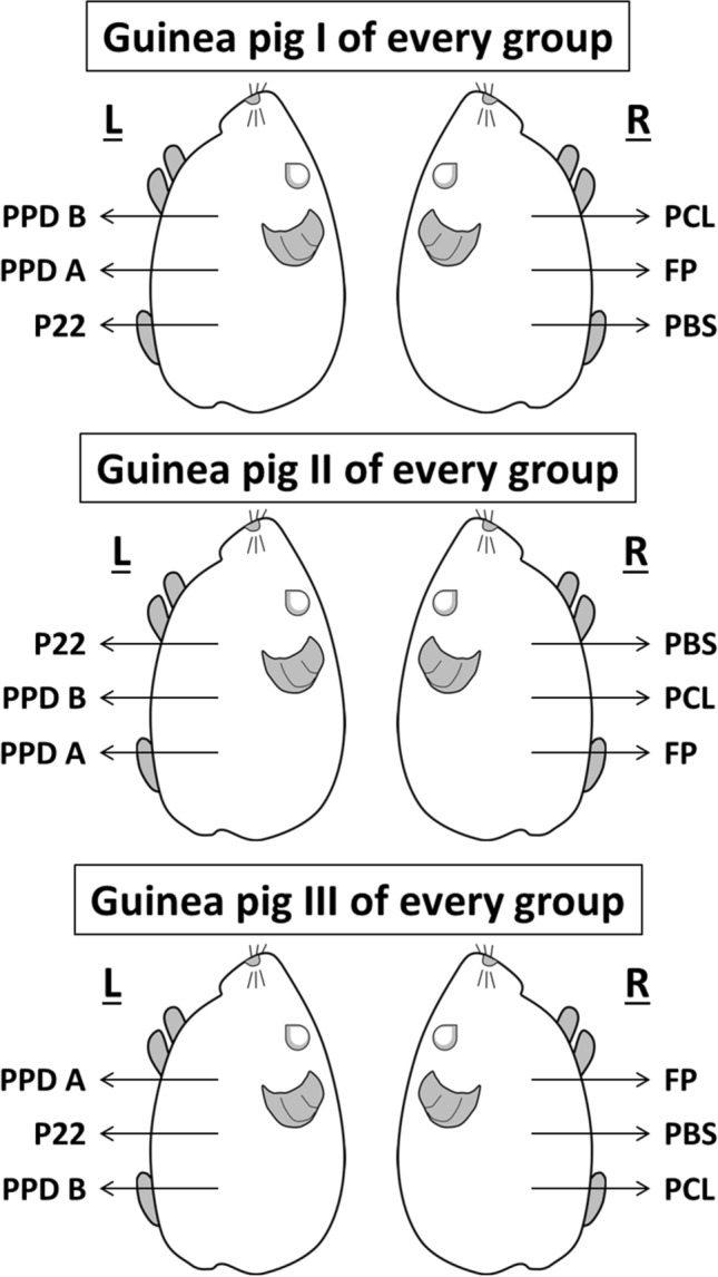
The single and comparative intradermal tuberculin tests (SITT and CITT) are official in vivo tests for bovine tuberculosis (TB) diagnosis using bovine and avian purified protein derivatives (PPD-B and PPD-A). Infection with bacteria other than Mycobacterium tuberculosis complex (MTC) can result in nonspecific reactions to these tests. We evaluated the performance of the skin test with PPDs and new defined antigens in the guinea pig model. A standard dose (SD) of Rhodococcus equi, Nocardia sp., M. nonchromogenicum, M. monacense, M. intracellulare, M. avium subsp. paratuberculosis, M. avium subsp. avium, M. avium subsp. hominissuis, M. scrofulaceum, M. persicum, M. microti, M. caprae and M. bovis, and a higher dose (HD) of M. nonchromogenicum, M. monacense, M. intracellulare, M. avium subsp. paratuberculosis were tested using PPD-B, PPD-A, P22, ESAT-6-CFP-10-Rv3615c peptide cocktail long (PCL) and fusion protein (FP). The SD of R. equi, Nocardia sp., M. nonchromogenicum, M. monacense, M. intracellulare and M. avium subsp. paratuberculosis did not cause any reactions. The HD of M. nonchromogenicum, M. monacense, M. intracellulare, and M. avium subsp. paratuberculosis and the SD of M. avium subsp. hominissuis, M. scrofulaceum and M. persicum, caused nonspecific reactions (SIT). A CITT interpretation would have considered M. avium complex and M. scrofulaceum groups negative, but not all individuals from M. nonchromogenicum HD, M. monacense HD and M. persicum SD groups. Only animals exposed to M. bovis and M. caprae reacted to PCL and FP. These results support the advantage of complementing or replacing PPD-B to improve specificity without losing sensitivity.
© 2023. The Author(s).
Conflict of interest statement
The authors declare that the research was conducted in the absence of any competing interests. G.J.J. and H.M.V. are employed by the Animal and Plant Health Agency (APHA) who holds 3 patents for the use of Rv3615c in diagnostic tests for bovine TB (WO/2009/060184, WO/2011/135369 and WO/2012/010875).
Figures
References
-
- Schiller I, et al. Bovine tuberculosis: A review of current and emerging diagnostic techniques in view of their relevance for disease control and eradication. Transbound. Emerg. Dis. 2010;57:205–220. - PubMed
Publication types
MeSH terms
Substances
LinkOut - more resources
Full Text Sources
Miscellaneous
