. 2023 Feb 17;14(1):903.
doi: 10.1038/s41467-023-36481-5.

Watching the release of a photopharmacological drug from tubulin using time-resolved serial crystallography

Affiliations

Watching the release of a photopharmacological drug from tubulin using time-resolved serial crystallography

Maximilian Wranik et al. Nat Commun. .

Abstract

The binding and release of ligands from their protein targets is central to fundamental biological processes as well as to drug discovery. Photopharmacology introduces chemical triggers that allow the changing of ligand affinities and thus biological activity by light. Insight into the molecular mechanisms of photopharmacology is largely missing because the relevant transitions during the light-triggered reaction cannot be resolved by conventional structural biology. Using time-resolved serial crystallography at a synchrotron and X-ray free-electron laser, we capture the release of the anti-cancer compound azo-combretastatin A4 and the resulting conformational changes in tubulin. Nine structural snapshots from 1 ns to 100 ms complemented by simulations show how cis-to-trans isomerization of the azobenzene bond leads to a switch in ligand affinity, opening of an exit channel, and collapse of the binding pocket upon ligand release. The resulting global backbone rearrangements are related to the action mechanism of microtubule-destabilizing drugs.


Conflict of interest statement

The authors declare no competing interests.

Figures

Fig. 1
Fig. 1. Tracking ligand release from tubulin using a photochemical affinity switch.
A The cis-azo-CA4 (yellow sticks) binds to the colchicine site that is formed by secondary structural elements stemming from both the α (blue) and β (cyan) subunits of the αβ-tubulin heterodimer. Illumination with a flash of a laser light triggers the release of trans-azo-CA4 (colored in orange, diffusion indicated by including several molecules, cylindrical arrows designate conformational changes of the protein) from its binding pocket. B Difference electron density maps (Fobslight − Fobsdark) from time-resolved XFEL data were integrated and subjected to Pearson correlation analysis. Three areas of elevated correlation can be identified. The box in the nanoseconds (1) corresponds to the relaxed conformation of the ligand observed as a metastable binding pose. Correlations between maps in the microseconds (2) are less homogeneous and stretch from 1 μs to the opening of the exit pathway at 1 ms. The final snapshots in the milliseconds (3) correspond to the predominantly unliganded protein. C Plot showing negative density integrated around azo-CA4 with a strong increase after 1 ms indicating release of the ligand. D Time-resolved structural snapshots of azo-CA4 release. The time arrow depicts the investigated time regime. The panels from left to right show the isomorphous difference maps obtained at 100 ns with changes centered on the ligand, 100 μs with changes centered on the binding pocket, and 100 ms with conformational changes propagating throughout the protein. All panels show isomorphous difference maps in red (negative) and green (positive) at 3 σ. The structure in the given time range (colored in orange) is compared to that of the previous time range (colored in gray). A movie showing the electron changes within the binding pocket over all nine time delays is available as Supplementary material.
Fig. 2
Fig. 2. Photoisomerization leads to the formation of a metastable binding state.
A The two chemical structures of azo-CA4 show the wavelength-dependent conformational change between the cis (top) and trans (bottom) conformations. Central to the switch is the N=N azo bond that allows to switch azo-CA4 reversibly and with high efficiency between its high- and low-affinity stereoisomers, respectively. B Before laser-induced isomerization, cis-azo-CA4 (yellow sticks) is bound in the colchicine site located between the α (blue) and β (cyan) subunits close to the βT7 loop (green) of β-tubulin. Hydrophobic interactions (green lines) between tubulin residues and the A and B rings of cis-azo-CA4 as well as hydrophilic interactions (blue dashed lines) toward the ring substituents anchor the ligand within the colchicine site. C Within nanoseconds after illumination, trans-azo-CA4 (orange sticks) has relaxed into a metastable binding pose with an altered interaction network and reduced affinity. For clarity, only interacting residues are shown.
Fig. 3
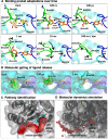
Fig. 3. Molecular changes in the βT7 gating loop allow ligand release.
A Within nanoseconds, the light-induced isomerization and relaxation repositions the A ring of azo-CA4 closer to the βT7 loop (green), which acts like a lid on the colchicine site. Further reorganizations (black arrows) in the microseconds open a channel between the βH8 helix and the βT7 loop. In the millisecond range, the βT7 loop is folded back and packed against residues of the empty binding pocket. B Opening of a release pathway through relocation of the βT7 loop in the 1 ms structure is illustrated by the position of βLeu246 (green sticks and surface) at the indicated time delays. C The release pathway (red spheres) plotted onto the 1 ms structure. D The experimentally deduced unbinding pathway of azo-CA4 is confirmed by MD simulations. The small red spheres depict the centers of masses of ligand atoms, plotted every 50 frames along the simulation.
Fig. 4
Fig. 4. Correlation between colchicine-site dynamics and global tubulin backbone rearrangements.
A Relaxation of azo-CA4: dark to 100 ns. B Movements of βT7 loop: 100 ns to 1 ms. C Collapse of the colchicine site: 1 ms to 100 ms. The three rows depict conformational changes at different spatial levels: Binding pocket in the upper row, tubulin backbone in the middle row, and longitudinally aligned tubulin dimers in the lower row. Starting structures are shown in gray. Backbone movements within tubulin (middle row) are represented by modevectors between consecutive time delays. Every second Cα-backbone atom in the main secondary structural elements with a displacement of at least 0.5 Å is shown. The N-terminal nucleotide-binding domains, the C-terminal domains, and the intermediate domains for both α and β tubulin are shown in blue, red, and green, respectively. The release pathway (red spheres) is indicated to illustrate the directional movements of the tubulin subunits. The global conformational changes (lower row) are visualized by three longitudinally aligned tubulin dimers (“Straight” tubulin dimers as found in the main shaft of a microtubule are shown in surface representation (dark gray, α-tubulin; light gray, β-tubulin; PDB ID 5SYE); “Curved” tubulin dimers (α-tubulin in blue and β-tubulin in cyan) at the indicated time delay and in relation to the previous state are shown in black). Deviations demonstrate the conformational plasticity of tubulin during azo-CA4 relaxation in its binding pocket and βT7 loop movements of β-tubulin, as well as a directed curvature adjustment after azo-CA4 release. Arrows indicate the main directions of movements in the indicated time delays.

References

    1. Hull K, Morstein J, Trauner D. In vivo photopharmacology. Chem. Rev. 2018;118:10710–10747. doi: 10.1021/acs.chemrev.8b00037. - DOI - PubMed
    1. Beharry AA, Woolley GA. Azobenzene photoswitches for biomolecules. Chem. Soc. Rev. 2011;40:4422–4437. doi: 10.1039/c1cs15023e. - DOI - PubMed
    1. Borowiak M, et al. Photoswitchable inhibitors of microtubule dynamics optically control mitosis and cell death. Cell. 2015;162:403–411. doi: 10.1016/j.cell.2015.06.049. - DOI - PubMed
    1. Gaspari R, Prota AE, Bargsten K, Cavalli A, Steinmetz MO. Structural basis of cis- and trans-combretastatin binding to tubulin. Chem. 2017;2:102–113. doi: 10.1016/j.chempr.2016.12.005. - DOI
    1. Brouhard GJ, Rice LM. Microtubule dynamics: an interplay of biochemistry and mechanics. Nat. Rev. Mol. Cell Biol. 2018;19:451–463. doi: 10.1038/s41580-018-0009-y. - DOI - PMC - PubMed

Publication types