Case Reports
. 2023 Mar;29(3):583-587.
doi: 10.1038/s41591-023-02213-x. Epub 2023 Feb 20.

In-depth virological and immunological characterization of HIV-1 cure after CCR5Δ32/Δ32 allogeneic hematopoietic stem cell transplantation

Affiliations
Case Reports

In-depth virological and immunological characterization of HIV-1 cure after CCR5Δ32/Δ32 allogeneic hematopoietic stem cell transplantation

Björn-Erik Ole Jensen et al. Nat Med. 2023 Mar.

Abstract

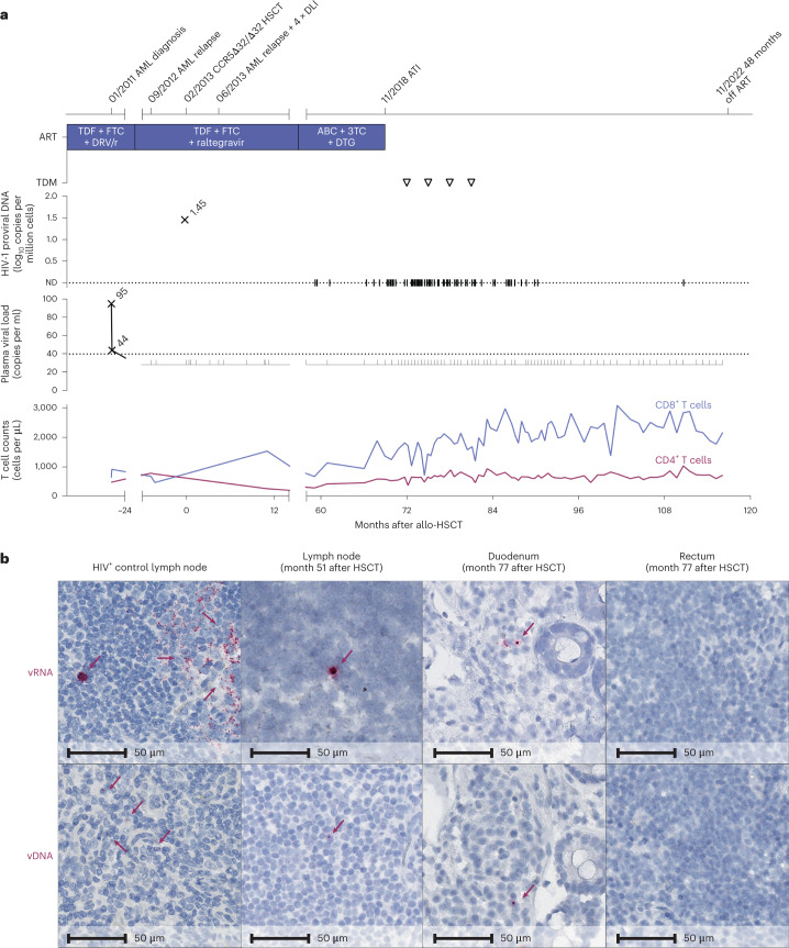
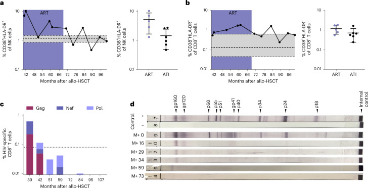
Despite scientific evidence originating from two patients published to date that CCR5Δ32/Δ32 hematopoietic stem cell transplantation (HSCT) can cure human immunodeficiency virus type 1 (HIV-1), the knowledge of immunological and virological correlates of cure is limited. Here we characterize a case of long-term HIV-1 remission of a 53-year-old male who was carefully monitored for more than 9 years after allogeneic CCR5Δ32/Δ32 HSCT performed for acute myeloid leukemia. Despite sporadic traces of HIV-1 DNA detected by droplet digital PCR and in situ hybridization assays in peripheral T cell subsets and tissue-derived samples, repeated ex vivo quantitative and in vivo outgrowth assays in humanized mice did not reveal replication-competent virus. Low levels of immune activation and waning HIV-1-specific humoral and cellular immune responses indicated a lack of ongoing antigen production. Four years after analytical treatment interruption, the absence of a viral rebound and the lack of immunological correlates of HIV-1 antigen persistence are strong evidence for HIV-1 cure after CCR5Δ32/Δ32 HSCT.


Conflict of interest statement

B.-E.O.J. received honoraria for presentations from Gilead, ViiV Healthcare, MSD and Janssen (unrelated to the submitted work) and served on advisory boards for ViiV Healthcare and Gilead (unrelated to the submitted work). N.L. received honoraria for presentations from Gilead, MSD, Abbvie and ViiV Healthcare and served on the advisory boards for ViiV Healthcare and Theratechnologies (all unrelated to the submitted work). C. Münk received a research grant from Gilead (unrelated to the submitted work). I.H. and J.H. are cofounders and shareholders of PROVIREX Genome Editing Therapies GmbH, a start-up company focusing on the excision of HIV-1 by genome editing (unrelated to this work). J.S.z.W. received lecture fees from Gilead (unrelated to the submitted work). A.M.J.W. received an unrestricted research grant from Gilead and did consultancy for Gilead, GlaxoSmithKline and ViiV Healthcare (unrelated to the submitted work). J.M.-P. received institutional grants and educational/consultancy fees from AbiVax, AstraZeneca, Gilead, Grifols, Janssen, Merck Sharp & Dohme and ViiV Healthcare (unrelated to the submitted work). The other authors declare no competing interests.

Figures

Fig. 1
Fig. 1. Clinical course and HIV-1 reservoir before and after ATI.
a, Clinical events, ART regimen, HIV-1 plasma viral load, proviral load from PBMC and CD4+ and CD8+ counts. The triangles indicate negative results in the therapeutic drug monitoring (TDM) 3, 6, 9 and 12 months after ATI. ABC, abacavir; ND, not detectable. b, In situ hybridization assays for viral DNA (vDNA) and viral RNA (vRNA) revealed HIV-1 DNA and RNA traces in lymph node tissue (month 51) and duodenum but not rectum biopsies (month 77). Left panels: untreated HIV+ control patient.
Fig. 2
Fig. 2. Waning HIV-1-specific cellular and humoral immune responses before and after ATI.
a,b, NK cells (a) and CD8+ T cells (b) both showed decreasing and stable immune activation (coexpression of CD38 and HLA-DR). See also data from months 41–59 after HSCT for IciStem no. 19 as published in ref. . Data are shown as the mean with s.d. and a reference range from a healthy cohort (n = 8) in gray as the median with interquartile range (IQR). c, HIV-1-specific CD8+ T cell responses (production of IFNγ, TNFα, IL-2 and/or expression of CD107a) against HIV-1 Gag (burgundy), Nef (dark purple) and Pol (light purple) peptide pools waned after HSCT. See also the data from months 39 and 42 after HSCT for IciStem no. 19 as published in ref. . The dotted line represents the average background signal. d, Full-virus lysate immunoblot for antibodies against HIV-1 antigens revealed waning HIV-1-specific antibody responses after HSCT and prolonged weakening of gp160- and gp120-specific antibodies. M+, months after HSCT. Source data
Extended Data Fig. 1
Extended Data Fig. 1. Donor chimerism and CCR5 genotype and phenotype in PBMC after HSCT.
a, The donor chimerism after HSCT was genotypically assessed from a set of 13 genes. Full (100%) donor chimerism was first achieved 34 days after HSCT then dropped to 72% during the second relapse of AML but returned to 100% after successful therapy with 5-azacitidine and donor lymphocytes, and was sustained afterwards. HSCT, hematopoietic stem cell transplantation; AML, acute myeloid leukemia. b, In CCR5 genotyping with agarose gel electrophoresis of CCR5-PCR products, PBMC of the patient from month 35 after HSCT as well as in vitro generated HIV-1-specific CTL lines showed the homozygous CCR5Δ32 mutation. PBMC and B-cell lines from CCR5wt/wt, CCR5Δ32/wt, and CCR5Δ32/Δ32 individuals and a 50 bp DNA ladder served as controls. PBMC, peripheral blood mononuclear cells; wt, wildtype; RT, reverse transcriptase. c, CCR5 expression was lost from peripheral blood CD4+ T cells after HSCT in flow cytometric analyses. See also data from months 41–59 after HSCT for IciStem no. 19 as published in. A reference range from a healthy cohort (n = 8) is given in grey as median with IQR. ART, antiretroviral therapy. Source data
Extended Data Fig. 2
Extended Data Fig. 2. Timeline of viral outgrowth assays.
a, Timeline of the viral outgrowth assays. Orange arrows indicate assays performed at the Heinrich Heine University Duesseldorf, green arrows indicate assays performed at the Leibniz Institute of Virology, and blue arrows assays at the IrsiCaixa AIDS Research Institute. All qVOA showed negative results and an in vivo viral rescue assay in Balb/c Rag2−/−γc−/− mice showed negative results in the immunohistochemical stainings for HIV-1 p24 Ag in the spleens, livers, and lymph nodes (Supplementary Fig. 1). HSCT, hematopoietic stem cell transplantation; ATI, analytical treatment interruption; qVOA, quantitative viral outgrowth assay; mVOA, murine viral outgrowth assay. b, A mVOA in n = 5 biologically independent NOD-scid mice from month 42 after HSCT showed good engraftment of the transferred CD4+ T cells and undetectable plasma HIV-1 RNA and cellular HIV-1 DNA in the blood of the mice. Filled symbols indicate detectable values and open symbols indicate the limit of detection for undetectable values. After six weeks, HIV-1 DNA in the spleen of the mice was undetectable when infused with the IciStem no. 19 patient cells (blue) compared to an HIV-1 positive control patient under ART (grey) (two-tailed Mann-Whitney test; P = 0.0079). Data are shown as mean with standard deviation.
Extended Data Fig. 3
Extended Data Fig. 3. Immunological profiling before and after ATI.
a–b, The distribution of memory subsets within the CD4+ and CD8+ T cell compartments showed markedly reduced frequencies of the early differentiated naïve T cells and elevated frequencies of the late differentiated Tem and TemRA subsets. Tnaive, naïve T cells (CD45RA+ CCR7+); Tcm, central memory T cells (CD45RA– CCR7+); Ttm, transitional memory T cells (CD45RA– CCR7– CD27+); Tem, effector memory T cells (CD45RA– CCR7– CD27–); TemRA, late effector memory T cells (CD45RA+ CCR7–); HD, healthy donors. c, Within the NK cell compartment, elevated frequencies of CD56neg NK cells were observed. For all subsets, the mean frequency from a healthy cohort (n = 8) is given.
Extended Data Fig. 4
Extended Data Fig. 4. Immune reconstitution of the tissue-resident immune system after HSCT and tissue inflammation.
a, The CD4+ T cell density is normal in lymphoid tissue. Slices were counterstained for macrophages (CD68/CD163) to allow the differentiation of CD4+ T cells from myeloid lineage cells by masking the faint CD4 expressed on these cells. MX1 (as a marker of type I IFN activation and inflammation) was not found to be strongly expressed in the lymphoid tissue slices. The slices of the lymphoid tissue are not sequential, but within 50 μm or less. MX1, Interferon-induced GTP-binding protein MX1; IFN, interferon. b, MX1 and MPO expression is not increased in gut tissue slices. MPO expression is indicative of PMN cell infiltration and thus of microbial translocation and gut barrier damage. For all immunohistochemical stainings of IciStem no. 19, the lymph node tissue was collected in month 51 after HSCT, and the gut biopsies were obtained in month 77 after HSCT. For comparison, tissue slices of HIV-1 negative and HIV-1 positive control patients are shown. The slices of the HIV-1 negative rectal tissue, duodenal and rectal tissue of the patient are not sequential, but within 50 μm or less. All slices were counterstained with hematoxylin. Scale bars represent 100 μm. MPO, myeloperoxidase; PMN, polymorphnuclear; HSCT, hematopoietic stem cell transplantation.
Extended Data Fig. 5
Extended Data Fig. 5. Cellular and humoral immune responses before and after ATI.
a, No substantial frequencies of HIV-1 (RT-YV9; YQYMDDLYV) specific CD8+ T cells were found before or after ATI after magnetic bead MHC class I tetramer enrichment despite sustained CMV-specific responses ex vivo (performed at the University Medical Center Hamburg). Shown are representative plots from month 75 after HSCT. b, IFN-γ ELISpot assays showed waning HIV-1-specific T-cell responses of in vitro expanded polyclonal T-cell lines specific for the gag07-121 (KELYPLASLRSLFGN) and RT-YV9 (YQYMDDLYV) peptides despite maintained EBV-specific responses (performed at the Friedrich-Alexander University Erlangen-Nuremberg). IFN-γ, interferon-gamma; ELISpot, enzyme-linked immune absorbent spot; EBV, Epstein Barr Virus. c, Strong CMV-specific CD8+ T-cell responses were maintained before and after ATI as assessed by the production of IFN-γ, TNF-α, IL-2, and/or expression of CD107a in the ICS (performed at the Institut Pasteur Paris). The dotted line shows the average background signal. CMV, cytomegalovirus; ATI, analytical treatment interruption; ICS, intracellular cytokine staining; HSCT, hematopoietic stem cell transplantation. d, In month 39, the HIV-1-specific antibody levels and avidity were below the cut-off for PLWH (n = 10 viremic and n = 10 ART treated) and comparable to HIV-1 negative patients (n = 4) as measured by a detuned low-sensitive version of the HIV-1 VITROS assay and the limiting antigen avidity assay (performed at IrsiCaixa AIDS Research Institute Barcelona). Data are shown as median with IQR.

Comment in

References

    1. Henrich TJ, et al. Long-term reduction in peripheral blood HIV type 1 reservoirs following reduced-intensity conditioning allogeneic stem cell transplantation. J. Infect. Dis. 2013;207:1694–1702. doi: 10.1093/infdis/jit086. - DOI - PMC - PubMed
    1. Salgado M, et al. Mechanisms that contribute to a profound reduction of the HIV-1 reservoir after allogeneic stem cell transplant. Ann. Intern. Med. 2018;169:674–683. doi: 10.7326/M18-0759. - DOI - PubMed
    1. Buzon MJ, et al. HIV-1 persistence in CD4+ T cells with stem cell-like properties. Nat. Med. 2014;20:139–142. doi: 10.1038/nm.3445. - DOI - PMC - PubMed
    1. Henrich TJ, et al. Antiretroviral-free HIV-1 remission and viral rebound after allogeneic stem cell transplantation: report of 2 cases. Ann. Intern. Med. 2014;161:319–327. doi: 10.7326/M14-1027. - DOI - PMC - PubMed
    1. Verheyen J, et al. Rapid rebound of a preexisting CXCR4-tropic human immunodeficiency virus variant after allogeneic transplantation with CCR5 Δ32 homozygous stem cells. Clin. Infect. Dis. 2019;68:684–687. doi: 10.1093/cid/ciy565. - DOI - PubMed

Publication types

LinkOut - more resources