. 2023 Jun;78(6):587-595.
doi: 10.1136/thorax-2022-219434. Epub 2023 Feb 20.

ABCA3-related interstitial lung disease beyond infancy

Affiliations

ABCA3-related interstitial lung disease beyond infancy

Yang Li et al. Thorax. 2023 Jun.

Abstract

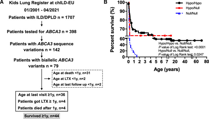
Background: The majority of patients with childhood interstitial lung disease (chILD) caused by pathogenic variants in ATP binding cassette subfamily A member 3 (ABCA3) develop severe respiratory insufficiency within their first year of life and succumb to disease if not lung transplanted. This register-based cohort study reviews patients with ABCA3 lung disease who survived beyond the age of 1 year.

Method: Over a 21-year period, patients diagnosed as chILD due to ABCA3 deficiency were identified from the Kids Lung Register database. 44 patients survived beyond the first year of life and their long-term clinical course, oxygen supplementation and pulmonary function were reviewed. Chest CT and histopathology were scored blindly.

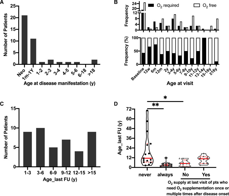
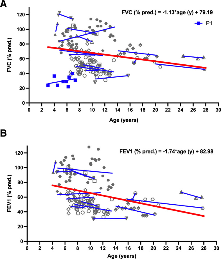
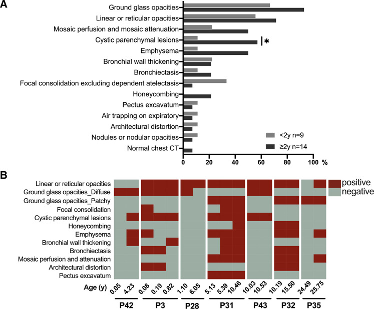
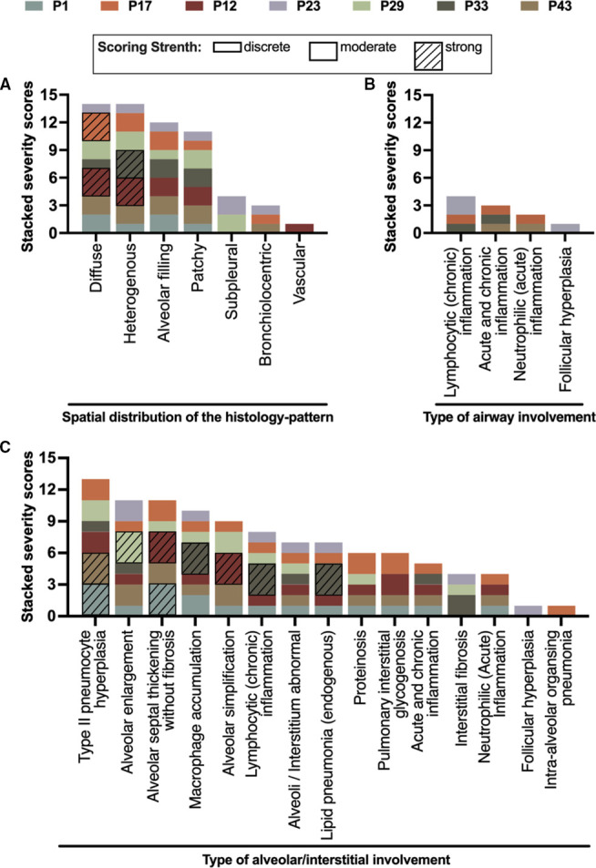
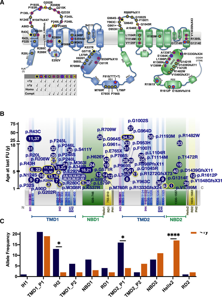
Results: At the end of the observation period, median age was 6.3 years (IQR: 2.8-11.7) and 36/44 (82%) were still alive without transplantation. Patients who had never received supplemental oxygen therapy survived longer than those persistently required oxygen supplementation (9.7 (95% CI 6.7 to 27.7) vs 3.0 years (95% CI 1.5 to 5.0), p=0.0126). Interstitial lung disease was clearly progressive over time based on lung function (forced vital capacity % predicted absolute loss -1.1% /year) and on chest CT (increasing cystic lesions in those with repetitive imaging). Lung histology pattern were variable (chronic pneumonitis of infancy, non-specific interstitial pneumonia, and desquamative interstitial pneumonia). In 37/44 subjects, the ABCA3 sequence variants were missense variants, small insertions or deletions with in-silico tools predicting some residual ABCA3 transporter function.

Conclusion: The natural history of ABCA3-related interstitial lung disease progresses during childhood and adolescence. Disease-modifying treatments are desirable to delay such disease course.

Keywords: ABCA3; paediatric interstitial lung disease; rare lung diseases.


Conflict of interest statement

Competing interests: None declared.

Figures

Figure 1
Figure 1
Flow chart of patient inclusion and survival analysis of patients with two ABCA3 variants in KLR. (A) All patients were enrolled from Kids Lung Register. Numbers in dashed frame: excluded patients. Numbers in solid line frame: included patients. LTX: lung transplantation. (B) For patients who went through genetic tests and diagnosed with biallelic ABCA3 variants, their survival rate was analysed according to different genotypes: hypo/hypo, hypo/null and null/null. chILD,childhood interstitial lung disease; ILD, interstitial lung disease.
Figure 2
Figure 2
Clinical follow-up of patients. (A) The age at the first manifestation of ABCA3-related chILD. Neo: first manifestation in neonatal period. 1m-1Y: from age 1-month-old to 1-year-old. (B) Absolute frequency (upper panel) and percentage of frequency (low panel) of patients’ requirement of oxygen at different visit age. Columns are cumulative frequency of patients during the indicated period. Baseline indicated patients in the neonatal period. 6w: 6 weeks; 12 m: 12 months; 2y: follow-up age from 1 to 2 years; 3-4y (and so forth): follow-up age from 3 to 4 years. (C) The distribution of patients according to the age of their last clinical follow-up (year). (D) O2 supplementation since onset of disease. According to oxygen supplementation changes from onset of disease to the last time of follow-up, patients were divided into four groups. From left to right, the columns represented the number of patients who never required oxygen since onset of disease; who always needed oxygen since onset of disease; who need oxygen once or multiple times after disease onset, but not at the last visit and who need oxygen once or multiple times after disease onset until the last follow-up time. *p<0.05 with Tukey’s test; **p<0.001. chILD, childhood interstitial lung disease.
Figure 3
Figure 3
Pulmonary function results of patients with repeated tests 14 patients preformed repeated pulmonary function test. (A) Predicted forced vital capacity (FVC%). (B) Predicted forced expiratory volume (FEV1%). The individual patients’ measurements were indicated by different symbols consistent for each patient in (A) and (B) (P1 in blue; due to short expiration no FEV1 value was available; P1 thus not included in overall regression analysis. Other patients in grey). For illustration of individual courses, linear regression lines were given (in blue). For overall regression, a mixed model for repeated measures was calculated for FVC (% predicted) and FEV1 (% predicted) against time (red line in bold).
Figure 4
Figure 4
Thorax HR-CT results. (A) HR-CT results of patients younger (n=9) and older (n=14) than 2 years old (total n=23). (B) Seven patients had repetitive HR-CT. Repeated results of each patient were presented as conjunct bars to observe CT patterns at different ages. Red blocks indicate the presence of a pattern in the CT scan at the age, grey blocks indicate the absence of the pattern. *p<0.05 with Tukey’s test indicates differences between age groups. HR-CT, high-resolution CT.
Figure 5
Figure 5
Histology of seven patients (each patient has a unique and different colour) had a lung biopsy. One pathologist trained in chILD reviewed all the histology, and analysed the pattern scores by severity (0=none, 1=discrete, 2=moderate and 3=strong). The severity is graphically expressed by the height of each column element on the y-axis. (A) Spatial distribution of the histopattern. (B). Type of airway involvement. (C). Type of alveolar/interstitial involvement. chILD, childhood interstitial lung disease.
Figure 6
Figure 6
Domain organisation of ABCA3 sequence variations observed in the cohort collected by the Kids Lung Register. (A) Frameshift, nonsense, missense, splice site, in-frame insertion/deletion variants were plotted according to the amino acid site, intronic variants not indicated. Variants plotted in blue: carrier survived beyond 1 year; plotted in orange: patients’ death before 1 year; edged yellow or blue dots: homozygous variants; dots with asterisk: compound heterozygous mutations. The position of TMDs and NBDs of ABCA3 were referred to the prediction algorithms of Onnée et al. (B). Age of survival of the mutations’ carrier (y-axis) in dependency of amino acid position (x-axis). For variants occurring in multiple patients, median survival age of carriers was indicated. Blue dots with yellow edge: homozygous variants; flat dots without edge: compound heterozygous variants. Number(s) inside dots: No of patients as listed in online supplemental table S1. Asterisk inside p.E292V dot indicated this variant was carried by multiple patients. (C) Allele frequency of variants for patients surviving >1 year or <1 year was counted in each domain and compared by Fisher’s exact test (*p<0.05, ***p<0.001). IHs, intracellular helices. TMD1_P1: part 1 of transmembrane domain 1, from p.K21 to p.R289. TMD1_P2: part 2 of transmembrane domain 1, from p.S301 to p.G450. TMD2_P1: part 1 of transmembrane domain 2, from p.M925 to p.Q1126. TMD2_P2: part 2 of transmembrane domain 2, from p.S1134 to p.E1325 (NP_001080.2). EHs, exocytoplasmic helices; IHs, intracellular helices; PHs, pinning helix; RDs, regulatory domains.

References

    1. Shulenin S, Nogee LM, Annilo T, et al. . A mutation in the surfactant protein C gene associated with familial interstitial lung disease. N Engl J Med 2004;350:1296–303. 10.1056/NEJMoa032178. - DOI - PubMed
    1. Kröner C, Wittmann T, Reu S, et al. . Lung disease caused by ABCA3 mutations. Thorax 2017;72:213–20. 10.1136/thoraxjnl-2016-208649 - DOI - PubMed
    1. Wambach JA, Casey AM, Fishman MP, et al. . Genotype-Phenotype correlations for infants and children with ABCA3 deficiency. Am J Respir Crit Care Med 2014;189:1538–43. 10.1164/rccm.201402-0342OC - DOI - PMC - PubMed
    1. Beers MF, Knudsen L, Tomer Y, et al. . Aberrant lung remodeling in a mouse model of surfactant dysregulation induced by modulation of the Abca3 gene. Ann Anat 2017;210:135–46. 10.1016/j.aanat.2016.11.015 - DOI - PMC - PubMed
    1. Doan ML, Guillerman RP, Dishop MK, et al. . Clinical, radiological and pathological features of ABCA3 mutations in children. Thorax 2008;63:366–73. 10.1136/thx.2007.083766 - DOI - PubMed

Publication types

Substances