. 2023 May;531(7):814-835.
doi: 10.1002/cne.25463. Epub 2023 Feb 20.

Characterization of Fragile X Mental Retardation Protein expression in human nociceptors and their axonal projections to the spinal dorsal horn

Affiliations

Characterization of Fragile X Mental Retardation Protein expression in human nociceptors and their axonal projections to the spinal dorsal horn

Molly E Mitchell et al. J Comp Neurol. 2023 May.

Abstract

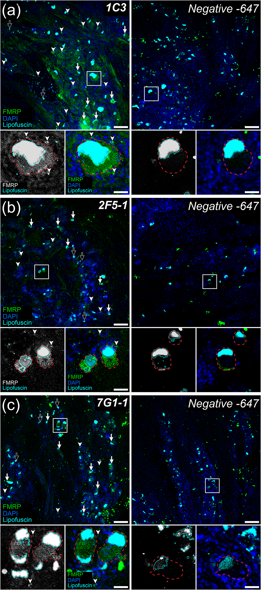
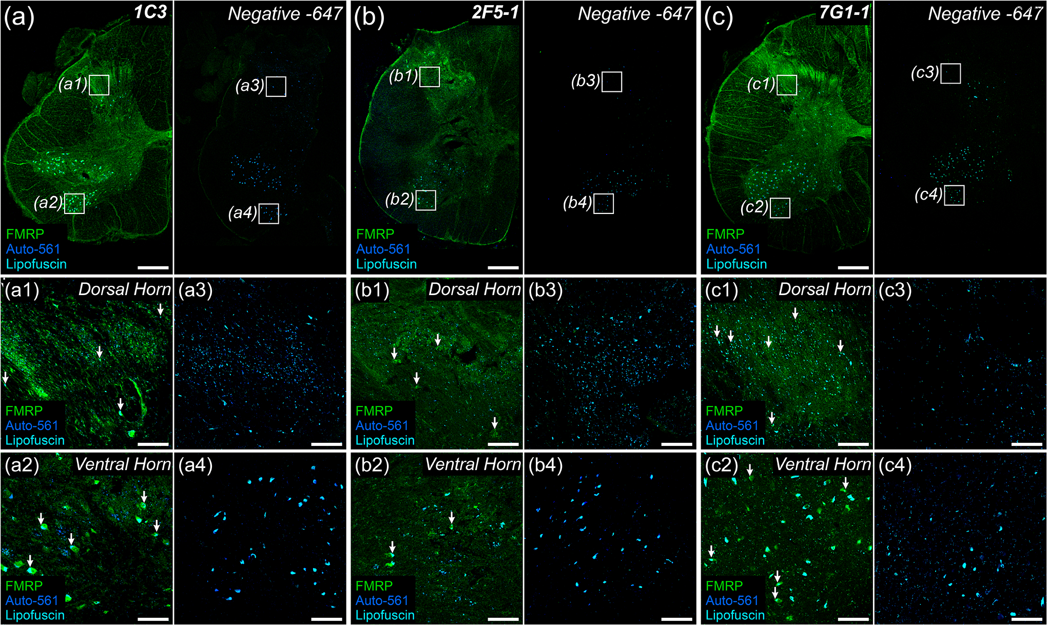
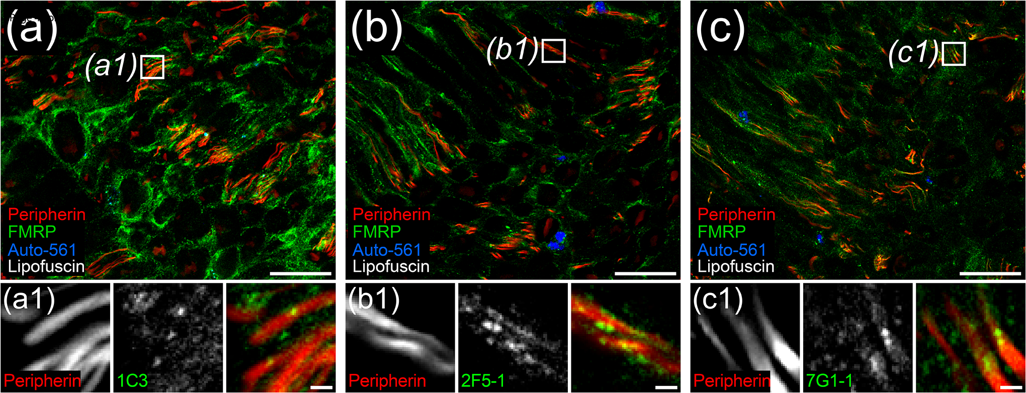
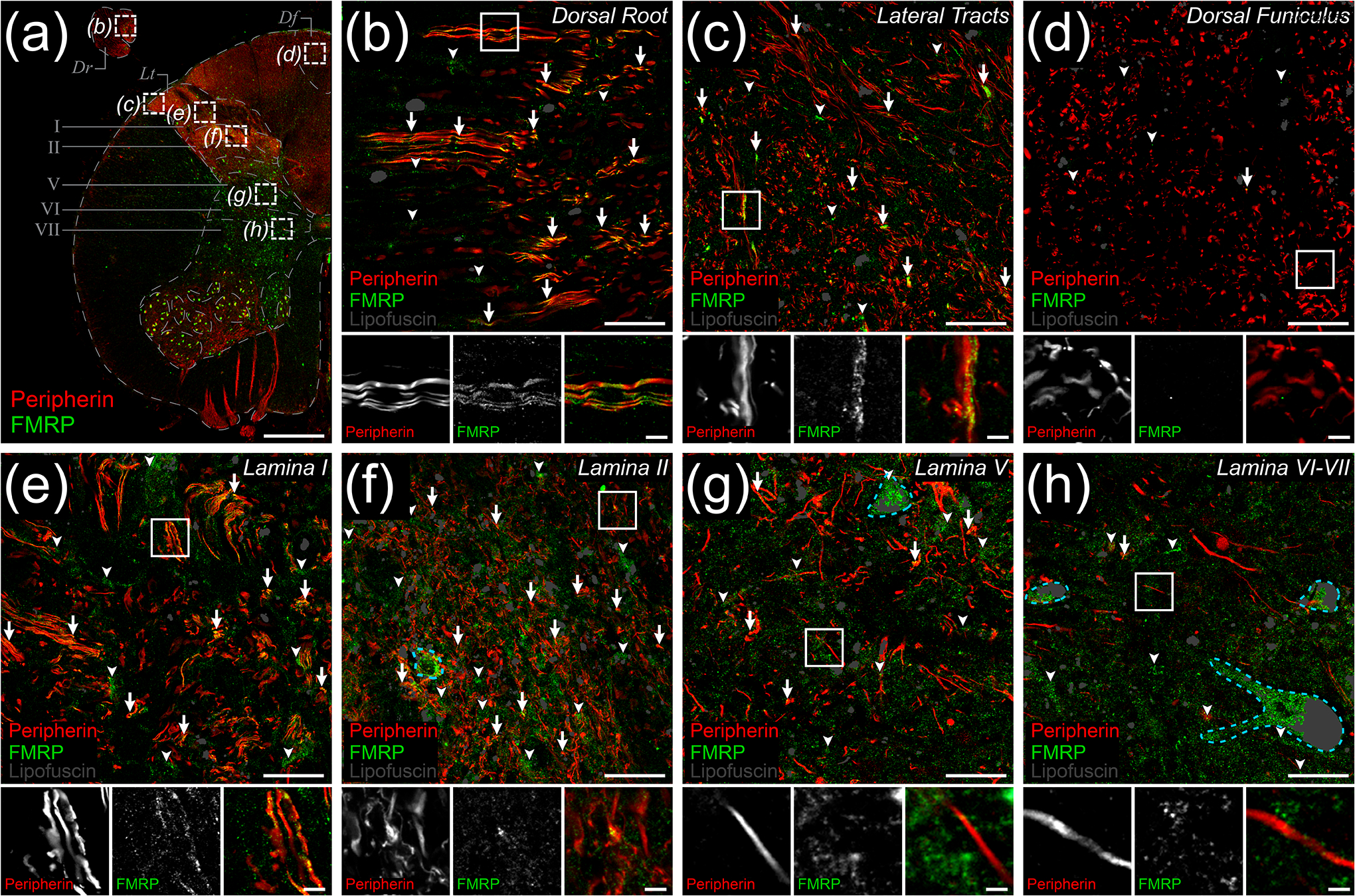
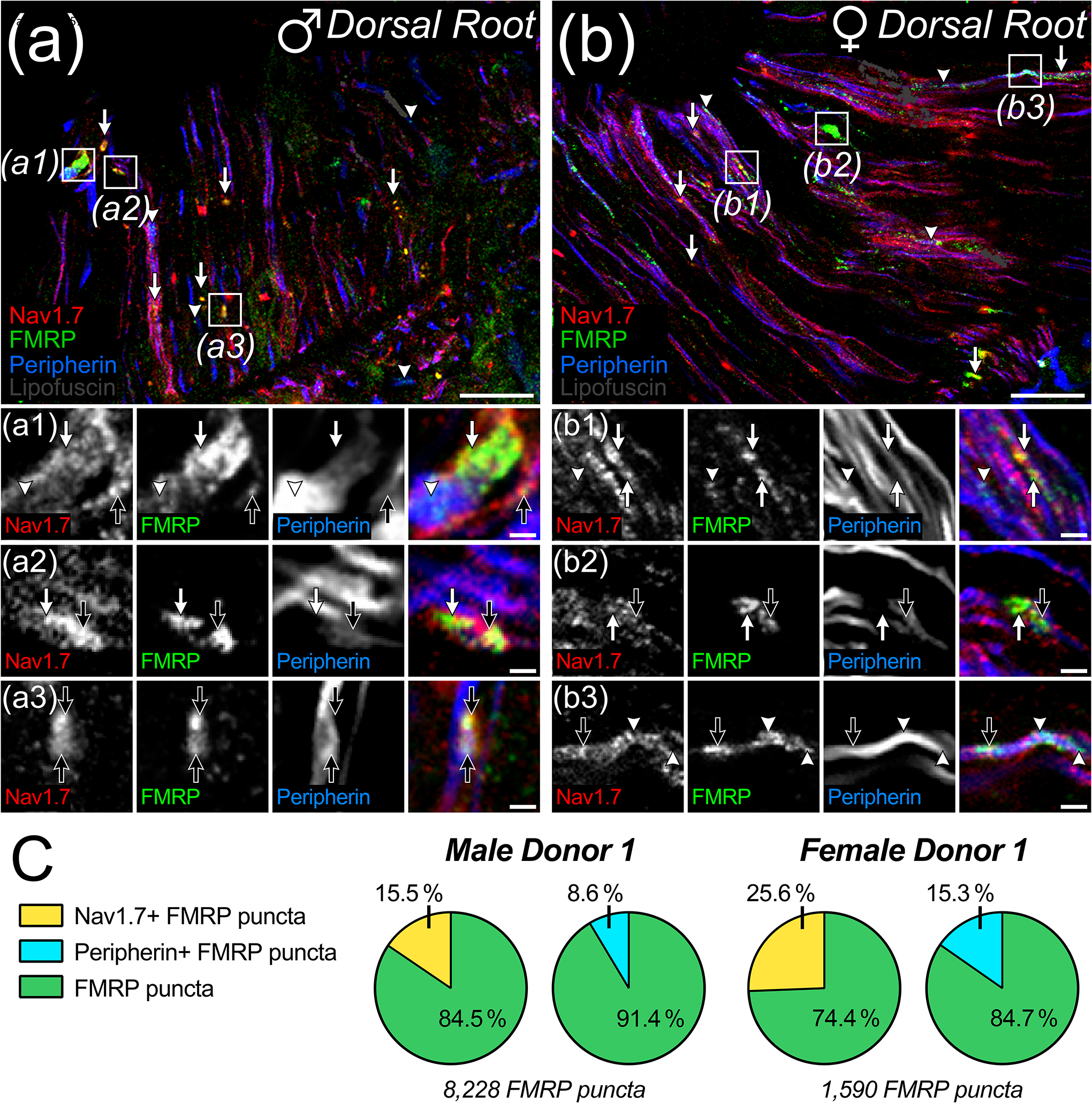
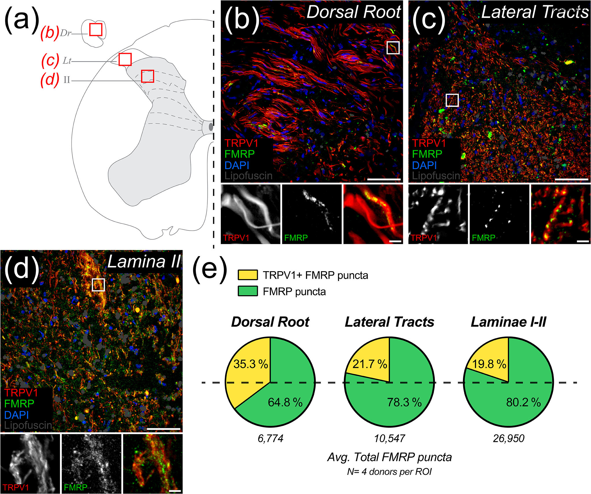
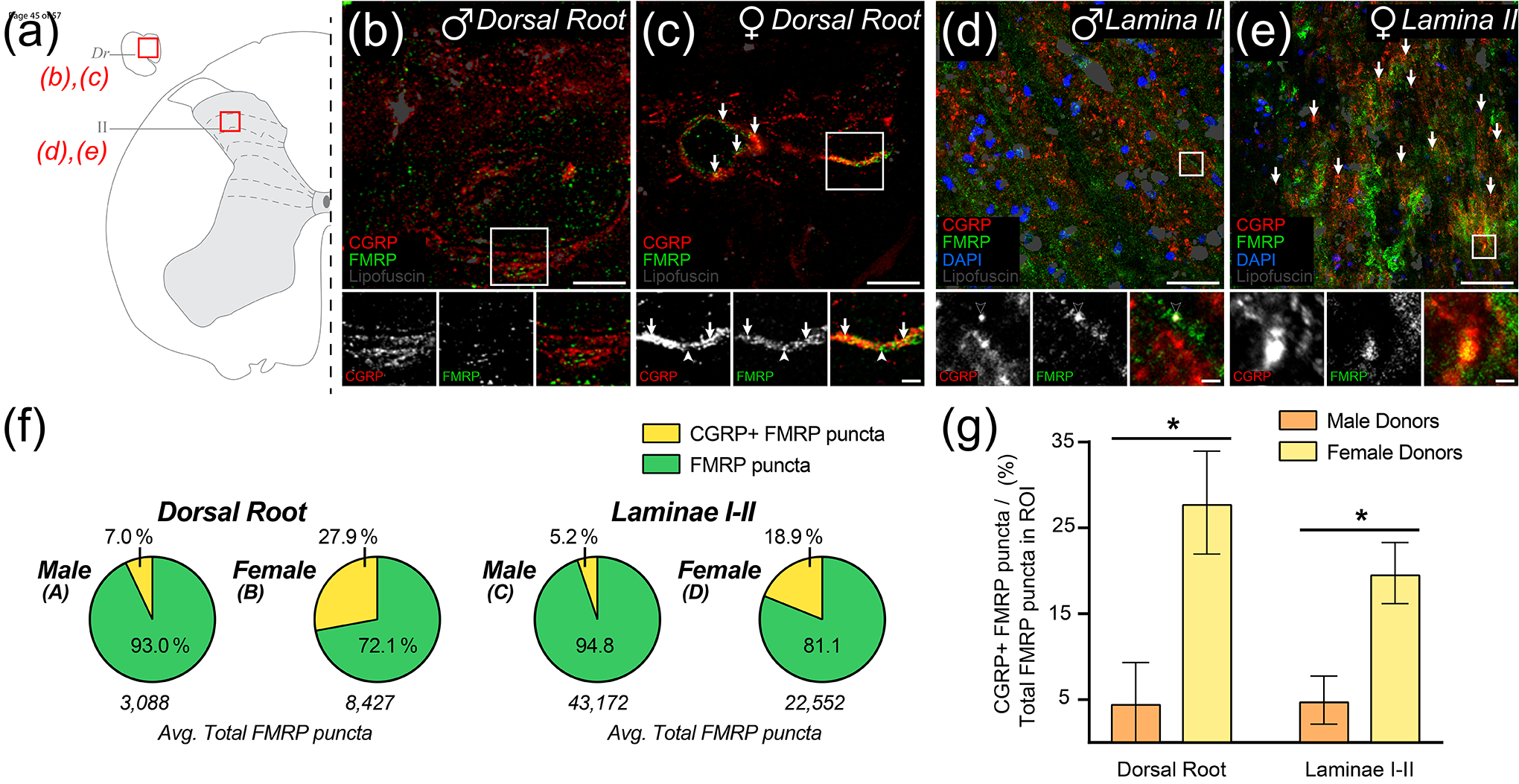
Fragile X Mental Retardation Protein (FMRP) regulates activity-dependent RNA localization and local translation to modulate synaptic plasticity throughout the central nervous system. Mutations in the FMR1 gene that hinder or ablate FMRP function cause Fragile X Syndrome (FXS), a disorder associated with sensory processing dysfunction. FXS premutations are associated with increased FMRP expression and neurological impairments including sex dimorphic presentations of chronic pain. In mice, FMRP ablation causes dysregulated dorsal root ganglion (DRG) neuron excitability and synaptic vesicle exocytosis, spinal circuit activity, and decreased translation-dependent nociceptive sensitization. Activity-dependent, local translation is a key mechanism for enhancing primary nociceptor excitability that promotes pain in animals and humans. These works indicate that FMRP likely regulates nociception and pain at the level of the primary nociceptor or spinal cord. Therefore, we sought to better understand FMRP expression in the human DRG and spinal cord using immunostaining in organ donor tissues. We find that FMRP is highly expressed in DRG and spinal neuron subsets with substantia gelatinosa exhibiting the most abundant immunoreactivity in spinal synaptic fields. Here, it is expressed in nociceptor axons. FMRP puncta colocalized with Nav1.7 and TRPV1 receptor signals suggesting a pool of axoplasmic FMRP localizes to plasma membrane-associated loci in these branches. Interestingly, FMRP puncta exhibited notable colocalization with calcitonin gene-related peptide (CGRP) immunoreactivity selectively in female spinal cord. Our results support a regulatory role for FMRP in human nociceptor axons of the dorsal horn and implicate it in the sex dimorphic actions of CGRP signaling in nociceptive sensitization and chronic pain.


Conflict of interest statement

Conflict of Interest Statement

The authors declare that they have no conflicts of interest related to this work.

Figures

Figure 1.
Figure 1.. FMRP immunofluorescence in adult human dorsal root ganglion
Representative confocal micrographs showing FMRP immunofluorescence (Green) in adult human dorsal root ganglion using the 1C3 (a), 2F5-1 (b), or 7G1-1 (c) antibodies. DAPI counterstain (Blue) shows glial nuclei for tissue organization. Lipofuscin were identified as outlined in methods (Cyan) and are present in all images. Negative controls were treated exactly as the matched FMRP immunostained tissue but were exposed only to Alexa 647-conjugated secondaries. DRG neuronal cell bodies (Insets: Red dotted lines) were identified using their large size, placement within circumscribing nuclei of local satellite glia by DAPI, and robust somatic lipofuscin. For all FMRP primary antibodies tested, most DRG neuron somata exhibited FMRP IF (Arrows) with only a few appearing negative for FMRP (Black Arrows). These antibodies also exhibited signal consistent with FMRP staining in a subset of glia (Arrowheads). High magnification insets (White Boxes) show FMRP signal in DRG somata (Red dotted line) with surrounding glial nuclei (Blue). (a) Robust 1C3 signal was detected in a large subset of neuronal somata and frequently in glial cells. (b) Strong 2F5-1 signal was observed in most neuronal cell bodies and in some glia. (c) Strong 7G1-1 signal was evident in neuronal somata and in few glia. Section thickness: 60 μm Scale bars: Outsets = 100 μm; Insets = 30 μm.
Figure 2.
Figure 2.. FMRP immunofluorescence in adult human spinal cord
Representative confocal micrographs of FMRP immunofluorescence (Green) in adult human spinal cord using the 1C3 (a), 2F5-1 (b), or 7G1-1 (c) antibodies. Autofluorescence (Blue) highlighted lipofuscin (Cyan). Negative controls were handled as described in Figure 1. 1.25x images of lumbar hemi-cross sections show grey matter regions with most intense FMRP signal per primary antibody. a1–c4: 10x insets (Boxes) highlighting the consistency of increased FMRP signal in substantia gelatinosa (Dorsal Horn) or in lower motor neuron pools (Ventral Horn) across primary antibodies. FMRP IF was observed in neuronal cell bodies as identified by their large size and robust lipofuscin (Arrows). (a) 1C3 exhibited the brightest signal overall with the strongest signal in substantia gelatinosa (a1) and motor neuron pools (a2). (b) 2F5-1 signal was increased in neuropil within substantia gelatinosa (b1) when compared to ventral grey (b2). Note the disrupted morphology due to antigen retrieval. (c) 7G1-1 signal in somata and neuropil was comparable to that of 2F5-1 in substantia gelatinosa (c1) and ventral grey (c2). Section thickness: 60 μm Scale bars: Outsets = 1 mm; Insets = 100 μm.
Figure 3.
Figure 3.. FMRP immunofluorescence in human dorsal root axons
Representative confocal micrographs of peripherin (Red) and FMRP (Green) immunostaining with 1C3 (a), 2F5-1 (b), or 7G1-1 (c) antibodies in human dorsal root. Autofluorescence (Blue) highlighted lipofuscin (White). High magnification insets (Boxes) show punctate FMRP signal colocalization with peripherin+ DRG axons for all FMRP antibodies tested. (a) 1C3 signal was the most intense overall out the FMRP antibodies in dorsal root. (b) The brightest 2F5-1 signal was often punctate as previously documented in axons within human hippocampus sections (Akins et al., 2017). (c) 7G1-1 signal was similar to 2F5-1 signal. Section thickness: 60 μm. Scale bars: Magnified Images = 50 μm; Insets = 5 μm.
Figure 4.
Figure 4.. Peripherin and FMRP immunofluorescence in human dorsal horn
Representative confocal micrographs of immunostaining for peripherin (Red) and FMRP (Green) within dorsal horn in human spinal cord. Lipofuscin were identified as shown (Grey) to avoid false positives. (a) 1.25x image of lumbar hemi-cross section depicting dorsal horn ROIs [Dotted boxes (b)-(h)] for 40x images used to assess peripherin and FMRP signal overlap. Images were taken of the attached dorsal rootlet (Dr), lateral white matter tracts (Lt), dorsal funiculus (Df), superficial dorsal laminae (I-II), or deep dorsal laminae (V-VII). (b)-(h): Magnified 40x images representing ROIs containing DRG axons and branches as identified by peripherin IF. The frequency of FMRP puncta and peripherin IF colocalization (Arrows) differed between ROIs. The proportion of FMRP puncta that did not colocalize with peripherin+ fibers (Arrowheads) was notably increased in grey matter ROIs. In grey matter laminae, FMRP IF was present in neuronal cell bodies (Dotted cyan traces). Associated insets (Boxes) are further magnified to highlight the sub-axonal morphology of signal overlap. (b) Image of dorsal root shows FMRP puncta frequently colocalized with DRG axons. (c) Image of lateral tracts show FMRP puncta often colocalized with peripherin+ axons at the spinal border of the DREZ. (d) Image of dorsal funiculus shows virtually no colocalization between FMRP puncta and ascending peripherin+ arbors. (e) Image of lamina II neuropil showing frequent colocalization between FMRP puncta and peripherin+ fibers. (f) Image of lamina II neuropil shows FMRP puncta often colocalized with peripherin+ spinal branches in these synaptic fields. (g) Image of lamina V neuropil shows dramatically reduced colocalization between FMRP puncta and peripherin IF. (h) Image of lamina VI-VII neuropil shows FMRP puncta rarely colocalized with peripherin+ fibers in these synaptic fields. Section thickness: 60 μm. Scale bars: Outset = 1 mm; Magnified Images = 20 μm; Insets = 5 μm.
Figure 5.
Figure 5.. Colocalization of Nav1.7 and/or peripherin with FMRP immunofluorescence in dorsal root
(a)-(b): Representative confocal micrographs of immunostaining for Nav1.7 (Red), FMRP (Green), and peripherin (Blue) with lipofuscin (Grey) from attached dorsal root in male (a) and female (b) human spinal cord. Magnified 40x images representing dorsal root ROIs used to detect FMRP puncta colocalization with Nav1.7 (Arrows) and/or peripherin (Arrowheads) staining. a1–b3: Further magnified insets (Boxes) highlight the sub-axonal organization of these stains. and show varying colocalization amongst all three signals within an arbor (Black Arrows). (a) Colocalized FMRP puncta often overlapped with Nav1.7+ and peripherin+ loci in dorsal root arbors from male spinal cord. (b) Colocalized FMRP puncta often overlapped with Nav1.7+ or peripherin loci in dorsal root arbors from female spinal cord. (c) Pie charts showing the average proportion of FMRP puncta in dorsal root per male or female donor that colocalized with Nav1.7 and/or peripherin IF. Section thickness: 60 μm. Scale bars: Magnified Images = 10 μm; Insets = 3 μm.
Figure 6.
Figure 6.. Colocalization of TRPV1 and FMRP immunofluorescence in human nociceptive circuits
Representative confocal micrographs of human nociceptive circuitry as identified by TRPV1 staining (Red) with FMRP IF (Green), DAPI (Blue), and identified lipofuscin (Grey) in human spinal cord. (a) Schematic of lumbar hemi-section depicting ROIs from attached dorsal root, Dr (b); lateral tracts, Lt (c); or laminae II (d). (b)-(d): Magnified 40x images represent nociceptive circuit ROIs used to detect the proportions of FMRP puncta present that colocalized with TRPV1+ fibers. Associated insets (Boxes) are further magnified to highlight the sub-axonal morphology of this colocalization. (b) Image of dorsal root shows FMRP puncta frequently colocalized with TRPV1+ axons. Glial nuclei rarely colocalized with FMRP. (c) Image of lateral tracts shows FMRP puncta often colocalized with these TRPV1+ arbors. (d) Image of lamina I-II neuropil showing notable colocalization between FMRP puncta and TRPV1+ fibers in these synaptic fields. (e) Pie charts showing the average proportion of FMRP puncta per nociceptive circuit ROI the did or did not colocalize with peripherin IF. Section thickness: 60 μm. Scale bars: Magnified Images = 50 μm; Insets = 5 μm.
Figure 7.
Figure 7.. Colocalization of CGRP and FMRP immunofluorescence in dorsal root and laminae I-II
Representative confocal micrographs of CGRP (Red) and FMRP (Green) immunofluorescence with lipofuscin (Grey) in dorsal root and laminae I-II in male (b), (d) and female (c), (e) human spinal cord. (a) Schematic of lumbar hemi-section exemplifying ROIs from attached dorsal root, Dr (b)-(c) or laminae II (d)-(e). (b)-(c): Magnified 40x images representing ROIs used to detect colocalization between CGRP and FMRP from attached dorsal root. Colocalization was notably rare in tissues from male donors but was notably frequent in tissues from female donors (Arrows). Associated insets (Boxes) are further magnified highlighting the sub-axonal organization of these stains. (b) Image from a male donor showing that almost all FMRP puncta are spatially separated from CGRP+ loci. Few puncta outside our detection parameters rarely colocalize with CGRP staining (Black Arrow). (c) Image from a female donor showing FMRP puncta frequently colocalized with CGRP+ loci. (d)-(e): Magnified 40x images of laminae I-II were counterstained with DAPI (Blue) to show neuropil and somatic organization. (d) Image from a male donor showing that almost all FMRP puncta are spatially separated from CGRP+ loci as in dorsal root. Potential colocalized puncta were often identified as lipofuscin (Black Arrowhead). (e) Image from a female donor showing frequent colocalization between CGRP and FMRP puncta. (f) Pie charts showing the average proportion of FMRP puncta per ROI the did or did not colocalize with CGRP+ loci. (g) Comparisons showed that colocalization between FMRP puncta and CGRP+ loci was significantly increased in females when compared to males for both dorsal root (p= 0.0375) and laminae I-II (p= 0.0310) analyses. Values expressed as mean +/− SEM. T-test: *p < 0.05 (N = 3 per group). Section thickness: 60 μm. Scale bars: Dorsal root images = 15 μm; Laminae I-II images = 50 μm; All insets = 5 μm.

Similar articles

Cited by

References

    1. Abboud C, Duveau A, Bouali-Benazzouz R, Massé K, Mattar J, Brochoire L, Fossat P, Boué-Grabot E, Hleihel W, & Landry M (2021). Animal models of pain: Diversity and benefits. Journal of Neuroscience Methods, 348, 108997. 10.1016/j.jneumeth.2020.108997 - DOI - PubMed
    1. Akins MR, Berk-Rauch HE, Kwan KY, Mitchell ME, Shepard KA, Korsak LIT, Stackpole EE, Warner-Schmidt JL, Sestan N, Cameron HA, & Fallon JR (2017). Axonal ribosomes and mRNAs associate with fragile X granules in adult rodent and human brains. Human Molecular Genetics, 26(1), 192–209. 10.1093/hmg/ddw381 - DOI - PMC - PubMed
    1. Akins MR, LeBlanc HF, Stackpole EE, Chyung E, & Fallon JR (2012). Systematic mapping of fragile X granules in the mouse brain reveals a potential role for presynaptic FMRP in sensorimotor functions. Journal of Comparative Neurology, 520(16), 3687–3706. 10.1002/cne.23123 - DOI - PMC - PubMed
    1. Alelú-Paz R, Iturrieta-Zuazo I, Byne W, Haroutunian V, García-Villanueva M, Rábano A, García-Amado M, Prensa L, & Giménez-Amaya JM (2008). A New Antigen Retrieval Technique for Human Brain Tissue. PLOS ONE, 3(10), e3378. 10.1371/journal.pone.0003378 - DOI - PMC - PubMed
    1. An JW, Koh JC, Sun JM, Park JY, Choi JB, Shin MJ, & Lee YW (2016). Clinical Identification of the Vertebral Level at Which the Lumbar Sympathetic Ganglia Aggregate. The Korean Journal of Pain, 29(2), 103–109. 10.3344/kjp.2016.29.2.103 - DOI - PMC - PubMed

Publication types

Substances