Review
. 2022 Oct-Dec;63(4):599-606.
doi: 10.47162/RJME.63.4.01.

Radiographic methods for locating impacted maxillary canines

Affiliations
Review

Radiographic methods for locating impacted maxillary canines

Ştefania Dinu et al. Rom J Morphol Embryol. 2022 Oct-Dec.

Abstract

Maxillary canine impaction is a fairly common phenomenon in dental practice. Most studies indicate its palatal position. For a successful orthodontic and∕or surgical therapy, it is necessary to correctly identify the impacted canine in the depth of the maxillary bone, using conventional and digital radiological investigations, each with their advantages and disadvantages. Dental practitioners must indicate the most "targeted" radiological investigation. This paper aims to review the various radiographic techniques available for determining the location of the impacted maxillary canine.


Conflict of interest statement

The authors declare that they have no conflict of interests.

Figures

Figure 1
Figure 1
(A) Periapical radiograph of the central incisors, highlighting the crown of the included canine over the distal half of the root of the left incisor; (B) Rotated X-ray 30° to the left shows the overlap of the canine only on the root of the central incisor; (C) Rotating the X-rays more than 30°, the palatal inclusion of the canine is observed, without any overlap [22]. (Modified image).
Figure 2
Figure 2
(A) The first periapical film taken from a correct angle to the crown highlights the overlap of the right maxillary canine over the root of the incisor; (B) The second radiograph is placed in the same position, except that the X-ray beam is moved distally, 20°, and the crown of the canine appears to move with it, as it is found closer to the film than the other teeth [23]. (Modified image).
Figure 3
Figure 3
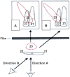
(A and B) Graphic representation of the “parallax” method. If the observer’s eye looks along the axis of the X-ray beam, in each case, the image on the film can be easily reconstructed [22, 23]. (Modified image).
Figure 4
Figure 4
Vertical angulation between orthopantomogram (OPT) and maxillary anterior occlusal.
Figure 5
Figure 5
Upper occlusal radiograph of a buccally impacted canine [19]. (Modified image).
Figure 6
Figure 6
Panoramic radiograph showing the impaction of the left maxillary canine. (Personal case).
Figure 7
Figure 7
Increasing the distance between the tooth and the film increases the magnification [19]. (Modified image).
Figure 8
Figure 8
(A) Canines begin eruption with their long axis aligned with the distal surface of the lateral incisor as they descend from the floor of the nasal orifice (yellow line). Bony crypt of the left maxillary canine (blue arrow); (B) The panoramic radiograph shows an apparent mesial inclination of the maxillary canine (yellow arrows), requiring caution because the panoramic is not a frontal view, but an average of radiographic images of lateral and frontal projections [29]. (Modified image).
Figure 9
Figure 9
Canine direction on the lateral cephalogram versus perpendicular to the Frankfurt plane [5]. (Modified image).
Figure 10
Figure 10
Palatal position of the impacted maxillary canine cusp. (Personal case).
Figure 11
Figure 11
3D CT scan: coronal, sagittal and axial section. (Personal case). 3D: Three-dimensional; CT: Computed tomography.
Figure 12
Figure 12
(A) CBCT aspect of the palatal impacted maxillary left canine, axial section; (B) The left upper maxilla, palatal view: 3D reconstruction on CBCT. (Personal case). 3D: Three-dimensional; CBCT: Cone-beam computed tomography.

References

    1. Akan S, Oktay H. Cone beam tomography and panoramic radiography in localization of impacted maxillary canine and detection of root resorption. Stoma Edu J. 2021;8(2):106–112.
    1. Alassiry A. Radiographic assessment of the prevalence, pattern and position of maxillary canine impaction in Najran (Saudi Arabia) population using orthopantomograms – a cross-sectional, retrospective study. Saudi Dent J. 2020;32(3):155–159. - PMC - PubMed
    1. Alamri A, Alshahrani N, Al-Madani A, Shahin S, Nazir M. Prevalence of impacted teeth in Saudi patients attending dental clinics in the Eastern Province of Saudi Arabia: a radiographic retrospective study. ScientificWorldJournal. 2020;2020:8104904–8104904. - PMC - PubMed
    1. El Beshlawy. Radiographic assessment of impacted maxillary canine position using CBCT: a comparative study of 2 methods. Egypt Dent J. 2019;65(4):3393–3402.
    1. Kumar S, Mehrotra P, Bhagchandani J, Singh A, Garg A, Kumar S, Sharma A, Yadav H. Localization of impacted canines. J Clin Diagn Res. 2015;9(1):ZE11–ZE14. - PMC - PubMed