

Cerebral blood flow pulsatility and cerebral artery stiffness acutely decrease during hemodialysis
- PMID: 36808481
- PMCID: PMC9937783
- DOI: 10.14814/phy2.15595
Cerebral blood flow pulsatility and cerebral artery stiffness acutely decrease during hemodialysis
Abstract
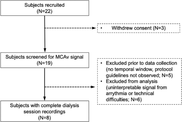
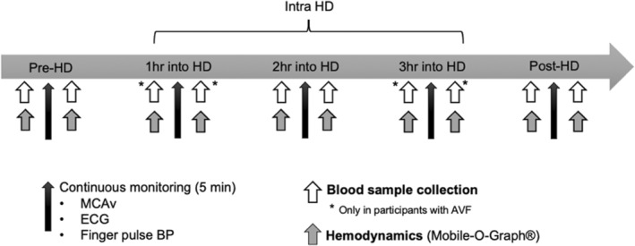
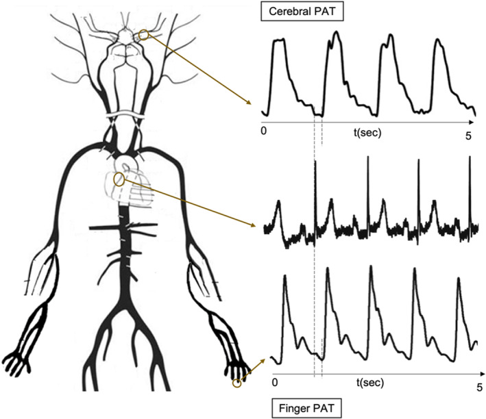
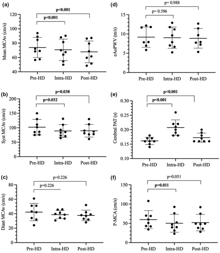
End-stage kidney disease (ESKD) is associated with increased arterial stiffness and cognitive impairment. Cognitive decline is accelerated in ESKD patients on hemodialysis and may result from repeatedly inappropriate cerebral blood flow (CBF). The aim of this study was to examine the acute effect of hemodialysis on pulsatile components of CBF and their relation to acute changes in arterial stiffness. In eight participants (age: 63 ± 18 years, men: 5), CBF was estimated using middle cerebral artery blood velocity (MCAv) assessed with transcranial Doppler ultrasound before, during, and after a single hemodialysis session. Brachial and central blood pressure, along with estimated aortic stiffness (eAoPWV) were measured using an oscillometric device. Arterial stiffness from heart to MCA was measured as the pulse arrival time (PAT) between electrocardiogram (ECG) and transcranial Doppler ultrasound waveforms (cerebral PAT). During hemodialysis, there was a significant reduction in mean MCAv (-3.2 cm/s, p < 0.001), and systolic MCAv (-13.0 cm/s, p < 0.001). While baseline eAoPWV (9.25 ± 0.80 m/s) did not significantly change during hemodialysis, cerebral PAT increased significantly (+0.027 , p < 0.001) and was associated with reduced pulsatile components of MCAv. This study shows that hemodialysis acutely reduces stiffness of arteries perfusing the brain along with pulsatile components of blood velocity.
Keywords: arterial stiffness; cerebral pulsatility index; end-stage kidney disease; hemodialysis; middle cerebral artery mean blood velocity; pulse wave velocity.
© 2023 The Authors. Physiological Reports published by Wiley Periodicals LLC on behalf of The Physiological Society and the American Physiological Society.
Conflict of interest statement
None.
Figures
References
-
- Ali, H. , Soliman, K. , Mohamed, M. M. , Daoud, A. , Shafiq, T. , Fülöp, T. , & Baharani, J. (2021). The effects of dialysis modality choice on cognitive functions in patients with end‐stage renal failure: A systematic review and meta‐analysis. International Urology and Nephrology, 53(1), 155–163. 10.1007/s11255-020-02603-x - DOI - PubMed
-
- Alwatban, M. R. , Aaron, S. E. , Kaufman, C. S. , Barnes, J. N. , Brassard, P. , Ward, J. L. , Miller, K. B. , Howery, A. J. , Labrecque, L. , & Billinger, S. A. (2021). Effects of age and sex on middle cerebral artery blood velocity and flow Pulsatility index across the adult lifespan. Journal of Applied Physiology, 130(6), 1675–1683. 10.1152/japplphysiol.00926.2020 - DOI - PMC - PubMed
-
- Balestrini, C. S. , Al‐Khazraji, B. K. , Suskin, N. , & Shoemaker, J. K. (2020). Does vascular stiffness predict white matter Hyperintensity burden in ischemic heart disease with preserved ejection fraction? American Journal of Physiology. Heart and Circulatory Physiology, 318(6), H1401–H1409. 10.1152/ajpheart.00057.2020 - DOI - PubMed
Publication types
MeSH terms
LinkOut - more resources
Full Text Sources
Medical
