

Tumor-associated macrophage-derived GDNF promotes gastric cancer liver metastasis via a GFRA1-modulated autophagy flux
- PMID: 36808605
- PMCID: PMC10060314
- DOI: 10.1007/s13402-022-00751-z
Tumor-associated macrophage-derived GDNF promotes gastric cancer liver metastasis via a GFRA1-modulated autophagy flux
Abstract
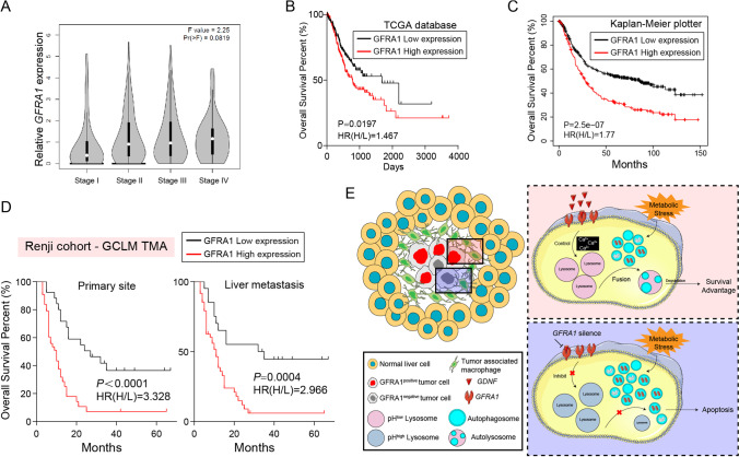
Purpose: Liver metastasis, a lethal malignancy of gastric cancer (GC) patients, execrably impairs their prognosis. As yet, however, few studies have been designed to identify the driving molecules during its formation, except screening evidence pausing before their functions or mechanisms. Here, we aimed to survey a key driving event within the invasive margin of liver metastases.
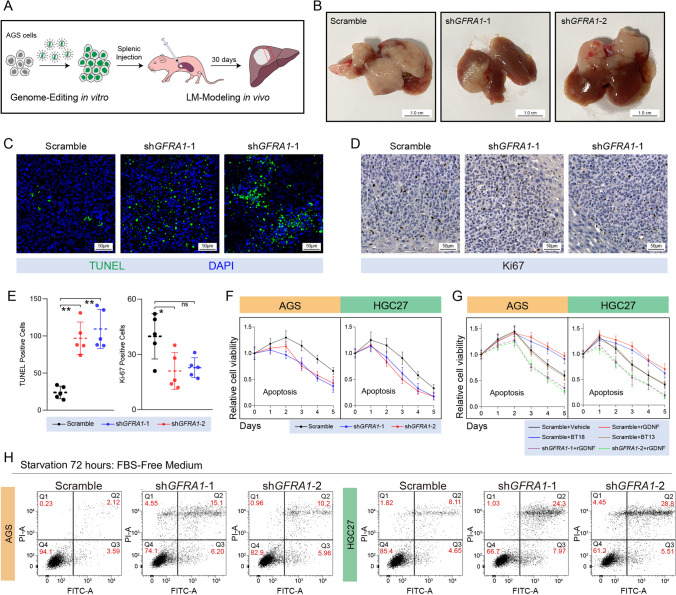
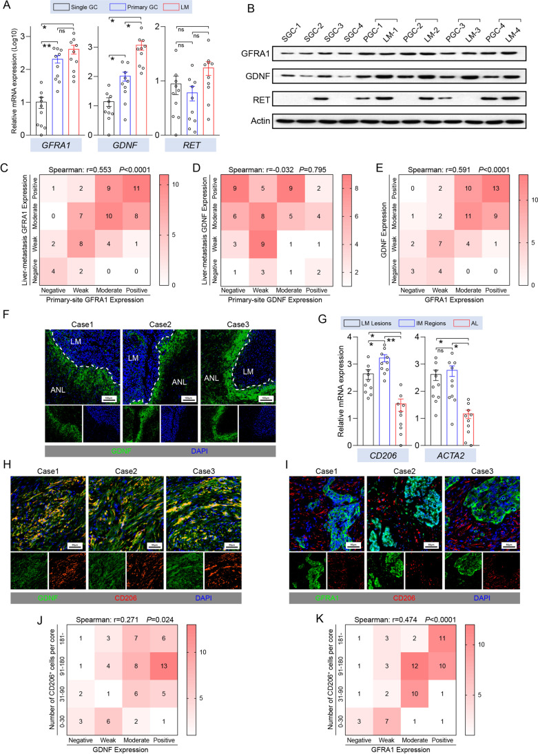
Methods: A metastatic GC tissue microarray was used for exploring malignant events during liver-metastasis formation, followed by assessing the expression patterns of glial cell-derived neurotrophic factor (GDNF) and GDNF family receptor alpha 1 (GFRA1). Their oncogenic functions were determined by both loss- and gain-of-function studies in vitro and in vivo, and validated by rescue experiments. Multiple cell biological studies were performed to identify the underlying mechanisms.
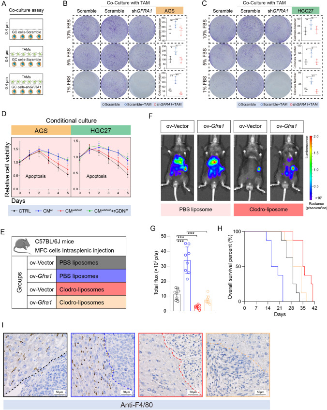
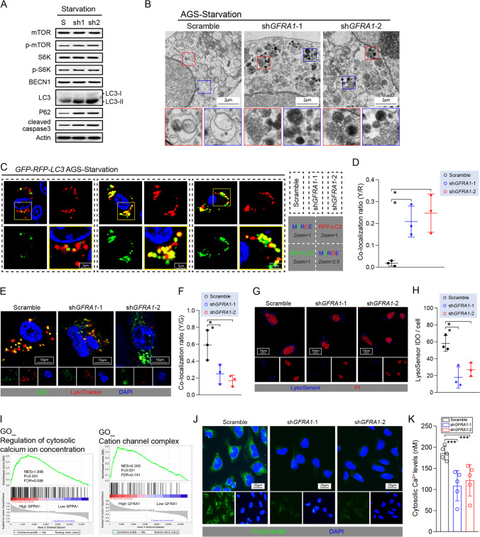
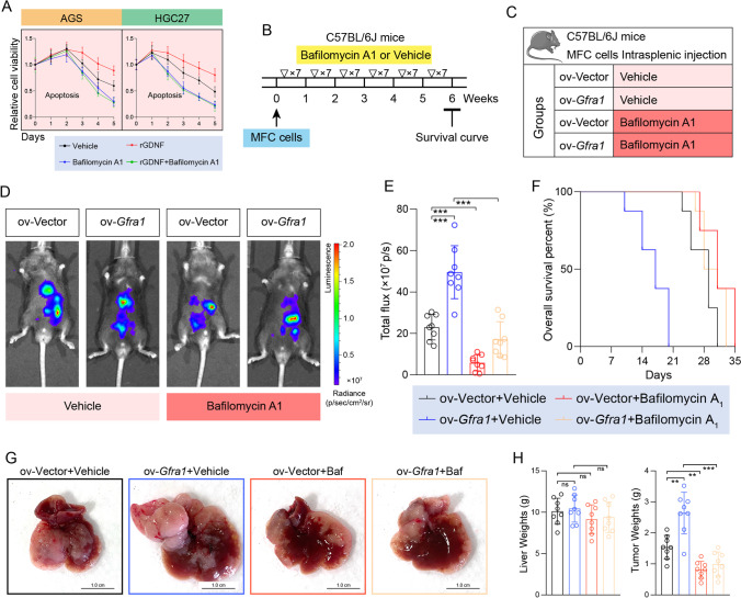
Results: In the invasive margin, GFRA1 was identified as a pivotal molecule involved in cellular survival during liver metastasis formation, and we found that its oncogenic role depends on tumor associated macrophage (TAM)-derived GDNF. In addition, we found that the GDNF-GFRA1 axis protects tumor cells from apoptosis under metabolic stress via regulating lysosomal functions and autophagy flux, and participates in the regulation of cytosolic calcium ion signalling in a RET-independent and non-canonical way.
Conclusion: From our data we conclude that TAMs, homing around metastatic nests, induce the autophagy flux of GC cells and promote the development of liver metastasis via GDNF-GFRA1 signalling. This is expected to improve the comprehension of metastatic pathogenesis and to provide a novel direction of research and translational strategies for the treatment of metastatic GC patients.
Keywords: Autophagy; GFRA1.; Gastric cancer; Liver metastasis; Tumor associated macrophage.
© 2023. The Author(s).
Conflict of interest statement
All authors disclose no competing interests.
Figures
References
-
- C. Allemani, T. Matsuda, V. Di Carlo, R. Harewood, M. Matz, M. Nikšić, A. Bonaventure, M. Valkov, C.J. Johnson, J. Estève, O.J. Ogunbiyi, G. Azevedo e Silva, W.-Q. Chen, S. Eser, G. Engholm, C.A. Stiller, A. Monnereau, R.R. Woods, O. Visser, G.H. Lim, J. Aitken, H.K. Weir, M.P. Coleman, S. Bouzbid, M. Hamdi-Chérif, Z. Zaidi, K. Meguenni, D. Regagba, S. Bayo, T. Cheick Bougadari, S.S. Manraj, K. Bendahhou, A. Fabowale, D. Bradshaw, N.I.M. Somdyala, I. Kumcher, F. Moreno, G.H. Calabrano, S.B. Espinola, B. Carballo Quintero, R. Fita, M.C. Diumenjo, W.D. Laspada, S.G. Ibañez, C.A. Lima, P.C.F. De Souza, K. Del Pino, C. Laporte, M.P. Curado, J.C. de Oliveira, C.L.A. Veneziano, D.B. Veneziano, M.R.D.O. Latorre, L.F. Tanaka, M.S. Rebelo, M.O. Santos, J.C. Galaz, M. Aparicio Aravena, J. Sanhueza Monsalve, D.A. Herrmann, S. Vargas, V.M. Herrera, C.J. Uribe, L.E. Bravo, L.S. Garcia, N.E. Arias-Ortiz, D. Morantes, D.M. Jurado, M.C. Yépez Chamorro, S. Delgado, M. Ramirez, Y.H. Galán Alvarez, P. Torres, F. Martínez-Reyes, L. Jaramillo, R. Quinto, J. Castillo, M. Mendoza, P. Cueva, J.G. Yépez, B. Bhakkan, J. Deloumeaux, C. Joachim, J. Macni, R. Carrillo, J. Shalkow Klincovstein, R. Rivera Gomez, E. Poquioma, G. Tortolero-Luna, D. Zavala, R. Alonso, E. Barrios, A. Eckstrand, C. Nikiforuk, G. Noonan, D. Turner, E. Kumar, B. Zhang, F.R. McCrate, S. Ryan, M. MacIntyre, N. Saint-Jacques, D.E. Nishri, C.A. McClure, K.A. Vriends, S. Kozie, H. Stuart-Panko, T. Freeman, J.T. George, J.T. Brockhouse, D.K. O'Brien, A. Holt, L. Almon, S. Kwong, C. Morris, R. Rycroft, L. Mueller, C.E. Phillips, H. Brown, B. Cromartie, A.G. Schwartz, F. Vigneau, G.M. Levin, B. Wohler, R. Bayakly, K.C. Ward, S.L. Gomez, M. McKinley, R. Cress, M.D. Green, K. Miyagi, L.P. Ruppert, C.F. Lynch, B. Huang, T.C. Tucker, D. Deapen, L. Liu, M.C. Hsieh, X.C. Wu, M. Schwenn, S.T. Gershman, R.C. Knowlton, G. Alverson, G.E. Copeland, S. Bushhouse, D.B. Rogers, J. Jackson-Thompson, D. Lemons, H.J. Zimmerman, M. Hood, J. Roberts-Johnson, J.R. Rees, B. Riddle, K.S. Pawlish, A. Stroup, C. Key, C. Wiggins, A.R. Kahn, M.J. Schymura, S. Radhakrishnan, C. Rao, L.K. Giljahn, R.M. Slocumb, R.E. Espinoza, F. Khan, K.G. Aird, T. Beran, J.J. Rubertone, S.J. Slack, L. Garcia, D.L. Rousseau, T.A. Janes, S.M. Schwartz, S.W. Bolick, D.M. Hurley, M.A. Whiteside, P. Miller-Gianturco, M.A. Williams, K. Herget, C. Sweeney, A.T. Johnson, M.B. Keitheri Cheteri, P. Migliore Santiago, S.E. Blankenship, S. Farley, R. Borchers, R. Malicki, J.R. Espinoza, J. Grandpre, R. Wilson, B.K. Edwards, A. Mariotto, Y. Lei, N. Wang, J.S. Chen, Y. Zhou, Y.T. He, G.H. Song, X.P. Gu, D. Mei, H.J. Mu, H.M. Ge, T.H. Wu, Y.Y. Li, D.L. Zhao, F. Jin, J.H. Zhang, F.D. Zhu, Q. Junhua, Y.L. Yang, C.X. Jiang, W. Biao, J. Wang, Q.L. Li, H. Yi, X. Zhou, J. Dong, W. Li, F.X. Fu, S.Z. Liu, J.G. Chen, J. Zhu, Y.H. Li, Y.Q. Lu, M. Fan, S.Q. Huang, G.P. Guo, H. Zhaolai, K. Wei, H. Zeng, A.V. Demetriou, W.K. Mang, K.C. Ngan, A.C. Kataki, M. Krishnatreya, P.A. Jayalekshmi, P. Sebastian, A. Nandakumar, R. Malekzadeh, G. Roshandel, L. Keinan-Boker, B.G. Silverman, H. Ito, H. Nakagawa, M. Sato, F. Tobori, I. Nakata, N. Teramoto, M. Hattori, Y. Kaizaki, F. Moki, H. Sugiyama, M. Utada, M. Nishimura, K. Yoshida, K. Kurosawa, Y. Nemoto, H. Narimatsu, M. Sakaguchi, S. Kanemura, M. Naito, R. Narisawa, I. Miyashiro, K. Nakata, S. Sato, M. Yoshii, I. Oki, N. Fukushima, A. Shibata, K. Iwasa, C. Ono, O. Nimri, K.W. Jung, Y.J. Won, E. Alawadhi, A. Elbasmi, A. Ab Manan, F. Adam, E. Sanjaajmats, U. Tudev, C. Ochir, A.M. Al Khater, M.M. El Mistiri, Y.Y. Teo, C.J. Chiang, W.C. Lee, R. Buasom, S. Sangrajrang, S. Kamsa-ard, S. Wiangnon, K. Daoprasert, D. Pongnikorn, A. Leklob, S. Sangkitipaiboon, S.L. Geater, H. Sriplung, O. Ceylan, I. Kög, O. Dirican, T. Köse, T. Gurbuz, F.E. Karaşahin, D. Turhan, U. Aktaş, Y. Halat, C.I. Yakut, M. Altinisik, Y. Cavusoglu, A. Türkköylü, N. Üçüncü, M. Hackl, A.A. Zborovskaya, O.V. Aleinikova, K. Henau, L. Van Eycken, Z. Valerianova, M.R. Yordanova, M. Šekerija, L. Dušek, M. Zvolský, H. Storm, K. Innos, M. Mägi, N. Malila, K. Seppä, J. Jégu, M. Velten, E. Cornet, X. Troussard, A.M. Bouvier, A.V. Guizard, V. Bouvier, G. Launoy, P. Arveux, M. Maynadié, M. Mounier, A.S. Woronoff, M. Daoulas, M. Robaszkiewicz, J. Clavel, S. Goujon, B. Lacour, I. Baldi, C. Pouchieu, B. Amadeo, G. Coureau, S. Orazio, P.M. Preux, F. Rharbaoui, E. Marrer, B. Trétarre, M. Colonna, P. Delafosse, K. Ligier, S. Plouvier, A. Cowppli-Bony, F. Molinié, S. Bara, O. Ganry, B. Lapôtre-Ledoux, P. Grosclaude, N. Bossard, Z. Uhry, F. Bray, M. Piñeros, R. Stabenow, H. Wilsdorf-Köhler, A. Eberle, S. Luttmann, I. Löhden, A.L. Nennecke, J. Kieschke, E. Sirri, K. Emrich, S.R. Zeissig, B. Holleczek, N. Eisemann, A. Katalinic, R.A. Asquez, V. Kumar, E. Petridou, E.J. Ólafsdóttir, L. Tryggvadóttir, K. Clough-Gorr, P.M. Walsh, H. Sundseth, G. Mazzoleni, F. Vittadello, E. Coviello, F. Cuccaro, R. Galasso, G. Sampietro, A. Giacomin, M. Magoni, A. Ardizzone, A. D'Argenzio, M. Castaing, G. Grosso, A.M. Lavecchia, A. Sutera Sardo, G. Gola, L. Gatti, P. Ricci, S. Ferretti, D. Serraino, A. Zucchetto, M.V. Celesia, R.A. Filiberti, F. Pannozzo, A. Melcarne, F. Quarta, A.G. Russo, G. Carrozzi, C. Cirilli, L. Cavalieri d'Oro, M. Rognoni, M. Fusco, M.F. Vitale, M. Usala, R. Cusimano, W. Mazzucco, M. Michiara, P. Sgargi, L. Boschetti, E. Borciani, P. Seghini, M.M. Maule, F. Merletti, R. Tumino, P. Mancuso, M. Vicentini, T. Cassetti, R. Sassatelli, F. Falcini, S. Giorgetti, A.L. Caiazzo, R. Cavallo, R. Cesaraccio, D.R. Pirino, M.L. Contrino, F. Tisano, A.C. Fanetti, S. Maspero, S. Carone, A. Mincuzzi, G. Candela, T. Scuderi, M.A. Gentilini, S. Piffer, S. Rosso, A. Barchielli, A. Caldarella, F. Bianconi, F. Stracci, P. Contiero, G. Tagliabue, M. Rugge, M. Zorzi, S. Beggiato, A. Brustolin, F. Berrino, G. Gatta, M. Sant, C. Buzzoni, L. Mangone, R. Capocaccia, R. De Angelis, R. Zanetti, A. Maurina, S. Pildava, N. Lipunova, I. Vincerževskiené, D. Agius, N. Calleja, S. Siesling, S. Larønningen, B. Møller, A. Dyzmann-Sroka, M. Trojanowski, S. Góźdź, R. Mężyk, T. Mierzwa, L. Molong, J. Rachtan, S. Szewczyk, J. Błaszczyk, K. Kępska, B. Kościańska, K. Tarocińska, M. Zwierko, K. Drosik, K.M. Maksimowicz, E. Purwin-Porowska, E. Reca, J. Wójcik-Tomaszewska, A. Tukiendorf, M. Grądalska-Lampart, A.U. Radziszewska, A. Gos, M. Talerczyk, M. Wyborska, J.A. Didkowska, U. Wojciechowska, M. Bielska-Lasota, G. Forjaz de Lacerda, R.A. Rego, J. Bastos, M.A. Silva, L. Antunes, J. Laranja Pontes, A. Mayer-da-Silva, A. Miranda, L.M. Blaga, D. Coza, L. Gusenkova, O. Lazarevich, O. Prudnikova, D.M. Vjushkov, A.G. Egorova, A.E. Orlov, L.A. Kudyakov, L.V. Pikalova, J. Adamcik, C. Safaei Diba, M. Primic-Žakelj, V. Zadnik, N. Larrañaga, A. Lopez de Munain, A.A. Herrera, R. Redondas, R. Marcos-Gragera, M.L. Vilardell Gil, E. Molina, M.J. Sánchez Perez, P. Franch Sureda, M. Ramos Montserrat, M.D. Chirlaque, C. Navarro, E.E. Ardanaz, M.M. Guevara, R. Fernández-Delgado, R. Peris-Bonet, M. Carulla, J. Galceran, C. Alberich, M. Vicente-Raneda, S. Khan, D. Pettersson, P. Dickman, I. Avelina, K. Staehelin, B. Camey, C. Bouchardy, R. Schaffar, H. Frick, C. Herrmann, J.L. Bulliard, M. Maspoli-Conconi, C.E. Kuehni, S.M. Redmond, A. Bordoni, L. Ortelli, A. Chiolero, I. Konzelmann, K.L. Matthes, S. Rohrmann, J. Broggio, J. Rashbass, D. Fitzpatrick, A. Gavin, D.I. Clark, A.J. Deas, D.W. Huws, C. White, L. Montel, B. Rachet, A.D. Turculet, R. Stephens, E. Chalker, H. Phung, R. Walton, H. You, S. Guthridge, F. Johnson, P. Gordon, K. D'Onise, K. Priest, B.C. Stokes, A. Venn, H. Farrugia, V. Thursfield, J. Dowling, D. Currow, J. Hendrix and C. Lewis, Global surveillance of trends in cancer survival 2000–14 (CONCORD-3): analysis of individual records for 37 513 025 patients diagnosed with one of 18 cancers from 322 population-based registries in 71 countries, The Lancet 391, 1023–1075 (2018) - PMC - PubMed
-
- Xiao Y, Liu S, Li J, Dai W, Tang W, Xiang L, Zhang W, Lin J, Wang J, Wu X, Liu G, Liu Y, Chen Y, Zhu H, Wang Y, Lin Z, Yang Q, Chen T, Sun Y, Li A, Xiong J, Wang J. The POU2F1/miR-4490/USP22 axis regulates cell proliferation and metastasis in gastric cancer. Cell Oncol. 2020;43:1017–1033. doi: 10.1007/s13402-020-00553-1. - DOI - PMC - PubMed
MeSH terms
Substances
LinkOut - more resources
Full Text Sources
Medical
Research Materials
Miscellaneous
