. 2023 Mar 29;89(3):e0204722.
doi: 10.1128/aem.02047-22. Epub 2023 Feb 21.

Engineered Bacterial Outer Membrane Vesicles with Lipidated Heterologous Antigen as an Adjuvant-Free Vaccine Platform for Streptococcus suis

Affiliations

Engineered Bacterial Outer Membrane Vesicles with Lipidated Heterologous Antigen as an Adjuvant-Free Vaccine Platform for Streptococcus suis

Quan Li et al. Appl Environ Microbiol. .

Abstract

Bacterial outer membrane vesicles (OMVs) are considered a promising vaccine platform for their high built-in adjuvanticity and ability to efficiently induce immune responses. OMVs can be engineered with heterologous antigens based on genetic engineering strategies. However, several critical issues should still be validated, including optimal exposure to the OMV surface, increased production of foreign antigens, nontoxicity, and induction of powerful immune protection. In this study, engineered OMVs with the lipoprotein transport machinery (Lpp) were designed to present SaoA antigen as a vaccine platform against Streptococcus suis. The results suggest that Lpp-SaoA fusions can be delivered on the OMV surface and do not have significant toxicity. Moreover, they can be engineered as lipoprotein and significantly accumulated in OMVs at high levels, thus accounting for nearly 10% of total OMV proteins. Immunization with OMVs containing Lpp-SaoA fusion antigen induced strong specific antibody responses and high levels of cytokines, as well as a balanced Th1/Th2 immune response. Furthermore, the decorated OMV vaccination significantly enhanced microbial clearance in a mouse infection model. It was found that antiserum against lipidated OMVs significantly promoted the opsonophagocytic uptake of S. suis in RAW246.7 macrophages. Lastly, OMVs engineered with Lpp-SaoA induced 100% protection against a challenge with 8× the 50% lethal dose (LD50) of S. suis serotype 2 and 80% protection against a challenge with 16× the LD50 in mice. Altogether, the results of this study provide a promising versatile strategy for the engineering of OMVs and suggest that Lpp-based OMVs may be a universal adjuvant-free vaccine platform for important pathogens. IMPORTANCE Bacterial outer membrane vesicles (OMVs) have become a promising vaccine platform due to their excellent built-in adjuvanticity properties. However, the location and amount of the expression of the heterologous antigen in the OMVs delivered by the genetic engineering strategies should be optimized. In this study, we exploited the lipoprotein transport pathway to engineer OMVs with heterologous antigen. Not only did lapidated heterologous antigen accumulate in the engineered OMV compartment at high levels, but also it was engineered to be delivered on the OMV surface, thus leading to the optimal activation of antigen-specific B cells and T cells. Immunization with engineered OMVs induced a strong antigen-specific antibodies in mice and conferred 100% protection against S. suis challenge. In general, the data of this study provide a versatile strategy for the engineering of OMVs and suggest that OMVs engineered with lipidated heterologous antigens may be a vaccine platform for significant pathogens.

Keywords: Streptococcus suis; heterologous antigens; outer membrane vesicles (OMVs); protective immunity; vaccine.


Conflict of interest statement

The authors declare no conflict of interest.

Figures

FIG 1
FIG 1
Construction, confirmation, and characterization of the engineered SaoA. (A) Schematic illustration of the pS-SaoA and pS-Lpp-SaoA construct. Heterologous antigen SaoA was synthesized as fusion proteins with the Bla-SS N-terminal β-lactamase signal sequence (pS-SaoA) or the Lpp lipoprotein leader sequence (pS-Lpp-SaoA). (B) PCR confirmation of the plasmids (pS-SaoA and pS-Lpp-SaoA). The correctness of the cloning was confirmed by PCR with two pairs of primers (P1/P3 and P2/P3) and verified by sequencing (P1/P3). Lane M, markers. (C) Western blot analysis of SaoA production in E. coli strain χ7213. (D) Western blot analysis of SaoA production in S. Choleraesuis strain rSC0016. Immunoblots are representative of three independent experiments. The optical density was quantified using ImageJ software. **, P < 0.01.
FIG 2
FIG 2
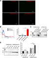
Surface exposure analysis of Lpp-SaoA fusion protein. (A) Immunofluorescence analysis of surface exposure of Lpp-SaoA. The rSC0016 strains were induced with 0.5 mM IPTG for 3 h. Cells were incubated with rabbit anti-SaoA serum and goat anti-rabbit IgG antibody. The blank control was incubated with PBS instead of rabbit anti-SaoA antibody. (B) Flow cytometry analysis of the surface localization of Lpp-SaoA. The mean fluorescence intensity (MFI) of rSC0016 cells incubated with anti-SaoA antibody is expressed as the percentage of the corresponding strain incubated with PBS. Flow cytometry profiles are representative of three independent experiments. (C) Subcellular localization of SaoA in supernatant, outer membrane, periplasmic, and cytoplasmic fractions of rSC0016(pS-SaoA) and rSC0016(pS-Lpp-SaoA). (D) Western blot analysis of rSC0016(pS-SaoA) and rSC0016(pS-Lpp-SaoA) expressing SaoA treated or not with proteinase K. Immunoblots are representative of three independent experiments. Quantification of the optical density was performed using ImageJ software. **, P < 0.01; n.s., no significance.
FIG 3
FIG 3
Immunofluorescence, flow cytometric, and Western blot analyses of SaoA production. (A) Immunofluorescence analysis of the SaoA protein levels on the bacterial surface following IPTG treatment for 0 h, 1 h, 3 h, 6 h, 17 h, and 24 h. Immunofluorescence profiles are representative of three independent experiments. The percentage of positive cells with green fluorescence is indicated. (B) Western blot analysis of the SaoA production levels of rSC0016(pS-SaoA) and rSC0016(pS-Lpp-SaoA) strains. GroEL was chosen as an internal reference protein. Immunoblots are representative of three independent experiments. (C) Relative SaoA expression of rSC0016(pS-SaoA) and rSC0016(pS-Lpp-SaoA) based on grayscale analysis with GroEL as a reference protein. Quantification of the optical density was performed using ImageJ software. (D) Flow cytometry analysis of the surface localization of SaoA after 3 h and 17 h of IPTG treatment. The MFI of rSC0016(pS-Lpp-SaoA) is expressed as the percentage of the rSC0016(pS-SaoA) strain. Flow cytometry profiles are representative of three independent experiments. *, P < 0.05; **, P < 0.01; ***, P < 0.001.
FIG 4
FIG 4
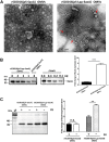
Expression and characterization of SaoA in engineered OMVs from rSC0016(pS-Lpp-SaoA). (A) OMVs derived from rSC0016(pS-SaoA) and rSC0016(pS-Lpp-SaoA) were evaluated under transmission electron microscopy. Red arrows indicate irregularly shaped OMVs. (B) The amount of foreign antigen SaoA expression in OMVs was evaluated by loading different quantities of rSaoA and engineered OMVs, and the corresponding band intensities were compared by Western blotting. (C) Western blot analysis of rSC0016(pS-SaoA) OMVs and rSC0016(pS-Lpp-SaoA) OMVs expressing SaoA treated or not with proteinase K. Immunoblots are representative of three independent experiments. Quantification of the optical density was performed using ImageJ software. **, P < 0.01; ***, P < 0.001.
FIG 5
FIG 5
Analysis of protein lipidation by Triton X-114 extraction of bacterial and OMV proteins. The whole-cell lysates and OMV samples of rSC0016(pS-SaoA) or rSC0016(pS-Lpp-SaoA) were dissolved at 4°C in 1% Triton X-114. Subsequently, the detergent phase and the aqueous phase were partitioned through centrifugation. Next, the proteins in the detergent and aqueous phases were concentrated through methanol-chloroform precipitation and then separated by SDS-PAGE for Western blot analysis.
FIG 6
FIG 6
Evaluation of engineered OMV toxicity using cell and mouse models. (A) Murine macrophage line RAW264.7 was stimulated with 10 μg χ3761 OMVs, rSC0016(pS-Lpp-SaoA) OMVs, rSC0016(pS-SaoA) OMVs, or PBS, and LDH release was measured using a cytotoxicity detection kit. The experiment was repeated independently three times. (B) Amounts of proinflammatory cytokines (including IL-1β, IL-6, and TNF-α) in serum samples from immunized mice 6 h after infection. The results are expressed as means ± SEM. (C) Mice (n = 8 per group) were immunized intraperitoneally with 10 μg of χ3761 OMVs, rSC0016(pS-SaoA) OMVs, rSC0016(pS-Lpp-SaoA) OMVs, or PBS. χ3761 OMVs led to a high mortality rate (62.5%) of the mice, whereas rSC0016(pS-SaoA) OMVs, rSC0016(pS-Lpp-SaoA) OMVs, and PBS did not. The survival data were analyzed through log rank test. **, P < 0.01; ***, P < 0.001.
FIG 7
FIG 7
Antibody responses to SaoA in immunized mice. (A) Immunization scheme used for the protection assay. (B to D) Anti-SaoA IgG (B), IgG1 (C), and IgG2a (D) antibody titers at 31 days postvaccination in different immunized mice. (E) Ratios of IgG2a/IgG1 to SaoA antigen at 31 days postvaccination. The results are expressed as means ± SEM. *, P < 0.05; **, P < 0.01; ***, P < 0.001.
FIG 8
FIG 8
Levels of IL-4 and IFN-γ cytokines in immunized mice. (A) Scheme of immunization regimen. (B and C) Amounts of IL-4 (B) and IFN-γ (C) in the serum samples at 6 h after boost immunization in different immunized mice. (D) Ratios of IFN-γ/IL-4 at 6 h after boost immunization. (E and F) IL-17A in the serum samples at 6 h (E) and in the spleen tissues at 7 days (F) after boost immunization of the immunized mice were evaluated by ELISA. (G) IL-4 and IFN-γ secretion by splenocytes isolated from the different immunized groups was evaluated by the ELISPOT assay. (H) Quantitative data of the ELISPOT assays. The data are expressed as means ± SEM. *, P < 0.05; **, P < 0.01; ***, P < 0.001.
FIG 9
FIG 9
Immunization and protective efficacy of mice challenged by S. suis. (A) The uptake of S. suis serotype 2 by RAW264.7 macrophages was examined after opsonization with sera from control and immunized mice. (B and C) Bacterial burden was evaluated in blood (B) and brain tissue (C) of different immunized mice. The data are expressed as means ± SEM. (D) At 35 days after the initial immunization, the mice were challenged with 8× the LD50 of S. suis serotype 2 intraperitoneally. (E) At 35 days after the initial immunization, the mice were challenged with 16× the LD50 of S. suis serotype 2 intraperitoneally. The mice were observed for 2 weeks after challenge. The survival data were analyzed through log rank test. *, P < 0.05; **, P < 0.01; ***, P < 0.001.

Similar articles

Cited by

References

    1. Scallan E, Hoekstra RM, Angulo FJ, Tauxe RV, Widdowson MA, Roy SL, Jones JL, Griffin PM. 2011. Foodborne illness acquired in the United States—major pathogens. Emerg Infect Dis 17:7–15. 10.3201/eid1701.p11101. - DOI - PMC - PubMed
    1. Rappuoli R, Mandl CW, Black S, De Gregorio E. 2011. Vaccines for the twenty-first century society. Nat Rev Immunol 11:865–872. 10.1038/nri3085. - DOI - PMC - PubMed
    1. Fair RJ, Tor Y. 2014. Antibiotics and bacterial resistance in the 21st century. Perspect Medicin Chem 6:25–64. 10.4137/PMC.S14459. - DOI - PMC - PubMed
    1. Schwechheimer C, Kuehn MJ. 2015. Outer-membrane vesicles from Gram-negative bacteria: biogenesis and functions. Nat Rev Microbiol 13:605–619. 10.1038/nrmicro3525. - DOI - PMC - PubMed
    1. Kulp A, Kuehn MJ. 2010. Biological functions and biogenesis of secreted bacterial outer membrane vesicles. Annu Rev Microbiol 64:163–184. 10.1146/annurev.micro.091208.073413. - DOI - PMC - PubMed

Publication types