. 2023 Apr 25;14(2):e0337222.
doi: 10.1128/mbio.03372-22. Epub 2023 Feb 21.

Ribavirin Treatment Failure-Associated Mutation, Y1320H, in the RNA-Dependent RNA Polymerase of Genotype 3 Hepatitis E Virus (HEV) Enhances Virus Replication in a Rabbit HEV Infection Model

Affiliations

Ribavirin Treatment Failure-Associated Mutation, Y1320H, in the RNA-Dependent RNA Polymerase of Genotype 3 Hepatitis E Virus (HEV) Enhances Virus Replication in a Rabbit HEV Infection Model

Bo Wang et al. mBio. .

Abstract

Chronic hepatitis E virus (HEV) infection has become a significant clinical problem that requires treatment in immunocompromised individuals. In the absence of an HEV-specific antiviral, ribavirin (RBV) has been used off-label, but treatment failure may occur due to mutations in the viral RNA-dependent RNA polymerase (RdRp), including Y1320H, K1383N, and G1634R. Chronic hepatitis E is mostly caused by zoonotic genotype 3 HEV (HEV-3), and HEV variants from rabbits (HEV-3ra) are closely related to human HEV-3. Here, we explored whether HEV-3ra, along with its cognate host, can serve as a model to study RBV treatment failure-associated mutations observed in human HEV-3-infected patients. By utilizing the HEV-3ra infectious clone and indicator replicon, we generated multiple single mutants (Y1320H, K1383N, K1634G, and K1634R) and a double mutant (Y1320H/K1383N) and assessed the role of mutations on replication and antiviral activity of HEV-3ra in cell culture. Furthermore, we also compared the replication of the Y1320H mutant with the wild-type HEV-3ra in experimentally infected rabbits. Our in vitro analyses revealed that the effects of these mutations on rabbit HEV-3ra are altogether highly consistent with those on human HEV-3. Importantly, we found that the Y1320H enhances virus replication during the acute stage of HEV-3ra infection in rabbits, which corroborated our in vitro results showing an enhanced viral replication of Y1320H. Taken together, our data suggest that HEV-3ra and its cognate host is a useful and relevant naturally occurring homologous animal model to study the clinical relevance of antiviral-resistant mutations observed in human HEV-3 chronically-infected patients. IMPORTANCE HEV-3 causes chronic hepatitis E that requires antiviral therapy in immunosuppressed individuals. RBV is the main therapeutic option for chronic hepatitis E as an off-label use. Several amino acid changes, including Y1320H, K1383N, and G1634R, in the RdRp of human HEV-3 have reportedly been associated with RBV treatment failure in chronic hepatitis E patients. In this study, we utilized an HEV-3ra from rabbit and its cognate host to investigate the effect of these RBV treatment failure-associated HEV-3 RdRp mutations on viral replication efficiency and antiviral susceptibility. The in vitro data using rabbit HEV-3ra was highly comparable to those from human HEV-3. We demonstrated that the Y1320H mutation significantly enhanced HEV-3ra replication in cell culture and enhanced virus replication during the acute stage of HEV-3ra infection in rabbits. The rabbit HEV-3ra infection model should be useful in delineating the role of human HEV-3 RBV treatment failure-associated mutations in antiviral resistance.

Keywords: chronic hepatitis E (CHE); genotype 3 HEV (HEV-3); hepatitis E virus (HEV); rabbit HEV-3ra; ribavirin (RBV) treatment failure-associated mutations.


Conflict of interest statement

The authors declare no conflict of interest.

Figures
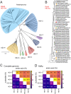
FIG 1
FIG 1
Phylogeny of rabbit HEV-3ra and sequence identities between rabbit HEV-3ra and four major HEV genotypes. (A) A neighbor-joining tree of maximum likelihood distances was generated based on HEV genomic sequences of representative members within the genus Paslahepevirus. Clusters of eight distinct HEV genotypes and the rabbit HEV-3ra were highlighted with different colors. (B) A maximum-likelihood tree was generated based on 52 genomic sequences of rabbit HEV-3ra. The HEV-3 reference Meng strain (GenBank accession no. AF082843) served as an outgroup. Virus designations include GenBank accession number, country origin, and host. HEV-3ra sequences used in this study and those detected in humans are highlighted with red- and orange-filled circles, respectively. Evolutionary analyses were conducted in molecular evolutionary genetics analysis 11 (MEGA 11) based on the multiple sequence alignment in Geneious Prime. General time reversible (GTR) + gamma distributed (G) + invariable sites (I) nucleotide substitution model with the lowest Bayesian information criterion (BIC) score was selected based on the find best-fit substitution model (ML) in MEGA 11. Bootstrap values (>90%) are presented at specific nodes. Scale bars indicate the estimated number of nucleotide substitutions per site. (C) Comparisons of nucleotide sequences of complete genomes and amino acid sequences of concatenated ORF1 and ORF2 among four major HEV genotypes and HEV-3ra. (D) Comparisons of nucleotide and amino acid sequences of RNA-dependent RNA polymerase (RdRp) among four major HEV genotypes and HEV-3ra. Representative viral genomes of eight HEV genotypes and HEV-3ra were included for analyses.
FIG 2
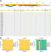
FIG 2
Locations of three RBV treatment failure-associated mutations in HEV-3 RdRp and epidemiological prevalence of the mutations at different amino acid positions among the eight HEV genotypes and HEV-3ra. (A) Three mutations in the RdRp (Y1320H, K1383N, and G1634R) that are associated with RBV treatment failure reported in a clinical CHE case are shown in the HEV-3 reference Meng strain (GenBank accession no. AF082843). The putative functional domains within ORF1 are depicted: Met, methyltransferase; Y, Y domain; PCP, papain-like cysteine protease; HVR, hypervariable region; PPR, poly proline region; X, macro domain; Hel, helicase; RdRp, RNA-dependent RNA polymerase. The viral genome in nucleotide bases is shown on the top. The RdRp region within ORF1 of 52 rabbit HEV-3ra genomes is aligned with the HEV-3 Meng strain, and the locations of these three mutations are mapped and highlighted in the genomes of HEV-3ra. (B) Epidemiological prevalence of the three RBV treatment failure-associated HEV-3 RdRp mutations at different amino acid positions among eight HEV genotypes and HEV-3ra. HEV full-length genomes were retrieved (as of October 2022) from the GenBank database and aligned for comprehensive sequence analyses. The numbers of viral genomes analyzed for each genotype are 82 for HEV-1, 2 for HEV-2, 644 for HEV-3, 52 for HEV-3ra, 225 for HEV-4, 2 for HEV-5, 2 for HEV-6, 3 for HEV-7, and 6 for HEV-8. The amino acid residues of each mutation are presented in different colors.
FIG 3
FIG 3
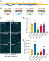
Effect of RBV treatment failure-associated individual HEV-3 RdRp mutations on in vitro replication efficiency of HEV-3ra. (A) Schematic representation of the rabbit HEV-3ra infectious clone LR. The putative functional domains within ORF1 are depicted: Met, methyltransferase; Y, Y domain; PCP, papain-like cysteine protease; HVR, hypervariable region; PPR, poly proline region; X, macro domain; Hel, helicase; RdRp, RNA-dependent RNA polymerase. The three selected RBV treatment failure-associated HEV-3 RdRp mutations (Y1320H, K1383N, and K1634G/R) are indicated. The genome of the HEV-3ra LR in nucleotide bases is shown on the top. (B) Construction of the following four HEV-3ra LR-derived mutants: Y1320H, K1383N, K1634G, and K1634R. Chromatograms of nucleotide and amino acid substitutions compared with the parental wild-type LR strain are highlighted. (C) Representative immunofluorescence staining of HEV ORF2-positive foci of Huh7-S10-3 cells at 7 days posttransfection of HEV-3ra LR wild type and mutants. HEV ORF2-positive foci are shown in green (anti-ORF2 polyclonal antibody raised from rabbit and goat anti-rabbit monoclonal antibody Alexa Fluor 488), and cell nuclei are shown in blue (DAPI) (scale bar, 200 μm). (D) Number of HEV-3ra ORF2-positive cells at 7 days posttransfection of HEV-3ra LR wild type and mutants. (E) HEV RNA copy numbers were quantified by RT-qPCR from the culture supernatant of Huh7-S10-3 cells at 7 days posttransfection with respective HEV-3 LR wild type and mutants. Values represent means plus standard deviations (SDs) (error bars) from independent experiments (n = 3). Statistical differences were determined with one-way ANOVA. *, P < 0.05; ***, P < 0.001; ns, not statistically significant.
FIG 4
FIG 4
Construction of rabbit HEV-3ra indicator replicon LRG and comparative analyses of replication efficiency of LRG wild type and mutants containing each single RdRp mutation. (A) A schematic representation of the HEV-3ra indicator replicon LRG. The putative functional domains within ORF1 are depicted as follows: Met, methyltransferase; Y, Y domain; PCP, papain-like cysteine protease; HVR, hypervariable region; PPR, poly proline region; X, macro domain; Hel, helicase; RdRp, RNA-dependent RNA polymerase. The three selected RBV treatment failure-associated RdRp mutations (Y1320H, K1383N, and K1634G/R) are indicated, and the Gaussia luciferase (GLuc) gene is indicated in green. The genome of the HEV-3ra LRG in nucleotide bases is shown on the top. (B) Growth kinetics of LRG wild type as measured by Gluc expression activity. Cell culture media of Huh7-S10-3 cells were collected at serial time points posttransfection with LRG wild type, and the Gluc activity was monitored. Values represent means plus SD (error bars) from independent experiments (n = 4). (C) Comparative analyses of replication efficiency of LRG wild type and mutants. At 4- and 8-days posttransfection with LRG wild type and mutants, respectively, cell culture media of Huh7-S10-3 cells were harvested, and the Gluc expression activity was measured and compared. Values represent means plus SD (error bars) from independent experiments (n = 4). Statistical differences were determined with one-way ANOVA. **, P < 0.01; ***, P < 0.001; ns, not statistically significant.
FIG 5
FIG 5
The Y1320H mutation compensated for the K1383N mutation-associated replication defects, and the Y1320H/K1383N double mutant has a markedly higher sensitivity to RBV. (A) Comparative analyses of replication efficiency in Huh7-S10-3 cells of HEV-3ra LRG wild type and mutants containing a single RdRp mutation or a double mutation (Y1320H/K1383N). (B) Comparative analyses of RBV sensitivity to HEV-3ra LRG wild type and mutants. Huh7-S10-3 cells were cultured without RBV or with 1 μM RBV or 10 μM RBV. At 7 days posttransfection with LRG wild-type and mutants, respectively, cell culture media of Huh7-S10-3 cells were collected, and the Gluc expression activity was measured and compared. Values represent means plus SDs (error bars) from independent experiments (n = 4). Statistical differences were determined with one-way ANOVA. *, P < 0.05; ***, P < 0.001; ns, not statistically significant.
FIG 6
FIG 6
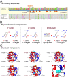
Physicochemical and structural comparisons of RBV treatment failure-associated HEV-3 RdRp mutations. (A) Alignment of the RdRp sequences of four major human-infecting HEV genotypes and rabbit HEV-3ra. The eight motifs in RdRp and three selected RdRp mutations (Y1320H, K1383N, and K1634G/R) are indicated. The RBV treatment failure-associated HEV-3 RdRp mutation K1383N located at motif I is highlighted. Locations of functional domains and motifs within HEV ORF1 are according to the HEV prototype Burma strain (GenBank accession no. M73218) (66). (B) Physicochemical comparisons of the three RBV treatment failure-associated HEV-3 RdRp mutations. The impact of these mutations on in vitro replication of HEV-3ra is indicated in red arrows. The physicochemical properties and structures of relevant proteinogenic amino acids in this study are illustrated and adapted from https://commons.wikimedia.org/wiki/File:Proteinogenic_Amino_Acid_Table.png created by Thomas Ryckmans (2022). (C) Structural comparisons of RdRp of wild-type HEV-3ra LR or that with each single RBV treatment failure-associated HEV-3 RdRp mutation. The three-dimensional structures of RdRp are predicted with AlphaFold (68) and visualized and annotated in Geneious Prime software version 2022.2.2. The alpha helices and beta sheets are indicated in red and blue, respectively. The positions of three RBV treatment failure-associated HEV-3 RdRp mutations (114, 177, and 428) with original amino acid residues are shown in orange atoms, and with amino acid changes are highlighted in cyan atoms. The figures only aim to present the locations of the three RBV treatment failure-associated HEV-3 RdRp mutations based on the three-dimensional structure of the RdRp of the HEV-3ra LR strain and do not necessarily represent its true three-dimensional structure.
FIG 7
FIG 7
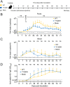
Comparisons of replication and infection dynamics of HEV-3ra LR_WT and LR_Y1320H in rabbits. (A) Illustration of intravenous inoculation via the ear vein of specific-pathogen-free female New Zealand White rabbits with HEV-3ra LR_WT (n = 5, yellow, round), LR_Y1320H (n = 5, blue, square), and PBS for mock infection (n = 5, gray, triangle). Fecal samples were collected from each individual rabbit twice weekly, and serum samples were collected weekly after inoculation. All rabbits were necropsied at 11 weeks (77 days) postinoculation. (B) Kinetics of viral RNA loads during the course of infection in the fecal samples of HEV-3ra LR_WT- and LR_Y1320H-infected rabbits. (C) Kinetics of viral RNA loads during the course of infection in the serum samples of HEV-3ra LR_WT and LR_Y1320H-infected rabbits. (D) Kinetics of anti-HEV-3ra IgG antibody response in rabbits experimentally inoculated with HEV-3ra LR_WT and LR_Y1320H. Seroconversion to anti-HEV IgG antibodies in HEV-3ra LR_WT- and LR_Y1320H-infected rabbits during the course of infection was detected by ELISA with a correct S/N ratio cutoff of 0.35 for positive samples. Values represent means plus SD (error bars) from five animals (n = 5) in each group. Statistical differences were determined with a two-sided, unpaired, multiple Student's t test without adjustments. P value is indicated when less than 0.05; ns, no statistical significance.

Similar articles

Cited by

References

    1. Nimgaonkar I, Ding Q, Schwartz RE, Ploss A. 2018. Hepatitis E virus: advances and challenges. Nat Rev Gastroenterol Hepatol 15:96–110. doi:10.1038/nrgastro.2017.150. - DOI - PMC - PubMed
    1. Velavan TP, Pallerla SR, Johne R, Todt D, Steinmann E, Schemmerer M, Wenzel JJ, Hofmann J, Shih JWK, Wedemeyer H, Bock CT. 2021. Hepatitis E: an update on One Health and clinical medicine. Liver Int 41:1462–1473. doi:10.1111/liv.14912. - DOI - PubMed
    1. Sooryanarain H, Meng XJ. 2019. Hepatitis E virus: reasons for emergence in humans. Curr Opin Virol 34:10–17. doi:10.1016/j.coviro.2018.11.006. - DOI - PMC - PubMed
    1. Purdy MA, Drexler JF, Meng XJ, Norder H, Okamoto H, Van der Poel WHM, Reuter G, de Souza WM, Ulrich RG, Smith DB. 2022. ICTV virus taxonomy profile: Hepeviridae 2022. J Gen Virol 103:1778. doi:10.1099/jgv.0.001778. - DOI - PubMed
    1. Kinast V, Klohn M, Nocke MK, Todt D, Steinmann E. 2022. Hepatitis E virus species barriers: seeking viral and host determinants. Curr Opin Virol 56:101274. doi:10.1016/j.coviro.2022.101274. - DOI - PubMed

Publication types