. 2023 Apr 1;324(4):L493-L506.
doi: 10.1152/ajplung.00285.2022. Epub 2023 Feb 21.

Mucociliary clearance augmenting drugs block SARS-CoV-2 replication in human airway epithelial cells

Affiliations

Mucociliary clearance augmenting drugs block SARS-CoV-2 replication in human airway epithelial cells

Javier Campos-Gómez et al. Am J Physiol Lung Cell Mol Physiol. .

Abstract

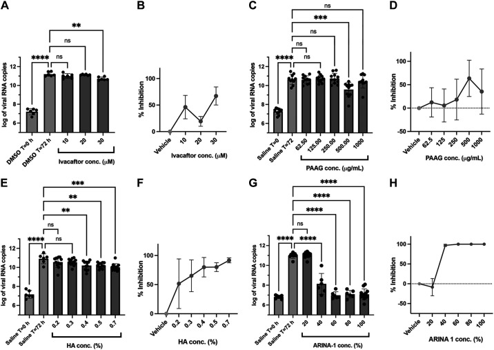
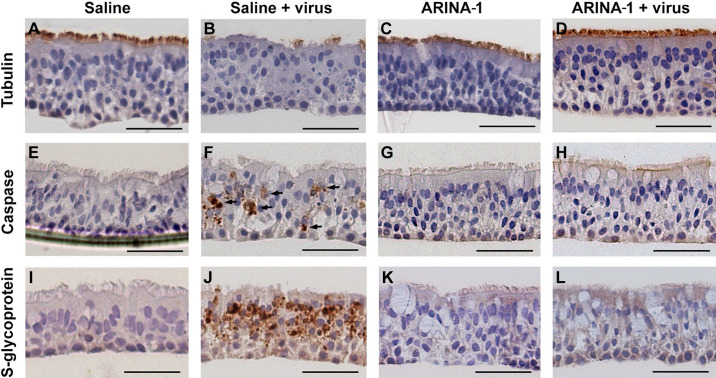
The coronavirus disease (COVID-19) pandemic, caused by SARS-CoV-2 coronavirus, is devastatingly impacting human health. A prominent component of COVID-19 is the infection and destruction of the ciliated respiratory cells, which perpetuates dissemination and disrupts protective mucociliary transport (MCT) function, an innate defense of the respiratory tract. Thus, drugs that augment MCT could improve the barrier function of the airway epithelium and reduce viral replication and, ultimately, COVID-19 outcomes. We tested five agents known to increase MCT through distinct mechanisms for activity against SARS-CoV-2 infection using a model of human respiratory epithelial cells terminally differentiated in an air/liquid interphase. Three of the five mucoactive compounds tested showed significant inhibitory activity against SARS-CoV-2 replication. An archetype mucoactive agent, ARINA-1, blocked viral replication and therefore epithelial cell injury; thus, it was further studied using biochemical, genetic, and biophysical methods to ascertain the mechanism of action via the improvement of MCT. ARINA-1 antiviral activity was dependent on enhancing the MCT cellular response, since terminal differentiation, intact ciliary expression, and motion were required for ARINA-1-mediated anti-SARS-CoV2 protection. Ultimately, we showed that the improvement of cilia movement was caused by ARINA-1-mediated regulation of the redox state of the intracellular environment, which benefited MCT. Our study indicates that intact MCT reduces SARS-CoV-2 infection, and its pharmacologic activation may be effective as an anti-COVID-19 treatment.

Keywords: SARS-CoV-2; antioxidant; cilia movement; mucoactive drug; mucociliary transport.


Conflict of interest statement

S.M.R. reports grant support from Renovion for research studies conducted through university grants/contracts and personal fees including stock options for consulting services on the design and conduct of clinical trials. None of the other authors has any conflicts of interest, financial or otherwise, to disclose.

Figures

None
Graphical abstract
Figure 1.
Figure 1.
HBEC-ALI-based antiviral assay and validation. A: schematic representation of the antiviral assay. Differentiated HBE cells grown at the ALI on transwells microporous filters are washed with PBS to remove mucus. Test compounds are added basolaterally (camostat, ivacaftor) or apically (PAAG, HA, ARINA-1). After 1 h of incubation, SARS-CoV-2 virus is added at an MOI = 0.24. Unattached viral particles are removed after an hour of incubation. At this point, the transwell data corresponding to time zero (before viral replication) was collected. The remainder of the transwells is incubated for 72 h with additional administrations of the apically delivered compounds (PAAG, HA, ARINA-1) at T = 0, 24, and 48 h. Cells from each time point (0 and 72 h) are collected from the filters and used for RNA purification. Finally, the collected RNA is used to measure the viral load by RT-qPCR. B: validation of the assay using camostat mesylate compound. A graph of the viral copy number measured by the RT-qPCR is shown for a representative experiment with three filter replicates per condition. Camostat and vehicle control were added basolaterally. DMSO was used as the diluent for camostat and the vehicle control. Data were logarithmically transformed. C: data from two independent experiments, as the one shown in B, were converted to percentage of viral inhibition, averaged (three replicates/experiment) and compared using an ordinary one-way ANOVA (****P < 0.0001). ALI, air-liquid interface; HA, hyaluronic acid; HBEC, human bronchial epithelial cells; PAAG, poly-N(acetyl, arginyl) glucosamine.
Figure 2.
Figure 2.
Anti-SARS-CoV-2 activity of the mucoactive agents tested in well-differentiated primary HBE cells. A: effect on the viral copy number measured by the RT-qPCR caused by ivacaftor at the concentrations shown on the graph. B: data from graph in A were converted to percentage of viral inhibition. CH: as in A and B, but using the compounds PAAG, HA, and ARINA-1, respectively. DMSO was the vehicle used for ivacaftor (hydrophobic compound) and saline for PAAG, HA, and ARINA-1 (hydrophilic compounds). All experiments were performed at least in duplicate independent assays, each with at least three transwell filter replicates per condition. Treatments were compared using ordinary one-way ANOVA statistical analysis. Hydrophobic compounds (added basolaterally) and hydrophilic (added apically) are shown in green and red, respectively. For each compound, each independent experiment was done with primary HBEC from a different donor. HA, hyaluronic acid; HBEC, human bronchial epithelial cells; PAAG, poly-N(acetyl, arginyl) glucosamine; **P < 0.01, ***P < 0.001, ****P < 0.0001.
Figure 3.
Figure 3.
ARINA-1 inhibits the production of infectious virus by primary HBEC. Quantification of SARS-COV-2 virus using a luminescence TCID50 assay showed that ARINA-1-treated cells produced two orders of magnitude less infectious virus than the saline-treated controls (TCID50 = 1- vs. 131-fold dilution, respectively). Values are averages and SD of three independent experiments, each using primary HBEC from a different donor. For each experiment, every dilution of the virus was assessed in sextuplicate using Vero E6 cells as indicators of cytotoxicity using a luminescence assay that measures ATP as a proxy for cell viability. HBEC, human bronchial epithelial cells; TCID50, tissue culture infection dose.
Figure 4.
Figure 4.
Histopathology studies showed that ARINA-1 protects primary HBE cells from SARS-CoV-2-mediated cytopathology. AL: representative photo micrographs of HBEC cross-sections with the immunohistochemistry and treatments. Each row corresponds to the immunohistochemistry using the antibody against the cell marker shown at the left, and each column corresponds to the treatment shown above the upper pictures. SARS-CoV-2 caused cilia shortening and loss in saline-treated cells (B). However, ARINA-1 protected cilia from damage (D). Similarly, the virus induced significant apoptosis in the mock-treated HBE cells (F, apoptotic cells indicated with arrows), which was not observed in those treated with ARINA-1 (H). In addition, mock-treated cells showed significant immunostaining using an antibody against the viral S glycoprotein (J), which again was not observed in the ARINA-1-treated cells (L). Scale bars represent 100 µm. Fully differentiated primary HBEC from two donors were used for the histopathology study. n ≥ 10 pictures per donor and condition were taken and analyzed. HBEC, human bronchial epithelial cells.
Figure 5.
Figure 5.
ARINA-1 induces a hypernormal mucociliary transport (MCT) in well-differentiated primary HBEC. A and B: cilia beating frequency (CBF) and mucociliary transport (MCT) measurements using µOCT, respectively, in SARS-CoV-2-infected or uninfected, ARINA-1-treated or untreated HBEC. Comparisons were performed using an ordinary one-way ANOVA. MCT values were normalized against the MCT average of saline-treated baseline controls. CF: resliced images of µOCT captured videos (Supplemental Videos S1–S4) in which the slope of the diagonal streak (yellow arrow) indicates the vectorial transport of mucus particles over time. This allows visualization of MCT rate on still images, in which higher slope angles with respect to time vectors are indicative of faster MCT rates. HBEC, human bronchial epithelial cells; *P < 0.05, ****P < 0.0001.
Figure 6.
Figure 6.
ARINA-1 has no direct antiviral effect on SARS-CoV-2 virus. A: flow diagram showing the procedure followed to test the direct antiviral activity of ARINA-1 on the virus. Briefly, a suspension of SARS-CoV-2 virus was exposed to ARINA-1 and then filtered through a 3,000 Da pore size membrane to remove the ARINA-1 components. The ARINA-1-free virus suspension was used to infect Vero E6 or HBE/ALI cells, and the viral load determined after 48 h post infection. Virus particles were treated in parallel with saline as control. Viral load of the ARINA-1-treated virus compared with the saline-treated virus measured in Vero E6 cells (B) or in the HBEC/ALI assay (C). RNA copy numbers were logarithmically transformed and compared using ordinary one-way ANOVA statistical analysis. ALI, air-liquid interface; HBEC, human bronchial epithelial cells; ns, nonsignificant.
Figure 7.
Figure 7.
ARINA-1 is not protective when cilia beating is inhibited with BAPTA/AM or cilia are not active or present in cells. A: viral loads in HBEC treated with ARINA-1, BAPTA/AM or ARINA-1 plus BAPTA/AM. Two independent experiments with three technical replicates for BAPTA/AM and BAPTA/AM plus ARINA-1 were performed. Viral loads of undifferentiated 16HBE (B) and primary HBEC (C), which lack cilia, were treated with ARINA-1 or saline as control. Two independent experiments with three technical replicates for 16HBE and one experiment with three replicates for HBEC were performed. Viral loads of ALI differentiated hNE cells from two WT donors (D) and from two PCD-suffering human donors with mutations in genes CCDC39 and DNAI1 (E), which encode proteins essential for the assembly of dynein arm complexes and for dynein protein itself, respectively, treated with ARINA-1 or saline as control. Two experiments with at least two technical replicates for PCD-hNE cells and one experiment with four replicates for each WT-hNE cell donor were performed. Data were logarithmically transformed and compared using ordinary one-way ANOVA. ALI, air-liquid interface; HBEC, human bronchial epithelial cells; PCD, primary ciliary dyskinesia; WT, wild type; **P < 0.01, ***P < 0.001, ****P < 0.0001.
Figure 8.
Figure 8.
A proper redox state of the cell is essential for the antiviral protection conferred by mucociliary transport. A: viral load in the presence of allopurinol (400 µM) compared with vehicle (DMSO) in the HBEC/ALI assay. B: viral load in the presence of the antioxidant agents N-acetylcysteine (NAC) and sulforaphane (SFN) at the specified concentrations. ALI, air-liquid interface; HBEC, human bronchial epithelial cells; *P < 0.05, ***P < 0.001, ****P < 0.0001.

Update of

References

    1. Lee ARYB, Wong SY, Chai LYA, Lee SC, Lee MX, Muthiah MD, Tay SH, Teo CB, Tan BKJ, Chan YH, Sundar R, Soon YY. Efficacy of covid-19 vaccines in immunocompromised patients: systematic review and meta-analysis. BMJ 376: e068632, 2022. doi:10.1136/BMJ-2021-068632. - DOI - PMC - PubMed
    1. Mehrabi Nejad MM, Shobeiri P, Dehghanbanadaki H, Tabary M, Aryannejad A, Haji Ghadery A, Shabani M, Moosaie F, SeyedAlinaghi SA, Rezaei N. Seroconversion following the first, second, and third dose of SARS-CoV-2 vaccines in immunocompromised population: a systematic review and meta-analysis. Virol J 19: 132, 2022. doi:10.1186/s12985-022-01858-3. - DOI - PMC - PubMed
    1. Koc HC, Xiao J, Liu W, Li Y, Chen G. Long COVID and its Management. Int J Biol Sci 18: 4768–4780, 2022. doi:10.7150/IJBS.75056. - DOI - PMC - PubMed
    1. Hoffmann M, Krüger N, Schulz S, Cossmann A, Rocha C, Kempf A, Nehlmeier I, Graichen L, Moldenhauer AS, Winkler MS, Lier M, Dopfer-Jablonka A, Jäck HM, Behrens GMN, Pöhlmann S. The Omicron variant is highly resistant against antibody-mediated neutralization: implications for control of the COVID-19 pandemic. Cell 185: 447–456.e11, 2022. doi:10.1016/J.CELL.2021.12.032. - DOI - PMC - PubMed
    1. Gavriatopoulou M, Ntanasis-Stathopoulos I, Korompoki E, Fotiou D, Migkou M, Tzanninis IG, Psaltopoulou T, Kastritis E, Terpos E, Dimopoulos MA. Emerging treatment strategies for COVID-19 infection. Clin Exp Med 21: 167–179, 2021. doi:10.1007/S10238-020-00671-Y. - DOI - PMC - PubMed

Publication types