. 2023 Feb 21;18(2):e0281876.
doi: 10.1371/journal.pone.0281876. eCollection 2023.

Merlin tumor suppressor function is regulated by PIP2-mediated dimerization

Affiliations

Merlin tumor suppressor function is regulated by PIP2-mediated dimerization

Robert F Hennigan et al. PLoS One. .

Abstract

Neurofibromatosis Type 2 is an inherited disease characterized by Schwann cell tumors of cranial and peripheral nerves. The NF2 gene encodes Merlin, a member of the ERM family consisting of an N-terminal FERM domain, a central α-helical region, and a C-terminal domain. Changes in the intermolecular FERM-CTD interaction allow Merlin to transition between an open, FERM accessible conformation and a closed, FERM-inaccessible conformation, modulating Merlin activity. Merlin has been shown to dimerize, but the regulation and function Merlin dimerization is not clear. We used a nanobody based binding assay to show that Merlin dimerizes via a FERM-FERM interaction, orientated with each C-terminus close to each other. Patient derived and structural mutants show that dimerization controls interactions with specific binding partners, including HIPPO pathway components, and correlates with tumor suppressor activity. Gel filtration experiments showed that dimerization occurs after a PIP2 mediated transition from closed to open conformation monomers. This process requires the first 18 amino acids of the FERM domain and is inhibited by phosphorylation at serine 518. The discovery that active, open conformation Merlin is a dimer represents a new paradigm for Merlin function with implications for the development of therapies designed to compensate for Merlin loss.


Conflict of interest statement

The authors have declared that no competing interests exist.

Figures
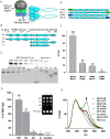
Fig 1
Fig 1. Merlin dimerizes via the FERM domain.
(A). A diagram depicting the dimerization assay using Merlin with either GFP or NanoLuc fused to its C-terminus. (B). A schematic diagram depicting the full-length Merlin (aa 1–595, WT) and the C-terminal deletion mutant (FH, aa 1–511), the FERM domain deletion mutant (HC, aa 315–595) and the dual FERM and CTD deletion (H, aa 315–511). (C). SDS-PAGE gels stained for total protein to access the quantity and purity of isolated probes for Merlin-NL (top) and GFP “bait” proteins (bottom) for GFP, Merlin-GFP, Merlin-FH-GFP, Merlin-HC-GFP and Merlin-H-GFP. (D). Merlin dimerization data quantified on a plate reader and presented as the bound luciferase normalized to 10% of the unbound input NL probe. The data is a mean of triplicate binding reactions with standard deviation and expressed as a percentage of the wild-type Merlin. Inset: an image of the light emitted from Merlin-NanoLuc bound to Merlin and Merlin deletion mutant-GFP bound magnetic beads, performed in triplicate, and dispensed into the wells of a 96-well plate (left). (E). A schematic diagram depicting the N- and C-terminal GFP fused “bait” and the N- and C-terminal fused Merlin-NanoLuc “Probe” constructs used in the BRET assays. (F). Triplicate Merlin dimerization assays for the N-and C-terminal Merlin constructs. The data is a ratio of bound to unbound NL probe, normalized to GFP fluorescence, (mean of triplicate binding reactions with standard deviation and expressed as a percentage of the peak value). (G). Emission spectrum from 400 nm to 600 nm of Merlin dimerization assays normalized to the 450 nm peak. The BRET emission peak at 510 nm is indicated by the arrow.
Fig 2
Fig 2. Merlin conformations.
(A) Fractionation of purified Merlin-NL (inset) on an SEC 650 gel filtration column, aliquots from 0.2 ml fractions were assayed for NanoLuc activity and plotted with the elution volume. The approximate Stokes radius of the peaks was calculated relative to the mobility of standards of known molecular weight and Stokes radius. (S2 Fig). The large peak at 12.4 ml has an approximate Stokes radius of 5.0 nm and the smaller peak at 15 ml has an approximate Stokes radius of 2.1 nm. (B) SDS-PAGE with BSA standards and an aliquot of the purified Merlin-NL stained for total protein (top). Fractions composing peaks 1 and 2 were immunoblotted and probed with antibodies for either total Merlin or P-S518-Merlin (bottom). (C). Gel fractionation of lysates from cells co-transfected with Merlin-NL and Merlin-GFP. NanoLuc activity from the fractions is shown as blue circles and depicted on the right-hand y-axis. Dimers represented by NanoLuc activity from GFP pulldowns of the fractions is shown as green squares and depicted on the left-hand y-axis. (D) Gel fractionation of purified Merlin-NL at in hypotonic (50 mM NaCl), isotonic (150 mM NaCl) and hypertonic (500 mM NaCl) conditions. (E) Images of dimerization assays dimerization assays performed at 50 mM, 75 mM, 150 mM, 300 mM, and 600 mM NaCl. The GFP-NanoLuc fusion protein used to control for the effects of salt on luminescence. (F) Quantitation of the dimerization data presented as a mean of triplicate binding reactions with standard deviation and expressed as a percentage of the peak value. (G) Immunoblot assays of iHSC-1λ immortalized Schwann cell lysates fractionated by gel filtration and probed for either total Merlin (top) or P-S518-Merlin (bottom).
Fig 3
Fig 3. Merlin mutant dimerization.
(A) A schematic diagram of Merlin mutants. Patient derived mutations: L64P, L360P and L535P, indicated by asterisks. ΔN18: deletion of the N-terminal 18 amino acids. N20: the first 20 amino acids fused to GFP. S518A/S518D: non-phosphorylatable and phosphomimetic mutants at S518. AR: closed conformation mutant. ΔEL: open conformation mutant. (B) Pulldown dimerization assays of patient derived Merlin mutants, L64P, Δ39–121, L360P, L535P and the lipid binding deficient mutant 6N. (C) Pulldown dimerization assays of Merlin N-terminal mutant, ΔN18, the unique N-terminal 20 amino acids fused directly to GFP, N20, the phosphorylation mutants S518A and S518D and the conformation mutants AR and ΔEL. GFP alone serves as the negative control. The data is a ratio of bound to unbound NL probe, normalized to GFP fluorescence, (mean of triplicate binding reactions with standard deviation and expressed as a percentage of the wild type Merlin. (D) Gel fractionation of lysates from cells transfected with plasmids expressing either wild type Merlin-NL (blue circles) Merlin-S518D-NL (red squares) or Merlin-S518A-NL (green triangles). (E) Gel fractionation of lysates from cell transfected with plasmids expressing either wild type Merlin-NL (blue circles) Merlin-AR-NL (red squares) or Merlin-ΔEL-NL (green triangles).
Fig 4
Fig 4. Interaction of dimerization mutants with Merlin targets.
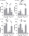
Normalized luciferase activity of GFP pulldowns from HEK 293T cells transfected with plasmids expressing wild type and mutant Merlin-NL and GFP fusions for (A) Angiomotin, (B) ASPP2, (C) YAP1, (D) Lats1. The data is a ratio of bound to unbound NL probe, normalized to GFP fluorescence shown as a mean of triplicate binding reactions with standard deviation and expressed as a percentage of the wild-type Merlin. Asterisks indicate significance by a two-way ANOVA test).
Fig 5
Fig 5. Effect of PIP2 and phosphorylation on dimerization.
(A) Gel fractionation of purified Merlin-NL in the absence (black circles) or presence (grey circles) of 200 μM PIP2-DiC8. (B) An image of the dimerization assay using 100 nM purified Merlin-GFP and Merlin-NL incubated with increasing concentrations of PIP2-DiC8. (C) Binding curve of Merlin dimerization data expressed in relative luciferase units (RLU) and fitted to a binding curve. (D) Dimerization assays using each of purified Merlin-GFP and Merlin-NL in with and without 200 μM PIP2-DiC8 and/or in vitro phosphorylation by recombinant PAK2. The data is presented as the bound luciferase normalized to 10% of the unbound input NL probe. The data is a mean of triplicate binding reactions with standard deviation and expressed as a percentage of the untreated control. (E) Western blots of proteins recovered from the dimerization assay showing total protein (top), probed with antibodies to Merlin (middle) or P-518 Merlin (bottom). (F) Gel fractionation of purified Merlin-ΔN18 NanoLuc in the absence (black circles) or presence (grey circles) of 200 μM PIP2-DiC8.
Fig 6
Fig 6. Merlin activation cycle.
(A) Model for PIP2 mediated Merlin dimerization with “closed” monomers transitioning to “open” monomers upon PIP2 binding the proceeding to dimerization. (B) Model for Merlin activation cycle. Inactive closed conformation associated with Angiomotin. PIP2 binding leading to open conformation, dimerization and binding to ASPP2/Lats1/YAP1. Active complexes are phosphorylated leading to dissolution of the complex followed by dephosphorylation to regenerate the Merlin-Angiomotin complex.

References

    1. Evans DG, Huson SM, Donnai D, Neary W, Blair V, Newton V, et al.. A clinical study of type 2 neurofibromatosis. The Quarterly journal of medicine. 1992;84(304):603–18. . - PubMed
    1. Gutmann DH, Aylsworth A, Carey JC, Korf B, Marks J, Pyeritz RE, et al.. The diagnostic evaluation and multidisciplinary management of neurofibromatosis 1 and neurofibromatosis 2. JAMA: the journal of the American Medical Association. 1997;278(1):51–7. . - PubMed
    1. Giovannini M, Robanus-Maandag E, van der Valk M, Niwa-Kawakita M, Abramowski V, Goutebroze L, et al.. Conditional biallelic Nf2 mutation in the mouse promotes manifestations of human neurofibromatosis type 2. Genes & development. 2000;14(13):1617–30. ; PubMed Central PMCID: PMC316733. - PMC - PubMed
    1. Gehlhausen JR, Park SJ, Hickox AE, Shew M, Staser K, Rhodes SD, et al.. A murine model of neurofibromatosis type 2 that accurately phenocopies human schwannoma formation. Human molecular genetics. 2015;24(1):1–8. doi: 10.1093/hmg/ddu414 ; PubMed Central PMCID: PMC4262489. - DOI - PMC - PubMed
    1. Morrison H, Sherman LS, Legg J, Banine F, Isacke C, Haipek CA, et al.. The NF2 tumor suppressor gene product, merlin, mediates contact inhibition of growth through interactions with CD44. Genes & development. 2001;15(8):968–80. doi: 10.1101/gad.189601 ; PubMed Central PMCID: PMC312675. - DOI - PMC - PubMed

Publication types

Substances