. 2023 Nov;209(6):907-928.
doi: 10.1007/s00359-023-01618-w. Epub 2023 Feb 21.

Neurons sensitive to non-celestial polarized light in the brain of the desert locust

Affiliations

Neurons sensitive to non-celestial polarized light in the brain of the desert locust

Marius Beck et al. J Comp Physiol A Neuroethol Sens Neural Behav Physiol. 2023 Nov.

Abstract

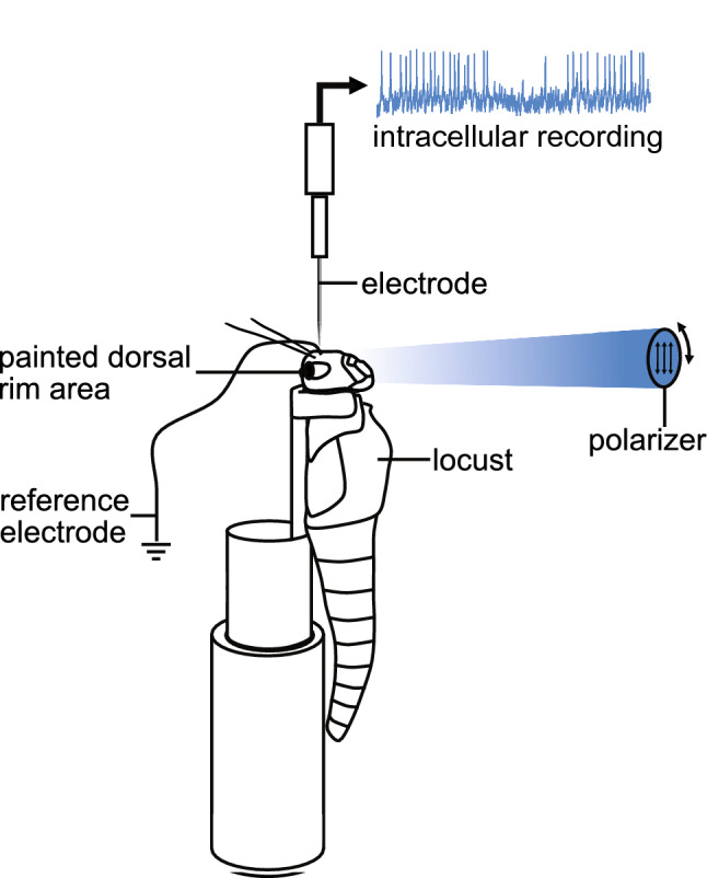
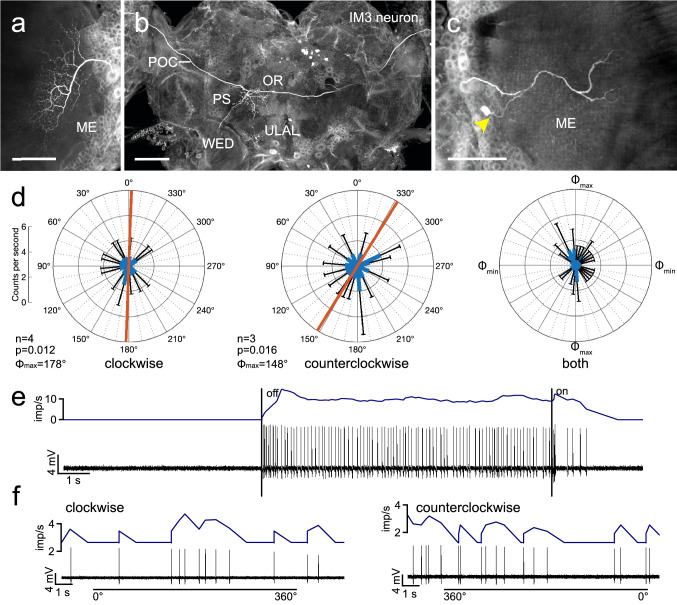
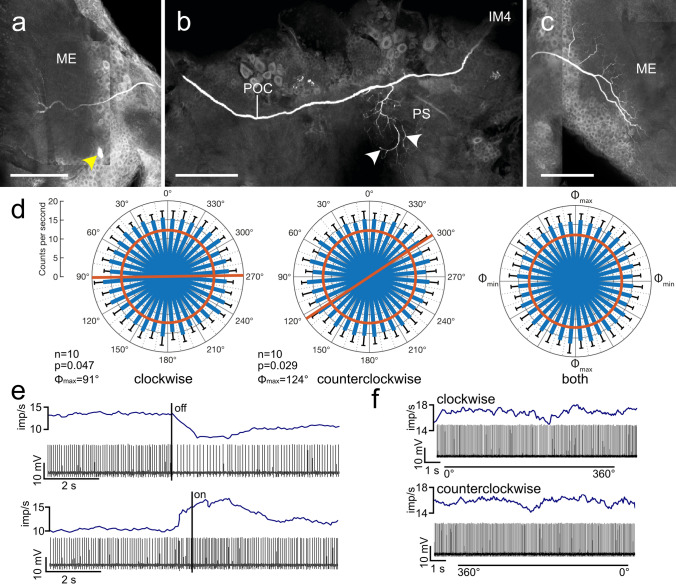
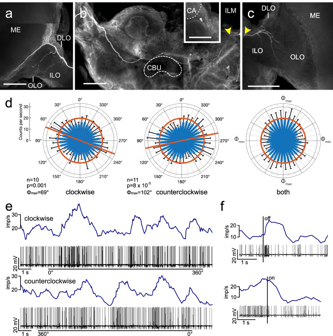
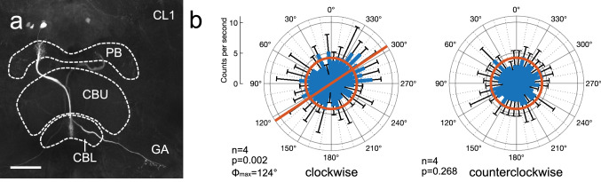
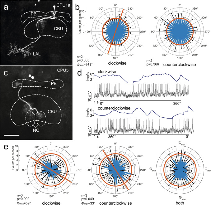
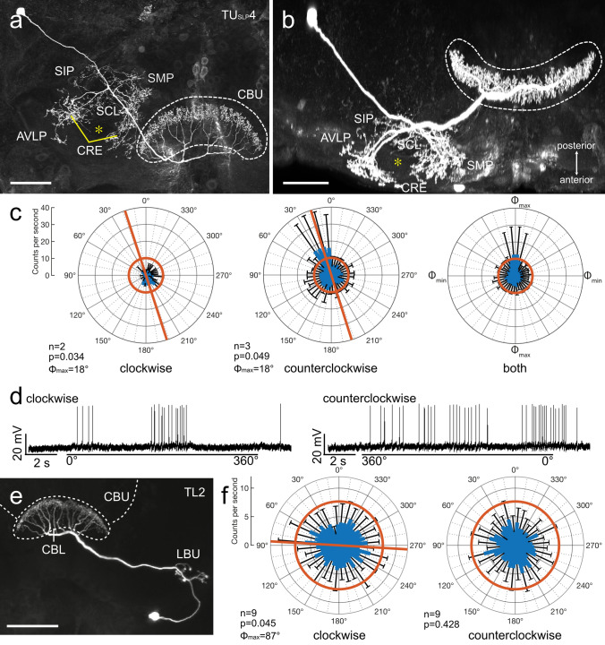
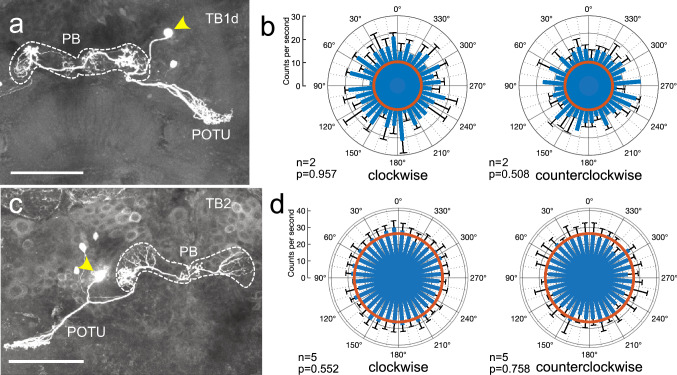
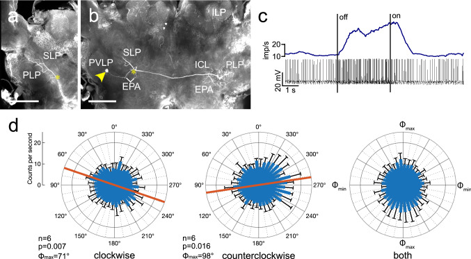
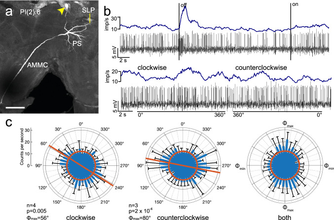
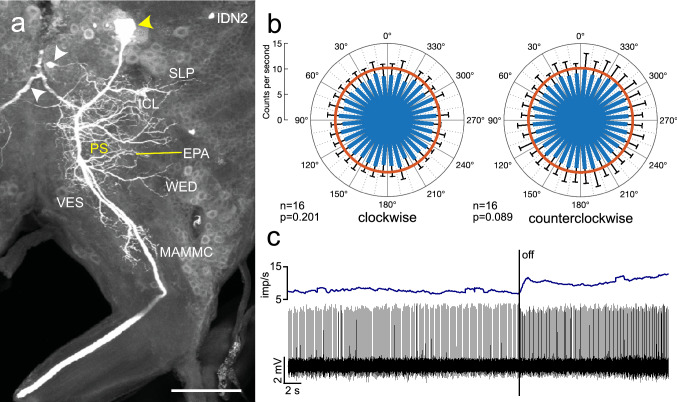
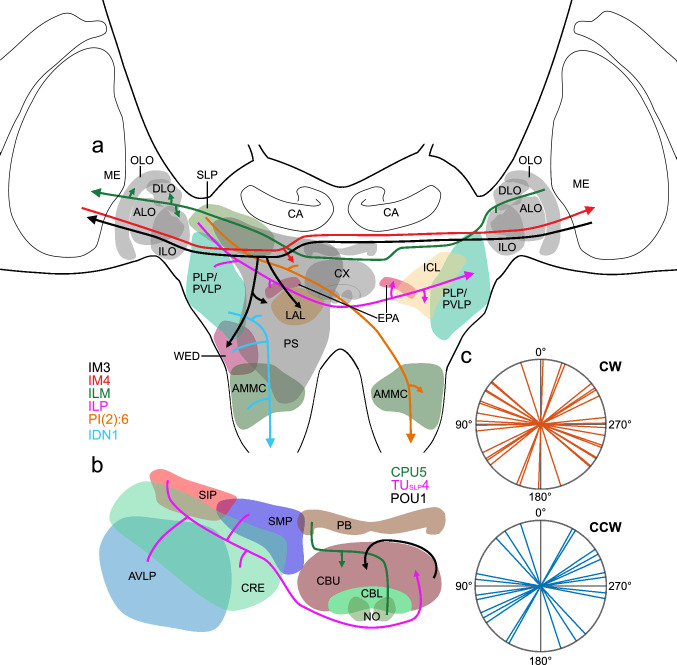
Owing to alignment of rhodopsin in microvillar photoreceptors, insects are sensitive to the oscillation plane of polarized light. This property is used by many species to navigate with respect to the polarization pattern of light from the blue sky. In addition, the polarization angle of light reflected from shiny surfaces such as bodies of water, animal skin, leaves, or other objects can enhance contrast and visibility. Whereas photoreceptors and central mechanisms involved in celestial polarization vision have been investigated in great detail, little is known about peripheral and central mechanisms of sensing the polarization angle of light reflected from objects and surfaces. Desert locusts, like other insects, use a polarization-dependent sky compass for navigation but are also sensitive to polarization angles from horizontal directions. In order to further analyze the processing of polarized light reflected from objects or water surfaces, we tested the sensitivity of brain interneurons to the angle of polarized blue light presented from ventral direction in locusts that had their dorsal eye regions painted black. Neurons encountered interconnect the optic lobes, invade the central body, or send descending axons to the ventral nerve cord but are not part of the polarization vision pathway involved in sky-compass coding.

Keywords: Central complex; Desert locust; Intracellular recordings; Non-celestial polarization vision; Sky compass coding.


Conflict of interest statement

The authors declare no conflict of interest.

Figures

Fig. 1
Fig. 1
Experimental setup. The locust was mounted to a metal holder after removing legs and wings. The dorsal rim areas of both eyes were occluded with black paint. Visual stimulation was provided by a blue light-emitting diode (ELJ-465-617, 465 nm; EPIGAP Optronic, Berlin, Germany) positioned ventrally from the locust head. The stimulus directed toward the locust head was polarized by a rotatable linear polarizer (HN38S, Polaroid, Cambridge, MA, USA)
Fig. 2
Fig. 2
Morphology and physiology of intermedulla neuron IM3. a–c Projection views of the Neurobiotin-labeled IM3 neuron. The neuron has ipsilateral ramifications in a small equatorial sector of an outer medulla (ME) layer. An axonal fiber crosses the brain midline in the posterior optic commissure (POC) and gives rise to beaded side branches in the posterior slope (PS), which partly extend into the wedge (WED), ocellar root (OR), and upper lateral accessory lobe (ULAL). The axon continues toward the contralateral optic lobe and gives rise to wide-field ramifications in an outer layer of the ME. Yellow arrowhead in c points at the soma near the accessory medulla. d Circular diagrams showing mean spiking activities (+ SD, black bars) plotted against polarization angle during 4 rotations in clockwise (left) and 3 rotations in counterclockwise (middle) direction. Red bars indicate preferred polarization angle (Φmax). Right diagram shows mean activities (+ SD) from all rotations with Φmax set at 0°. e Spike train (bottom trace) and mean spiking activity illustrating changes in activity when turning the light source (blue LED, polarizer at 0°) off and on. f Spike trains (bottom traces) and mean spike frequency (top, moving average with bin size of 0.5 s) during clockwise (0°–360°) and counterclockwise (360°–0°) rotation of the polarizer. Scale bars = 150 µm
Fig. 3
Fig. 3
Morphology and physiology of intermedulla neuron IM4. a-c Stacks of confocal images of the Neurobiotin-labeled IM4 neuron. a The neuron has ipsilateral ramifications in a small equatorial sector of an outer medulla (ME) layer. Yellow arrowhead points at the soma near the accessory medulla. b An axonal fiber crosses the brain midline in the posterior optic commissure (POC) and gives rise to beaded side branches in the posterior slope (PS, arrowheads). c The axon continues toward the contralateral optic lobe and gives rise to wide-field ramifications in an outer layer of the ME. d Circular diagrams showing mean spiking activities (+ SD, black bars) plotted against polarizer orientation during 10 rotations in clockwise (left) and counterclockwise (middle) direction. Red bars indicate preferred polarization angle (Φmax), red circles indicate background activity in the dark. Right diagram shows mean activities (+ SD) from all rotations with Φmax set at 0°. e Spike train (bottom trace) and mean spiking activity (top trace, moving average with bin size of 0.5 s) illustrating changes in activity when turning the light source (blue LED, polarizer set at 0°) off and on. f Spike trains (bottom traces) and mean spike frequency (top, moving average with bin size of 0.5 s) during clockwise (0°–360°) and counterclockwise (360°–0°) rotation of the polarizer. Scale bars = 200 µm
Fig. 4
Fig. 4
Morphology and physiology of the interlobula-medulla neuron (ILM). ac Stacks of confocal images of the neuron. c In the ipsilateral hemisphere, the neuron innervates the inner (ILO) and outer lobe (OLO) of the lobula complex. b Its axon crosses the brain midline in the superior arch commissure dorsally from the upper division of the central body (CBU). Inset shows cell body of the neuron near the calyx (CA) of the mushroom body. Yellow arrowheads in b and c point at the site where the cell body fiber joins the main neurite of the neuron. a In the contralateral optic lobe, the neuron targets the ILO, OLO, and dorsal lobe (DLO) of the lobula complex and has continuing fibers into the medulla (ME). d Circular plots showing mean spiking activity (+ SD, black bars) plotted in 10° bins during clockwise and counterclockwise rotation of the polarizer. Red bars indicate preferred polarization angles, red circles indicate background activity in the dark. Right diagram shows mean activities (+ SD) from all rotations with Φmax set at 0°. e Spike trains (bottom traces) and mean spike frequency (top, moving average with bin size of 0.5 s) during clockwise (0°–360°) and counterclockwise (360°–0°) rotation of the polarizer. f Change in spike rate when switching the light source (blue LED, polarizer at 0°) off and on. Scale bars = 200 µm
Fig. 5
Fig. 5
Tuning to polarization angle of light from ventral direction in a CL1 neuron of the central complex. a The neuron innervates column 4 in the right hemisphere of the protocerebral bridge (PB), the innermost column 1 in the right hemisphere of the lower division of the central body (CBL), and sends an axon to the gall (GA). b Circular plots showing mean spiking activity (+ SD, black bars) plotted in 10° bins during clockwise and counterclockwise rotation of the polarizer. Red bar in the left diagram indicates preferred polarization angle, red circles indicate background activity in the dark. No significant tuning to polarization angle was observed during counterclockwise rotation of the polarizer. Scale bar = 100 µm
Fig. 6
Fig. 6
Sensitivity to polarization angle in two types of columnar neuron of the upper division of the central body (CBU). a, b Morphology and physiology of a type 1a columnar neuron of the CBU (CPU1a). The neuron has columnar ramifications in the protocerebral bridge (PB) and the CBU and sends an axon with beaded terminals into the lateral accessory lobe (LAL). b Circular plots showing mean spiking activity (+ SD, black bars) plotted in 10° bins during clockwise and counterclockwise rotation of the polarizer. Red bar in b indicates preferred polarization angle. No tuning to polarization angle was found during counterclockwise polarizer rotation. Red circles indicate background activity in the dark. c–e Morphology and physiology of a type 5 columnar neuron of the CBU (CPU5). c The neuron has columnar ramifications in the PB and the CBU and in the upper unit of the contralateral nodulus (NO). d Spike trains (bottom traces) and mean spike frequency (top, moving average with bin size of 0.5 s) during clockwise (0°–360°) and counterclockwise (360° − 0°) rotation of the polarizer. e Circular plots showing mean spiking activity (+ SD, black bars) plotted in 10° bins during clockwise and counterclockwise rotation of the polarizer. Red bars indicate preferred polarization angles. Throughout both rotation directions spiking activity of the neuron is reduced compared to background activity in the dark (red circles). Diagram on the right shows mean activities (+ SD) from all rotations with Φmax set at 0°. Scale bars = 200 µm (a), 100 µm (c)
Fig. 7
Fig. 7
Sensitivity to polarization angle in two tangential neurons of the central body. a-d Morphology and physiology of a type 4 tangential neuron of the upper division of the central body (CBU) with soma near the superior lateral protocerebrum (TUSLP4). The neuron has wide ramifications in several brain areas, including the superior medial protocerebrum (SMP), the superior intermediate protocerebrum (SIP), the crepine (CRE), the anterior ventro-lateral protocerebrum (AVLP), and the superior clamp (SCL) encircling the vertical lobe of the mushroom body (asterisks in a and b). Its axon enters the central body through the anterior bundle and gives rise to beaded ramifications throughout layer IIa of the CBU. c Circular plots showing mean spiking activity (+ SD, black bars) plotted in 10° bins during clockwise (left) and counterclockwise rotation of the polarizer (middle). Red bars indicate preferred polarization angles. Red circles indicate background activity in the dark. Diagram on the right shows mean activities (+ SD) from all rotations with Φmax set at 0°. d Spike trains during clockwise (0°–360°) and counterclockwise (360° − 0°) rotation of the polarizer. e Morphology and physiology of a TL2 neuron connecting the lateral bulb (LBU) and lower division of the central body (CBL). f Circular plots showing mean spiking activity (+ SD, black bars) plotted in 10° bins during clockwise and counterclockwise rotation of the polarizer. Red bar indicates preferred polarization angle. Red circles indicate background activity in the dark. Scale bars = 100 µm
Fig. 8
Fig. 8
Sensitivity to polarization angle in two tangential neurons of the protocerebral bridge (PB). Both neurons connect the posterior optic tubercle (POTU) with the PB but differ in the innervated columns of the PB. Arrowheads in a and c point to soma position of the neurons. a Morphology of a TB1d tangential neuron with varicose ramifications in columns 5 in the ipsilateral and 4 in the contralateral hemisphere of the PB. b Circular plots from the neuron in a showing mean spiking activity (+ SD, black bars) plotted in 10° bins during clockwise rotation (left) and counterclockwise rotation of the polarizer (right). The neuron shows no angular preference to polarized light. Red circles indicate background activity in the dark. c Morphology of a TB2 tangential neuron with varicose ramifications in columns 1 and 8 in the ipsilateral and 1 in the contralateral hemisphere of the PB. d Circular plots from the neuron in c showing mean spiking activity (+ SD, black bars) plotted in 10° bins during clockwise rotation (left) and counterclockwise rotation of the polarizer (right). The neuron shows no angular preference. Red circles indicate background activity in the dark. Scale bars = 100 µm
Fig. 9
Fig. 9
Sensitivity to polarization angle in a POU1 pontine neuron of the upper division of the central body (CBU). a Stack of optical sections showing the morphology of the neuron. It has fine arborizations in outermost ipsilateral columns of the CBU and a second field of finely beaded processes in innermost contralateral columns. Yellow arrowhead indicates the position of the cell body. b Circular plots showing mean spiking activity (+ SD, black bars) plotted in 10° bins during clockwise rotation (left) and counterclockwise rotation of the polarizer (right). Red circles indicate background activity in the dark. The neuron shows increased spiking activity during polarizer rotation in both directions. Red bars indicate preferred polarization angles. c Spike trains during clockwise (0°–360°) and counterclockwise (360° − 0°) rotation of the polarizer. Scale bar = 150 µm
Fig. 10
Fig. 10
Morphology and physiology of the ILP interneuron, connecting the lateral protocerebrum of both brain hemispheres. a, Anterior and b, posterior stack of optical sections illustrating the morphology of the neuron, its connection is marked by yellow asterisks. Its soma lies in the soma rind between the antennal lobe and the anterior ventro-lateral protocerebrum of the brain (yellow arrowhead). Ramifications in the ipsilateral brain hemisphere are concentrated in the superior lateral protocerebrum (SLP), the epaulette (EPA), and the posterior lateral (PLP) and posterior ventro-lateral protocerebrum (PVLP). An axonal fiber projects contralaterally and gives rise to varicose ramifications in the inferior clamp (ICL), PLP and EPA. c Spike trains (bottom trace) and mean spike frequency (top, moving average with bin size of 0.5 s) illustrating inhibition of spiking during ventral light illumination (polarization angle at 0°). d Circular plots showing mean spiking activity (+ SD, black bars) plotted in 10° bins during clockwise rotation (left) and counterclockwise rotation of the polarizer (middle). Red bars indicate preferred polarization angles. Throughout both rotation directions spiking activity of the neuron is strongly reduced compared to background activity in the dark (25.7 imp s−1, not shown). Diagram on the right shows mean activities (+ SD) from all rotations with Φmax set at 0°. Scale bars = 200 µm
Fig. 11
Fig. 11
Morphology and physiology of a PI(2):6 descending neuron of the brain. a Stack of optical sections showing the morphology of the neuron. It has wide ramifications in the ipsilateral posterior slope (PS) and superior lateral protocerebrum (SLP), finely beaded processes in the contralateral antennal mechanosensory and motor center (AMMC), and an axonal fiber descending via the contralateral circumesophageal connective. Yellow arrowhead indicates the cell body of the neuron. b Spike trains (bottom traces) and mean spike frequency (top, moving average with bin size of 0.5 s) illustrating changes in spiking activity when ventral light (polarization angle at 0°) is turned off and on (upper part) and during clockwise and counterclockwise rotation of the polarizer (bottom). c Circular plots showing mean spiking activity (+ SD, black bars) plotted in 10° bins during clockwise rotation (left) and counterclockwise rotation of the polarizer (middle). The neuron shows increased spiking activity during polarizer rotation in both directions. Red bars indicate preferred polarization angles. Diagram on the right shows mean activities (+ SD) from all rotations with Φmax set at 0°. Red circles indicate background activity in the dark. Scale bar = 100 µm
Fig. 12
Fig. 12
Morphology and physiology of the ipsilaterally descending neuron 1 (IDN1) of the brain. a Composite image of several stacks of optical sections showing the morphology of IDN1. The neuron has wide ramifications in the ipsilateral posterior slope (PS), inferior clamp (ICL), posterior lateral protocerebrum (PLP), and wedge (WED) and an axonal fiber descending via the ipsilateral circumesophageal connective. White arrowheads point to a fiber of a second colabeled neuron. Inset shows sparse beaded side branches in the antennal mechanosensory and motor center (AMMC, yellow arrowheads). b Circular plots showing mean spiking activity (+ SD, black bars) plotted in 10° bins during clockwise rotation (top left) and counterclockwise rotation of the polarizer (top right). Red circles indicate background activity in the dark. Spiking activity of the neuron, inhibited throughout polarizer rotation, shows polarization-angle dependent modulation during polarizer rotation in both directions. Red bars indicate preferred polarization angles. Bottom diagram shows mean activities (+ SD) from all rotations with Φmax set at 0°. Scale bars = 200 µm (a), 25 µm (inset)
Fig. 13
Fig. 13
Morphology and physiology of the ipsilaterally descending neuron IDN2. a Stack of optical sections showing the morphology of the neuron. It has wide ramifications concentrated in the ipsilateral posterior slope (PS). Processes extend to adjacent areas including the superior lateral protocerebrum (SLP), the epaulette (EPA), the inferior clamp (ICL), the wedge (WED), the vest (VES), and the medial antennal mechanosensory and motor center (MAMMC). Yellow arrowhead points at the soma of the neuron, white arrowheads point at parts of other, colabeled neurons. b Circular plots showing mean spiking activity (+ SD, black bars) plotted in 10° bins during clockwise rotation (left) and counterclockwise rotation of the polarizer (right). Red circles indicate background activity in the dark. Neural activity shows no polarization-angle dependent modulation during polarizer rotation. c Spike train (bottom traces) and mean spike frequency (top, moving average with bin size of 0.5 s) illustrating changes in spiking activity when ventral light (polarization angle at 0°) is turned off. Scale bar = 200 µm
Fig. 14
Fig. 14
Summary diagrams illustrating areas in the locust brain innervated by neurons sensitive to polarized light from ventral direction (a, b) and preferred angles of polarization in those neurons (c). a Brain areas innervated by the different cell types excluding neurons of the central complex (CX), b neurons innervating the CX. Arrowheads point at presumed output areas of the neurons, blunt endings to presumed input sites. Only neurons sensitive to the angle of polarization during clockwise and counterclockwise rotation of the polarizer are included. c Circular plots showing mean Φmax values from all neurons studied during clockwise (CW) and counterclockwise (CCW) rotation of the polarizer. Rayleigh tests did not reveal evidence for a clustering of Φmax values at a particular angle (CW: p = 0.057; CCW: p = 0.103). ALO, anterior lobe of the lobula; AMMC, antennal mechanosensory and motor center; AVLP, anterior ventro-lateral protocerebrum; CA, calyx of the mushroom body; CBL, lower division of the central body; CBU, upper division of the central body; CPU5, type 5 columnar neuron of the PB and CPU: CRE, crepine; DLO, dorsal lobe of the lobula; EPA, epaulette; ICL, inferior clamp; IDN1, ipsilaterally descending neuron 1; ILM commissural interneuron of the LO and ME; ILO, inner lobe of the lobula; ILP, interneuron of the lateral protocerebrum; IM3, IM4, type 3, resp. 4 intermedulla neuron; LAL, lateral accessory lobe; ME, medulla; NO, noduli; OLO, outer lobe of the lobula; PB, protocerebral bridge; PI(2):6, descending neuron with soma in the pars intercerebralis; PLP, posterior lateral protocerebrum; POU1, type 1 pontine neurons of the CBU; PVLP, posterior ventro-lateral protocerebrum; PS, posterior slope; SIP, superior intermediate protocerebrum; SLP, superior lateral protocerebrum; SMP, superior medial protocerebrum; TUSLP4, type 4 tangential neuron of the CBU with cell body near the SLP; WED, wedge

References

    1. Baader A, Schäfer M, Rowell CHF. The perception of the visual flow field by flying locusts: a behavioural and neuronal analysis. J Exp Biol. 1992;165:137–160. doi: 10.1242/jeb.165.1.137. - DOI
    1. Batschelet E. Circular statistics in biology. London: Academic; 1981.
    1. Beetz MJ, el Jundi B, Heinze S, Homberg U. Topographic organization and possible function of the posterior optic tubercles in the brain of the desert locust Schistocerca gregaria. J Comp Neurol. 2015;523:1589–1607. doi: 10.1002/cne.23736. - DOI - PubMed
    1. Beetz MJ, Pfeiffer K, Homberg U. Neurons in the brain of the desert locust Schistocerca gregaria sensitive to polarized light at low stimulus elevations. J Comp Physiol A. 2016;202:759–781. doi: 10.1007/s00359-016-1116-x. - DOI - PubMed
    1. Bockhorst T, Homberg U. Amplitude and dynamics of polarization-plane signaling in the central complex of the locust brain. J Neurophysiol. 2015;113:3291–3311. doi: 10.1152/jn.00742.2014. - DOI - PMC - PubMed

LinkOut - more resources