Review
. 2023 Feb 21;22(1):37.
doi: 10.1186/s12943-023-01745-7.

Liquid biopsy at the frontier in renal cell carcinoma: recent analysis of techniques and clinical application

Affiliations
Review

Liquid biopsy at the frontier in renal cell carcinoma: recent analysis of techniques and clinical application

Mingyang Li et al. Mol Cancer. .

Abstract

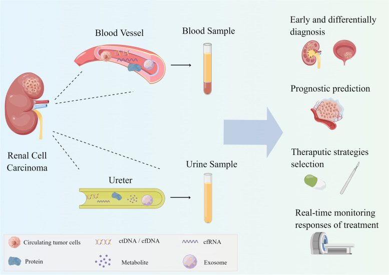
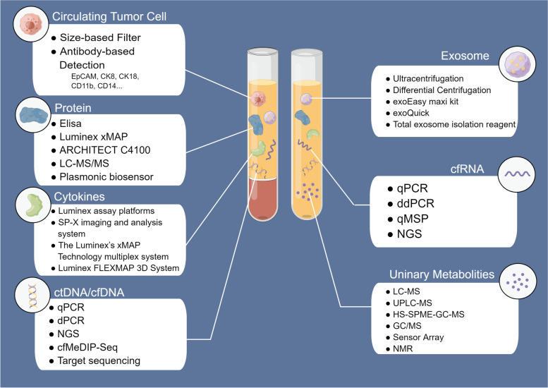
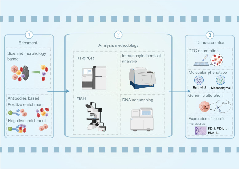
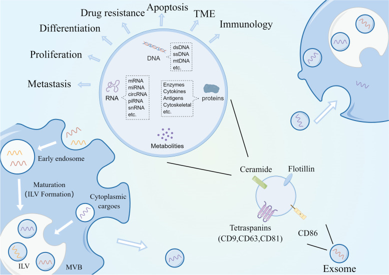
Renal cell carcinoma (RCC) is a major pathological type of kidney cancer and is one of the most common malignancies worldwide. The unremarkable symptoms of early stages, proneness to postoperative metastasis or recurrence, and low sensitivity to radiotherapy and chemotherapy pose a challenge for the diagnosis and treatment of RCC. Liquid biopsy is an emerging test that measures patient biomarkers, including circulating tumor cells, cell-free DNA/cell-free tumor DNA, cell-free RNA, exosomes, and tumor-derived metabolites and proteins. Owing to its non-invasiveness, liquid biopsy enables continuous and real-time collection of patient information for diagnosis, prognostic assessment, treatment monitoring, and response evaluation. Therefore, the selection of appropriate biomarkers for liquid biopsy is crucial for identifying high-risk patients, developing personalized therapeutic plans, and practicing precision medicine. In recent years, owing to the rapid development and iteration of extraction and analysis technologies, liquid biopsy has emerged as a low cost, high efficiency, and high accuracy clinical detection method. Here, we comprehensively review liquid biopsy components and their clinical applications over the past 5 years. Additionally, we discuss its limitations and predict its future prospects.

Keywords: Cell-free tumor DNA; Circulating tumor cells; Diagnosis; Liquid biopsy; Prognosis; Renal cell carcinoma; Treatment monitoring.


Conflict of interest statement

The authors declare no competing interests.

Figures

Fig. 1
Fig. 1
Samples and components for liquid biopsy of RCC. Liquid biopsy for RCC mainly uses blood and urine samples comprising CTCs, cfDNA/ctDNA, cfRNA, proteins, metabolites, and exosomes. Due to its non-invasiveness, liquid biopsy can be used to collect patient information continuously and plays multiple roles at different stages of disease. It can screen patients with RCC in the healthy population and identify urological masses for differential diagnosis. Before treatment, liquid biopsy can predict the risk of progression to identify high-risk patients and predict the response of patients to various treatments, which helps to select the appropriate treatment plan. After treatment, liquid biopsy allows real-time monitoring of patient outcomes and prevention of postoperative recurrence and metastasis
Fig. 2
Fig. 2
Commonly used techniques for extraction or analysis of RCC liquid biopsies in recent years. CTC isolation mainly includes size-based and antibody-based methods. cfDNA and ctDNA are separated by patients’ genomic alterations. PCR and sequencing are commonly used to detect and analyze mutations, size, expression, and methylation levels. ddPCR and targeting sequencing enable analysis of specific rare DNA with high sensitivity. With the development and diffusion of NGS, researchers can perform high-throughput analysis of cfDNA/ctDNA at a reduced cost. Similar to cfDNA/ctDNA, cfRNA is commonly analyzed by qPCR, ddPCR, methylation-specific quantitative PCR (qMSP), and NGS. Metabolite analysis is mainly performed using MS-based methods, while NMR and inductors have also been used in recent years. Protein analysis mainly depended on ELISA, the standard method for protein level measurements. Some automated analyzers with low cost and high efficiency are commercially available, which have potential for large-scale clinical applications. As an important field of proteomics, circulating cytokine assays use commercial detection platforms or technologies more frequently than Elisa, which enable a rapid detection of multiple cytokines in the blood. Up to now, there is no standard method for the extraction of exosomes. The most commonly used methods in recent years are ultracentrifugation and differential centrifugation. Meanwhile, some extraction reagents are used, such as exoQuick kit, exoEasy maxi kit and Total exosome isolation reagent
Fig. 3
Fig. 3
Isolation, detection, and characterization of CTCs and the associated clinical value. CTCs are firstly isolated from peripheral blood by size-, morphology-, and antibody-based methods. Then, several methods can be used to detect CTCs from isolating products or further characterize CTCs by analyzing cellular components or morphology. CTC analysis can provide a range of information such as CTC enumeration, molecular phenotypes, mutations, proteomics and metabolomics to analyze patient heterogeneity, drug resistance and risk of progression, which is widely used for early and differential diagnosis, prognostic assessment, treatment schedule selecting, and evaluation of treatment efficacy
Fig. 4
Fig. 4
Exosomal biogenesis and regulatory mechanisms. The cell membrane wraps specific cellular material inward to form EEs which mature into MVBs. Meanwhile, MVBs also receive specific cargo from the cytoplasm. Different materials within the MVBs are separated by the membrane to form ILVs. Low-cholesterol MVBs are degraded, while high-cholesterol MVBs fuse with the cell membrane to release ILVs into the circulation. The released ILVs are called exosomes. The exosomes are enriched with cell surface proteins on the membranes such as Tetraspanins (CD9, CD81, CD63), CD86, integrins and ceramide, which are used as exosome markers and recognized by target cells. It is reported that exosomes regulate tumor differentiation, proliferation, apoptosis, EMT, drug resistance, and the TME by transmitting intercellular messages

Similar articles

Cited by

References

    1. Sung H, et al. Global Cancer Statistics 2020: GLOBOCAN Estimates of Incidence and Mortality Worldwide for 36 Cancers in 185 Countries. CA Cancer J Clin. 2021;71(3):209–249. doi: 10.3322/caac.21660. - DOI - PubMed
    1. Kovacs G, et al. The Heidelberg classification of renal cell tumours. J Pathol. 1997;183(2):131–133. doi: 10.1002/(SICI)1096-9896(199710)183:2<131::AID-PATH931>3.0.CO;2-G. - DOI - PubMed
    1. Choueiri TK, Motzer RJ. Systemic Therapy for Metastatic Renal-Cell Carcinoma. N Engl J Med. 2017;376(4):354–366. doi: 10.1056/NEJMra1601333. - DOI - PubMed
    1. Hsieh JJ, et al. Renal cell carcinoma. Nat Rev Dis Primers. 2017;3:17009. doi: 10.1038/nrdp.2017.9. - DOI - PMC - PubMed
    1. Hahn AW, et al. First-line Treatment of Metastatic Renal Cell Carcinoma: A Systematic Review and Network Meta-analysis. Eur Urol Oncol. 2019;2(6):708–715. doi: 10.1016/j.euo.2019.09.002. - DOI - PubMed

Publication types