. 2023 Feb 21;11(1):30.
doi: 10.1186/s40168-023-01471-8.

Dimethyl itaconate ameliorates cognitive impairment induced by a high-fat diet via the gut-brain axis in mice

Affiliations

Dimethyl itaconate ameliorates cognitive impairment induced by a high-fat diet via the gut-brain axis in mice

Wei Pan et al. Microbiome. .

Erratum in

Abstract

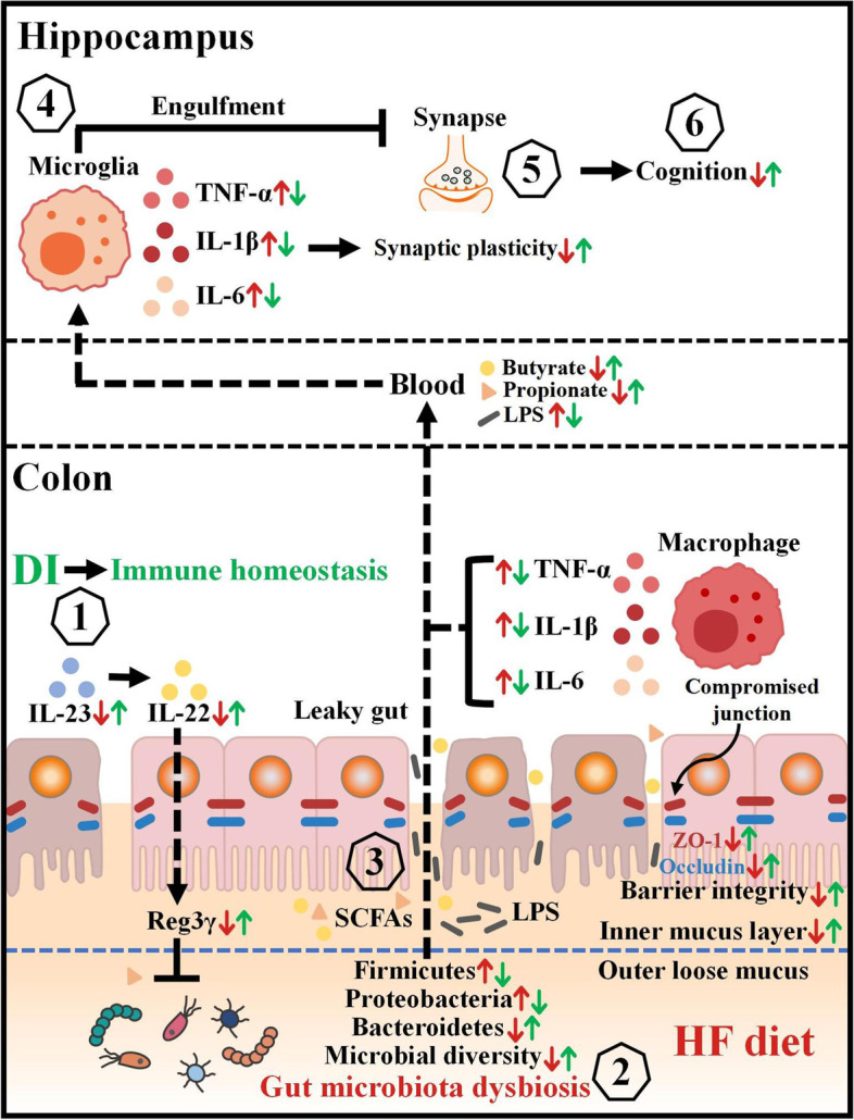
Background: Gut homeostasis, including intestinal immunity and microbiome, is essential for cognitive function via the gut-brain axis. This axis is altered in high-fat diet (HFD)-induced cognitive impairment and is closely associated with neurodegenerative diseases. Dimethyl itaconate (DI) is an itaconate derivative and has recently attracted extensive interest due to its anti-inflammatory effect. This study investigated whether intraperitoneal administration of DI improves the gut-brain axis and prevents cognitive deficits in HF diet-fed mice.

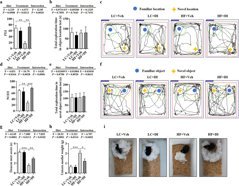
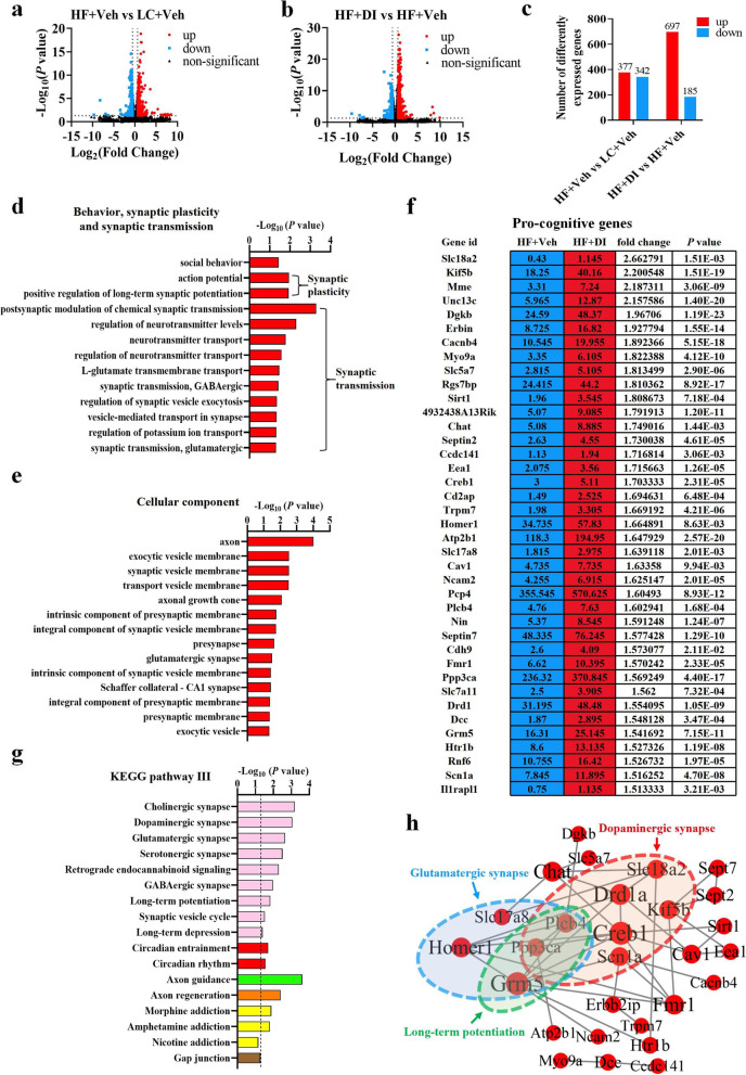
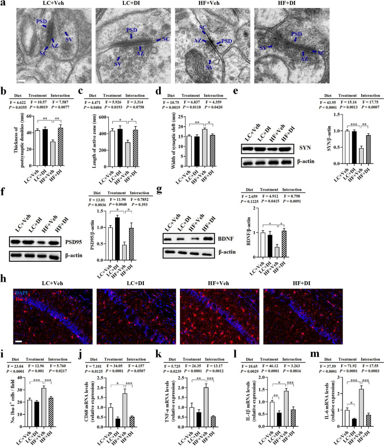
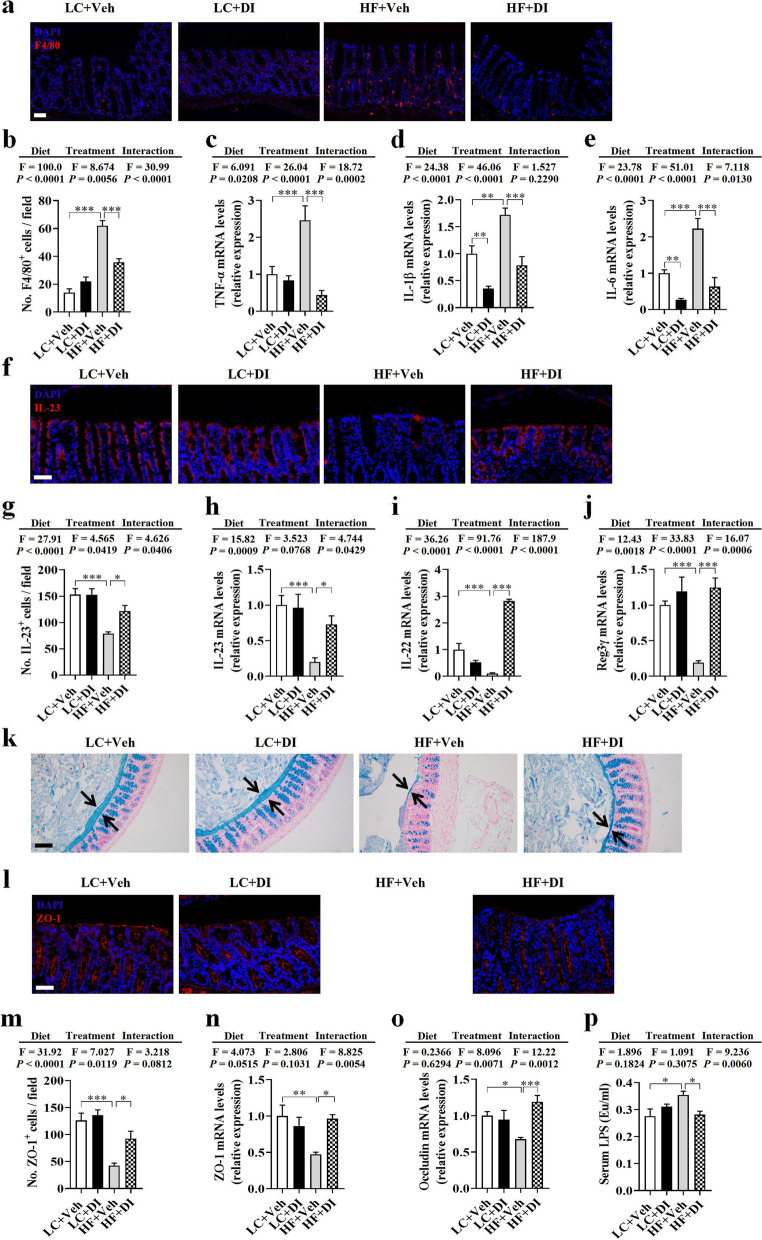
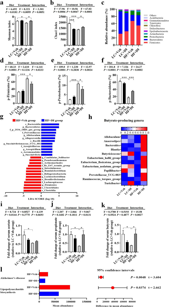
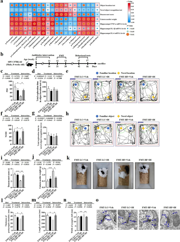
Results: DI effectively attenuated HFD-induced cognitive decline in behavioral tests of object location, novel object recognition, and nesting building, concurrent with the improvement of hippocampal RNA transcription profiles of genes associated with cognition and synaptic plasticity. In agreement, DI reduced the damage of synaptic ultrastructure and deficit of proteins (BDNF, SYN, and PSD95), the microglial activation, and neuroinflammation in the HFD-fed mice. In the colon, DI significantly lowered macrophage infiltration and the expression of pro-inflammatory cytokines (TNF-α, IL-1β, IL-6) in mice on the HF diet, while upregulating the expression of immune homeostasis-related cytokines (IL-22, IL-23) and antimicrobial peptide Reg3γ. Moreover, DI alleviated HFD-induced gut barrier impairments, including elevation of colonic mucus thickness and expression of tight junction proteins (zonula occludens-1, occludin). Notably, HFD-induced microbiome alteration was improved by DI supplementation, characterized by the increase of propionate- and butyrate-producing bacteria. Correspondingly, DI increased the levels of propionate and butyrate in the serum of HFD mice. Intriguingly, fecal microbiome transplantation from DI-treated HF mice facilitated cognitive variables compared with HF mice, including higher cognitive indexes in behavior tests and optimization of hippocampal synaptic ultrastructure. These results highlight the gut microbiota is necessary for the effects of DI in improving cognitive impairment.

Conclusions: The present study provides the first evidence that DI improves cognition and brain function with significant beneficial effects via the gut-brain axis, suggesting that DI may serve as a novel drug for treating obesity-associated neurodegenerative diseases. Video Abstract.

Keywords: Cognition; Gut microbiome; Gut-brain axis; Itaconate; Microglia; Obesity.


Conflict of interest statement

The authors declare that they have no competing interests.

Figures

Fig. 1
Fig. 1
DI supplementation improved cognitive decline in mice on HF diet. The object location test was performed to evaluate the spatial memory of mice (ac). a Percentage of time spent with the object in the novel place to total object exploration time. b The total object exploration time. c Representative track plots of LC+Veh, LC+DI, HF+Veh, and HF+DI groups recorded by the SMART video tracking system in the testing phase. Note that the LC+Veh mouse spent more time exploring the object in the novel place whereas the HF+Veh mouse did not show preference to the object in a novel place. The novel object recognition test was performed to evaluate the object recognition memory of mice (df). d Percentage of time spent with the novel object to total object exploration time. e The total object exploration time. f Representative track plots of LC+Veh, LC+DI, HF+Veh, and HF+DI groups recorded by the SMART video tracking system in the testing phase. The nest building test was used to assess the activities of daily living of mice (gi). g The nest score and h untorn nestlet weight (amount of untorn nesting material). i Representative nest result of LC+Veh, LC+DI, HF+Veh, and HF+DI groups. n = 12 mice for each group in behavior tests. Values are mean ± SEM. *P < 0.05, **P < 0.01, ***P < 0.001
Fig. 2
Fig. 2
DI supplementation improved the transcriptome profile associated with cognition in the hippocampus of mice on HF diet. a The volcano plot shows the distributions of differentially expressed genes (DEGs) between HF+Veh and LC+Veh mice. b The volcano plot shows the distributions of DEGs between HF+DI and HF+Veh mice. c The number of upregulated and downregulated DEGs. d The biological processes associated with behavior, synaptic plasticity, and transmission are upregulated in HF diet-fed mice after DI supplementation. e The cellular components associated with synapse are upregulated in the HF+DI group in comparison to HF+Veh group. f The fpkm value, fold change and P-value of pro-cognitive DEGs of HF+Veh and HF+DI groups. g The enriched KEGG pathways related to behavior, synapse, and brain development in HF diet-fed mice after DI supplementation. Columns with different colors represent different classification in level 2. The dotted line in the figure represents P = 0.05. h PPI network analysis of pro-cognitive genes based on STRING database and Cytoscape 3.9.1. Black lines denoted the interaction between two proteins. n = 3 mice for each group
Fig. 3
Fig. 3
DI supplementation mitigated synaptic impairment and neuroinflammation in the hippocampus of mice on HF diet. a Representative ultrastructure of synapses in the cornu ammonis 1 (CA1) region of mice on the electron micrograph (scale bar: 100 nm). bd Image analysis of the thickness of postsynaptic density (PSD), length of the active zone (AZ), and width of the synaptic cleft (SC) (n = 2, 8 images per mouse). eg The protein expression levels of SYN, PSD95, and BDNF in the hippocampus (n = 5). h The immunofluorescent staining of Iba-1 in CA1 of the hippocampus. i The quantification of Iba-1+ cells in CA1 of the hippocampus (n = 3, 5 images per mouse, scale bar: 50 μm). j–m The mRNA expression of CD68, TNF-α, IL-1β, and IL-6 in the hippocampus (n = 6). Values are mean ± SEM. *P < 0.05, **P < 0.01, ***P < 0.001
Fig. 4
Fig. 4
DI supplementation restored colonic immune homeostasis and alleviated colonic mucosa barrier impairment in mice on HF diet. a Immunofluorescence images of colonic sections stained with F4/80 (red) and DAPI (blue). b The quantification of F4/80+ cells (n = 3, 5 images per mouse, scale bar: 50 μm). ce The mRNA expression of TNF-α, IL-1β, and IL-6 in the colon (n = 6). f Immunofluorescence images of colonic sections stained with IL-23 (red) and DAPI (blue). g The quantification of IL-23+ cells (n = 3, 5 images per mouse, scale bar: 50 μm). h–j The mRNA expression of IL-23, IL-22, and Reg3γ in the colon (n = 6). k Alcian blue-stained colonic sections were showing the mucus layer (arrows). (scale bar: 100 μm). Opposing black arrows with shafts delineate the mucus layer measured. l Immunofluorescence images of colonic sections stained with ZO-1 (red) and DAPI (blue). m The quantification of ZO-1+ cells (n = 3, 5 images per mouse, scale bar: 50 μm). n, o The mRNA expression of ZO-1, and occludin in the colonic tissues (n = 6). p The serum levels of LPS (n = 7). Values are mean ± SEM *P < 0.05, **P < 0.01, ***P < 0.001
Fig. 5
Fig. 5
DI supplementation partially ameliorated gut microbiome alternation in mice on HF diet. Fecal microbiome composition was analyzed by 16S rRNA gene sequencing (n = 6). a Shannon index. b Chao1 index. c Composition of abundant bacterial phyla. d–f Relative abundance of Firmicutes, Proteobacteria, and Bacteroidetes. g Linear discriminant analysis (LDA) effect size (LEfSe) showing the most significantly abundant taxa enriched in microbiome from the HF+DI group compared to the HF+Veh group. h The heatmap of relative abundance of genera associated with the production of butyrate. The intensity of color in the heatmap (blue to red) indicates the normalized abundance score for each genus. ik The relative serum levels of acetate, propionate, and butyrate (n = 6). l Predicted KEGG functional pathways associated with Alzheimer’s disease and lipopolysaccharide biosynthesis at level 3 inferred from 16S rRNA gene sequences using PICRUSt. Values are mean ± SEM. *P < 0.05, **P < 0.01, ***P < 0.001. Abbreviations: p, phylum; c, class; o, order; f, family; g, genus
Fig. 6
Fig. 6
DI supplementation improved cognition and synaptic ultrastructure through the gut microbiome. a Pearson’s correlations among the effects of DI supplementation on gut microbiome, neuroinflammation of the hippocampus, and cognitive behaviors. b Schematic strategy for fecal microbiome transplantation. c Percentage of time spent with the object in the novel place to total object exploration time. d The total object exploration time in the object location test. e Representative track plots in object location test. f Percentage of time spent with the novel object to total object exploration time. g The total object exploration time in novel object recognition test. h Representative track plots in the novel object recognition test. i The nest score and j untorn nestlet weight. k Representative nest results in nesting building tests. n = 8 mice for each group in these behavior tests. ln Statistical analysis of the thickness of PSD, length of AZ, and width of SC in the CA1 region of the hippocampus (n = 2, 8 images per mouse). o Representative ultrastructure of synapses in the CA1 region of mouse hippocampus on the electron micrograph (scale bar: 100 nm). Values are mean ± SEM. *P < 0.05, **P < 0.01, ***P < 0.001
Fig. 7
Fig. 7
Schematic strategy for DI’s role in improving cognitive impairment induced by HF diet via the gut-brain axis. DI supplementation is proposed to alleviate the macrophage infiltration, upregulate the expression of IL-23, IL-22, and Reg3γ in the colon (1), which jointly prevent HF diet-induced microbiome shift (2). Then, the rescued gut microbiome can produce more butyrate and propionate to repair the compromised intestinal integrity (3). This decreases the levels of pro-inflammatory mediators in the blood and the brain, thereby mitigating neuroinflammation (4) and synaptic damage in the hippocampus (5), which ultimately improves cognition (6). Therefore, the supplementation of DI has a beneficial impact on cognition via the gut-brain axis. Red arrows represent altered indices induced by the HF diet, while green arrows show the protective effects of DI in HF diet-fed mice

References

    1. Livingston G, Sommerlad A, Orgeta V, Costafreda SG, Huntley J, Ames D, et al. Dementia prevention, intervention, and care. Lancet. 2017;390(10113):2673–2734. doi: 10.1016/s0140-6736(17)31363-6. - DOI - PubMed
    1. O'Brien PD, Hinder LM, Callaghan BC, Feldman EL. Neurological consequences of obesity. Lancet Neurol. 2017;16(6):465–477. doi: 10.1016/s1474-4422(17)30084-4. - DOI - PMC - PubMed
    1. Miller AA, Spencer SJ. Obesity and neuroinflammation: a pathway to cognitive impairment. Brain Behav Immun. 2014;42:10–21. doi: 10.1016/j.bbi.2014.04.001. - DOI - PubMed
    1. Morris MCED, Bienias JL, Tangney CC, Wilson RS. Dietary fat intake and 6-year cognitive change in an older biracial community population. Neurology. 2004;62(9):1573–1579. doi: 10.1212/01.wnl.0000123250.82849.b6. - DOI - PubMed
    1. Shi H, Yu Y, Lin D, Zheng P, Zhang P, Hu M, et al. beta-glucan attenuates cognitive impairment via the gut-brain axis in diet-induced obese mice. Microbiome. 2020;8(1):143. doi: 10.1186/s40168-020-00920-y. - DOI - PMC - PubMed

Publication types