. 2023 Feb;9(1):e002856.
doi: 10.1136/rmdopen-2022-002856.

NADPH oxidase 4 deficiency attenuates experimental osteoarthritis in mice

Affiliations

NADPH oxidase 4 deficiency attenuates experimental osteoarthritis in mice

Félix Renaudin et al. RMD Open. 2023 Feb.

Abstract

Objective: Low-grade inflammation plays a pivotal role in osteoarthritis (OA) through exposure to reactive oxygen species (ROS). In chondrocytes, NADPH oxidase 4 (NOX4) is one of the major ROS producers. In this study, we evaluated the role of NOX4 on joint homoeostasis after destabilisation of the medial meniscus (DMM) in mice.

Methods: Experimental OA was simulated on cartilage explants using interleukin-1β (IL-1β) and induced by DMM in wild-type (WT) and NOX4 knockout (NOX4-/-) mice. We evaluated NOX4 expression, inflammation, cartilage metabolism and oxidative stress by immunohistochemistry. Bone phenotype was also determined by micro-CT and histomorphometry.

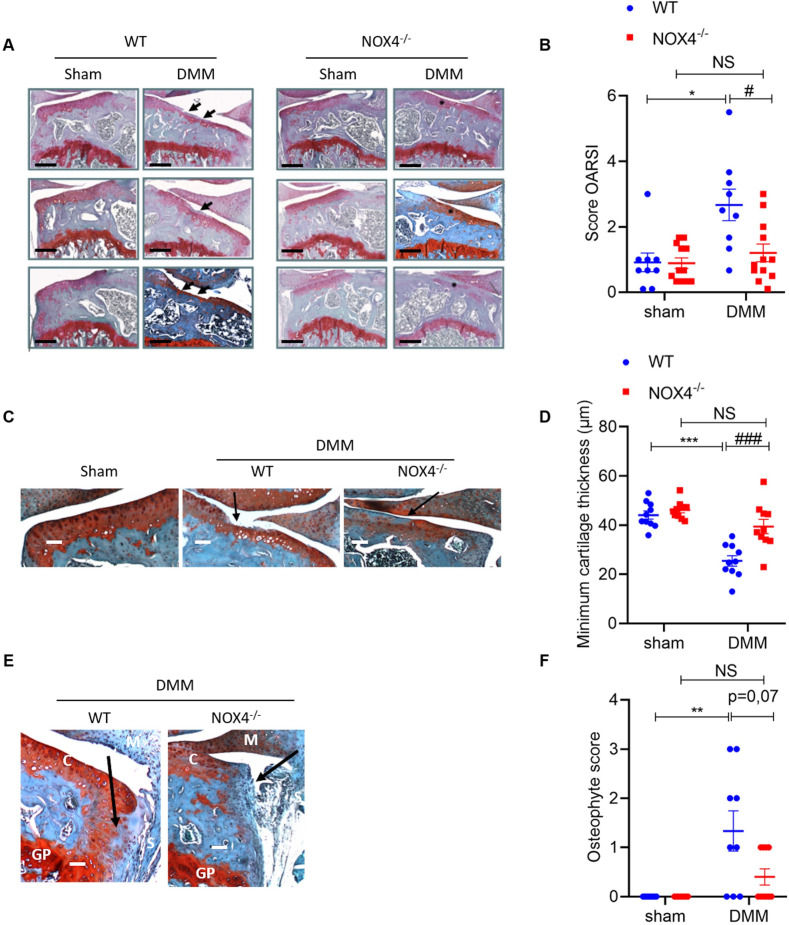
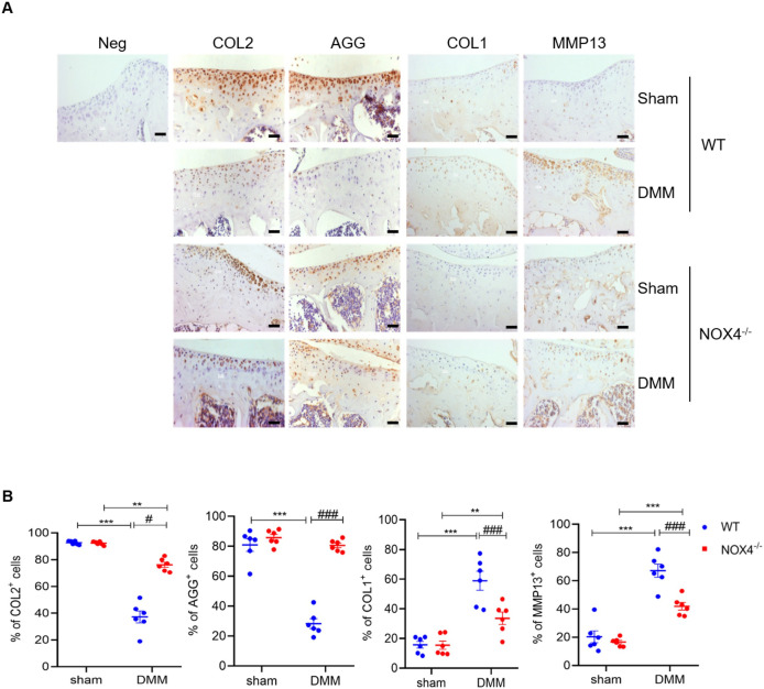
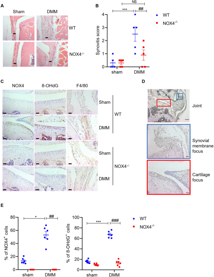
Results: Whole body NOX4 deletion attenuated experimental OA in mice, with a significant reduction of the OARSI score at 8 weeks. DMM increased total subchondral bone plate (SB.Th), epiphysial trabecular thicknesses (Tb.Th) and bone volume fraction (BV/TV) in both NOX4-/- and wild-type (WT) mice. Interestingly, DDM decreased total connectivity density (Conn.Dens) and increased medial BV/TV and Tb.Th only in WT mice. Ex vivo, NOX4 deficiency increased aggrecan (AGG) expression and decreased matrix metalloproteinase 13 (MMP13) and collagen type I (COL1) expression. IL-1β increased NOX4 and 8-hydroxy-2'-deoxyguanosine (8-OHdG) expression in WT cartilage explants but not in NOX4-/-. In vivo, absence of NOX4 increased anabolism and decreased catabolism after DMM. Finally, NOX4 deletion decreased synovitis score, 8-OHdG and F4/80 staining following DMM.

Conclusion: NOX4 deficiency restores cartilage homoeostasis, inhibits oxidative stress, inflammation and delays OA progression after DMM in mice. These findings suggest that NOX4 represent a potential target to counteract for OA treatment.

Keywords: Cytokines; Inflammation; Osteoarthritis.


Conflict of interest statement

Competing interests: None declared.

Figures

Figure 1
Figure 1
Deletion of NOX4 reduces cartilage degradation. (A) Knee sections stained with safranin-O from NOX4-/- (n=13 mice) and WT (n=11 mice). Cartilage lesions are shown with a black arrow. Scale bars: 250 µm. (B) OA lesions were assessed by OARSI score. RM-Two ways ANOVA on log transformed data test with FDR correction between WT and NOX4-/- (#) or between Sham and DMM (*). *p<0.05, **p<0.01, ***p<0.001. (C) Representative images of the minimum cartilage thickness and (D) its quantification using the Zen software (n=10). RM-Two ways ANOVA test with FDR correction between WT and NOX4-/- (#) or between Sham and DMM (*). *p<0.05, **p<0.01, ***p<0.001. Scale bars: 200 µm. (E) Representative images of osteophytes formation after DMM (M: meniscus, S: synovial membrane, C: cartilage and GP: growth plate). Osteophytes are showed with a black arrow. Scale bars: 200 µm. (F) Osteophytes formation were quantified with the osteophyte score (n=9). Kruskal-Wallis test with FDR correction between WT and NOX4-/- (#) or between sham and DMM (*). *p<0.05, **p<0.01, ***p<0.001. ANOVA, analysis of variance; FDR, false discovery rate; DMM, destabilisation of the medial meniscus; OARSI, OA Research Society International.
Figure 2
Figure 2
Ex vivo subchondral bone microarchitecture evaluation by micro-CT and histomorphometry. (A) Micro-CT images of a WT sham and DMM knee 8 weeks after DMM (SP: subchondral plate, GP: growth plate and O: osteophyte). (B) Quantification of the maximum Sb.Th of the lateral and medial part of the knee (WT: N=11 and NOX4-/-: N=13). (C) Representative images of the safranin-safranin-O stained sections used for histomorphometry. Black arrow shows the medial part of the joint. Scale bars: 250 µm. (D) Quantification of the different subchondral bone parameters on the medial part of the epiphysis: BV/TV, Tb.Th and Tb.Sp (WT: N=11 and NOX4-/-: N=13). RM-two-way ANOVA test on log transformed data with FDR correction between WT and NOX4-/- (#) or between sham and DMM (*). *p<0.05, **p<0.01, ***p<0.001. ANOVA, analysis of variance; DMM, destabilisation of the medial meniscus; FDR, false discovery rate; WT, wild type.
Figure 3
Figure 3
Deletion of NOX4 rescues the anabolic/catabolic balance. A) Representative IHC images of knee cartilage sections stained with COL2, AGG, COL1 and MMP13 antibody (N=6). Please note that DMM decreased AGG and COL2 expression and increase MMP13 and COL1 expression in the cartilage of WT mice but not in NOX4-/-. Scale bars: 40 µm. (B) Quantification of the proportions of positive cells for COL2, AGG, COL1 and MMP13 staining in the superficial cartilage. RM-two-way ANOVA test with FDR correction between WT and NOX4-/- (#) or between sham and DMM (*). *p<0.05, **p<0.01, ***p<0.001. ANOVA, analysis of variance; DMM, destabilisation of the medial meniscus; FDR, false discovery rate; WT, wild type. IHC: Immunohistochemistry.
Figure 4
Figure 4
Deletion of NOX4 protects cartilage in an IL-1β-induced osteoarthritis (OA) model. (A) Representative images of femoral heads explants, safranin-O staining and IHC of anabolism (COL2 and AGG) and catabolism (MMP13 and COL1). (B) NOX4 and 8-OHdG IHC. Please note that IL-1β treatment decreased AGG expression and increased MMP13 and COL1 expression in the cartilage of WT explants but not in NOX4-/-. Scale bars: 40 µm. (C) Quantification of the proportions of positive cells for COL2, AGG, COL1, MMP13 NOX4 and 8-OHdG staining in the superficial cartilage. RM-two-way ANOVA test with FDR correction between WT and NOX4-/- (#) or between sham and DMM (*). *p<0.05, **p<0.01, ***p<0.001. ANOVA, analysis of variance; WT, wild type. IHC: Immunohistochemistry.
Figure 5
Figure 5
Deletion of NOX4 protects from cartilage degradation and inflammation via inhibition of oxidative stress. (A) Knee sections focus on synovial membrane stained with H&E from NOX4-/- and WT (n=5 mice). Scale bars: 20 µm. (B) Quantification of the synovial Inflammation using the synovitis score. RM-two-way ANOVA test with FDR correction between WT and NOX4-/- (#) or between sham and DMM (*). (C) Representative IHC images of knee sections stained with NOX4, 8-OHdG and F4/80 antibody (N=6). Please note that DMM increased NOX4, 8OHdG and F4/80 expression in WT mice. There was no expression of 8OHdG and F4/80 in the NOX4-/- mice. Scale bars: 40 µm. (D) Representative IHC image of the whole Knee stained with NOX4 antibody (scale bar: 200 µm) and a focus on the synovial membrane (blue rectangle, scale bar: 40 µm) and the cartilage (red rectangle, scale bar: 40 µm). We observed an expression of NOX4 in the cartilage (red rectangle) and not the synovial membrane (blue rectangle) of the joints. (E) Quantification of the proportions of positive cells for NOX4 and 8-OHdG staining in the superficial cartilage. RM-two ways ANOVA test with FDR correction between WT and NOX4-/- (#) or between Sham and DMM (*). *p<0.05, **p<0.01, ***p<0.001. ANOVA, analysis of variance; DMM, destabilisation of the medial meniscus; FDR, false discovery rate; WT, wild type. IHC: Immunohistochemistry.

References

    1. Berenbaum F. Osteoarthritis as an inflammatory disease (osteoarthritis is not osteoarthrosis!). Osteoarthritis Cartilage 2013;21:16–21. 10.1016/j.joca.2012.11.012 - DOI - PubMed
    1. Bijlsma JWJ, Berenbaum F, Lafeber FPJG. Osteoarthritis: an update with relevance for clinical practice. Lancet 2011;377:2115–26. 10.1016/S0140-6736(11)60243-2 - DOI - PubMed
    1. Carr AJ, Robertsson O, Graves S, et al. . Knee replacement. Lancet 2012;379:1331–40. 10.1016/S0140-6736(11)60752-6 - DOI - PubMed
    1. Lohmander LS, Dahlberg L, Eyre D, et al. . Longitudinal and cross-sectional variability in markers of joint metabolism in patients with knee pain and articular cartilage abnormalities. Osteoarthritis Cartilage 1998;6:351–61. 10.1053/joca.1998.0134 - DOI - PubMed
    1. Nam J, Perera P, Liu J, et al. . Sequential alterations in catabolic and anabolic gene expression parallel pathological changes during progression of monoiodoacetate-induced arthritis. PLOS ONE 2011;6:e24320. 10.1371/journal.pone.0024320 - DOI - PMC - PubMed

Publication types