Review
. 2023 Jan 17;7(1):6.
doi: 10.3390/vision7010006.

An Overview of the Penguin Visual System

Affiliations
Review

An Overview of the Penguin Visual System

Peter W Hadden et al. Vision (Basel). .

Abstract

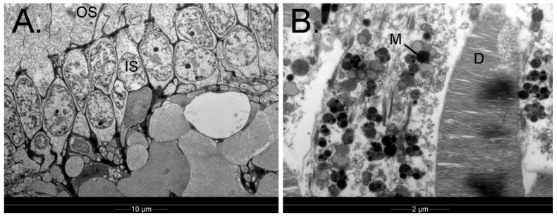
Penguins require vision that is adequate for both subaerial and submarine environments under a wide range of illumination. Here we provide a structured overview of what is known about their visual system with an emphasis on how and how well they achieve these goals. Amphibious vision is facilitated by a relatively flat cornea, the power in air varying from 10.2 dioptres (D) to 41.3 D depending on the species, and there is good evidence for emmetropia both above and below water. All penguins are trichromats with loss of rhodopsin 2, a nocturnal feature, but only deeper diving penguins have been noted to have pale oil droplets and a preponderance of rods. Conversely, the diurnal, shallow-diving little penguin has a higher ganglion cell density (28,867 cells/mm2) and f-number (3.5) than those that operate in dimmer light. In most species studied, there is some binocular overlap, but this reduces upon submergence. However, gaps in our knowledge remain, particularly with regard to the mechanism of accommodation, spectral transmission, behavioural measurements of visual function in low light, and neural adaptations to low light. The rarer species also deserve more attention.

Keywords: Spheniscidae; accommodation; amphibious; bird; eye; eyesight; nocturnal; underwater; vision.


Conflict of interest statement

The authors declare no conflict of interest.

Figures

Figure 1
Figure 1
A brown skua Stercorarius antarcticus predates a gentoo penguin Pygoscelis papua chick on land. Cuverville Island, Antarctic Peninsula. Credit: PWH.
Figure 2
Figure 2
A Galapagos penguin Spheniscus mendiculus forages in tropical waters above a rocky benthic substrate. Off Punta Vicente Roca, Galapagos Islands, Ecuador. Credit: PWH.
Figure 3
Figure 3
The orbit and ocular adnexae. (A) Skull of the Fiordland crested penguin Eudyptes pachyrhynchus, with the bony elements that border Orbita (the orbit) labelled (specimen AV1178, Otago Museum, Dunedin, New Zealand; Credit: Dane A. Gerneke, University of Auckland); (B) The orbital region of the little penguin Eudyptula minor, demonstrating the bones, muscles, and glands that surround Bulbus oculi (the eye). Reconstructed from publicly available raw data at https://figshare.com/search?q=10.17608/k6.auckland.c.5599341 (accessed on 9 June 2022) (Digital Science, London, UK) using Amira 2021.2 (Thermo Fisher Scientific, Waltham, MA, USA) software.
Figure 4
Figure 4
Transmission electron micrograph of Membrana nictitans of a gentoo penguin Pygoscelis papua; (A) Note the excrescences on the posterior surface (arrow); (B) There is a regular, parallel arrangement of collagen fibrils in the stroma (asterix). Credit: PWH.
Figure 5
Figure 5
Bulbus oculi (the eye) of selected penguins. (A) B-scan ultrasound, little penguin Eudyptula minor eye (Scanmate, DGH Technology Inc., Exton, PA, USA). In this eye, the axial length (blue) measures 16.1 mm and the distance from lens to retina (pink) is 10.18 mm. Credit: PWH; (B) Micro-CT of king penguin Aptenodytes patagonicus eye—note the aspheric shape of the eye and the bony elements within (beige). Reconstructed from publicly available raw data at https://figshare.com/articles/dataset/Hadden_King_penguin_micro_CT_stained_eye/12924741 (accessed on 23 August 2022) (Digital Science, London, UK) using CT Vox 3.3 (Bruker MicroCT, Kontich, Belgium) software.
Figure 6
Figure 6
Cornea of the king penguin Aptenodytes patagonicus. (A) Immediate post-mortem slit lamp photograph; note the transparent appearance and the translucent Membrana nictitans covering the cornea on the left-hand side (Credit: PWH); (B) Confocal micrographs of the king penguin cornea compared to those of the human (Heidelberg Retina Tomograph II Rostock Corneal Module, Heidelberg Engineering GmBH, Germany), which conforms to the description by Kafarnik et al. of other avian corneas. Note the absence of a subepithelial nerve plexus in Bowman’s layer; (C) Anterior segment optical coherence tomogram of the cornea (REVO NX, OPTOPOL Technology Sp. Z o.o., Zawierce, Poland). Credit: PWH.
Figure 7
Figure 7
Irides of selected species of penguin. (A) Yellow-eyed penguin Megadyptes antipodes; (B) Gentoo penguin Pygoscelis papua; (C) Little penguin Eudyptula minor—note that the pupil appears slightly octagonal in this photograph; (D) King penguin Aptenodytes patagonicus; (E) The intrinsic structure of the king penguin iris, as reconstructed from raw data available at https://figshare.com/search?q=10.17608/k6.auckland.c.5599341 (accessed on 31 May 2022) (Digital Science, London, UK) using CT Vox 3.3 (Bruker MicroCT, Kontich, Belgium). Credit: PWH.
Figure 8
Figure 8
The posterior segment of the eye of a 7-week-old gentoo penguin chick (Pygoscelis papua). (A) Haemotoxylin and eosin-stained frozen section adjacent to the optic disc. Note the layered structure of the retina (ONL = outer nuclear layer, INL = inner nuclear layer), underlain by the retinal pigment epithelium (RPE) and the cartilaginous component of the sclera, as well as the vascular pecten intruding into the vitreous cavity; (B) Optical coherence tomogram (OCT) of the central retina, showing many of the same layers: i. posterior vitreous base; ii. nerve fibre layer, iii. ganglion cell layer; iv. Inner nuclear layer; v. photoreceptors; vi. retinal pigment epithelium. No foveal dip was visible in any section either histologically or on OCT. Credit: PWH.
Figure 9
Figure 9
Retina of the king penguin Aptenodytes patagonicus, as imaged with transmission electron microscopy. (A) Both outer segments (OS) and inner segments (IS) of the retinal photoreceptors are approximately 6–7 mm apart; (B) The photoreceptor outer segments, which contain discs (D) wherein phototransduction takes place, are surrounded by melanosome-laden retinal pigment epithelial cells (M). Credit: PWH.
Figure 10
Figure 10
The colour of the fundus is predominantly a product of the choroid. No intrinsic retinal vasculature is present. (A) King penguin Aptenodytes patagonicus; (B) Gentoo penguin Pygoscelis papua. The king penguin has a pink fundus but that of the gentoo has a mostly dark brown colouration, with the exception of the inferior third. Credit: PWH.
Figure 11
Figure 11
There is limited binocular overlap in penguins because the eye faces more laterally than rostrally, although both pupils are visible at the same time in these penguins in air. (A) Gentoo penguin Pygoscelis papua; (B) King penguin Aptenodytes patagonicus. Credit: PWH.

Similar articles

References

    1. Cole T.L., Zhou C., Fang M., Pan H., Ksepka D.T., Fiddaman S.R., Emerling C.A., Thomas D.B., Bi X., Fang Q., et al. Genomic insights into the secondary aquatic transition of penguins. Nat. Commun. 2022;13:3912. doi: 10.1038/s41467-022-31508-9. - DOI - PMC - PubMed
    1. Martin G.R. The Sensory Ecology of Bird. Oxford University Press; Oxford, UK: 2017. pp. 27, 49–55, 122, 125, 191–196, 259–296.
    1. Cobb S. The size of the olfactory bulb in 108 species of birds. Auk. 1968;85:55–61. doi: 10.2307/4083624. - DOI
    1. First Penguin in New Zealand to Undergo Cataract Surgery Makes Full Recovery. [(accessed on 30 October 2022)]. Available online: https://www.stuff.co.nz/national/health/129758464/first-penguin-in-new-z....
    1. Church M.L., Priehs D.R., Denis H., Croft L., DiRocco S., Davis M. Technique, postoperative complications, and visual outcomes of phacoemulsification cataract surgery in 21 penguins (27 eyes): 2011–2015. Vet. Ophthalmol. 2018;21:612–621. doi: 10.1111/vop.12555. - DOI - PubMed

LinkOut - more resources