TRPS1 Is Differentially Expressed in a Variety of Malignant and Benign Cutaneous Sweat Gland Neoplasms
- PMID: 36810569
- PMCID: PMC9944056
- DOI: 10.3390/dermatopathology10010011
TRPS1 Is Differentially Expressed in a Variety of Malignant and Benign Cutaneous Sweat Gland Neoplasms
Abstract
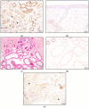
Neoplasms of sweat glands and the breast may be morphologically and immunophenotypically similar. A recent study showed that TRPS1 staining is a highly sensitive and specific marker for breast carcinoma. In this study, we analyzed TRPS1 expression in a spectrum of cutaneous sweat gland tumors. We stained five microcystic adnexal carcinomas (MACs), three eccrine adenocarcinomas, two syringoid eccrine carcinomas, four hidradenocarcinomas, six porocarcinomas, one eccrine carcinoma-NOS, 11 hidradenomas, nine poromas, seven cylindromas, three spiradenomas, and 10 syringomas with TRPS1 antibodies. All of the MACs and syringomas were negative. Every cylindroma and two of the three spiradenomas demonstrated intense staining in cells lining the ductular spaces, with negative to relatively weak expression in surrounding cells. Of the 16 remaining malignant entities, 13 were intermediate to high positive, one was low positive, and two were negative. From the 20 hidradenomas and poromas, intermediate to high positivity was revealed in 14 cases, low positivity in three cases, and negative staining in three cases. Our study demonstrates a very high (86%) expression of TRPS1 in malignant and benign adnexal tumors that are mainly composed of islands or nodules with polygonal cells, e.g., hidradenomas. On the other hand, tumors with small ducts or strands of cells, such as MACs, appear to be completely negative. This differential staining among types of sweat gland tumors may represent either differential cells of origin or divergent differentiation and has the potential to be used as a diagnostic tool in the future.
Keywords: TRPS1; eccrine gland; sweat gland tumors.
Conflict of interest statement
The authors declare no conflict of interest.
Figures


References
-
- Rollins-Raval M., Chivukula M., Tseng G.C., Jukic D., Dabbs D.J. An immunohistochemical panel to differentiate metastatic breast carcinoma to skin from primary sweat gland carcinomas with a review of the literature. Arch. Pathol. Lab. Med. 2011;135:975–983. doi: 10.5858/2009-0445-OAR2. - DOI - PubMed
-
- Wang Y., Lin X., Gong X., Wu L., Zhang J., Liu W., Li J., Chen L. Atypical GATA transcription factor TRPS1 represses gene expression by recruiting CHD4/NuRD(MTA2) and suppresses cell migration and invasion by repressing TP63 expression. Oncogenesis. 2018;7:96. doi: 10.1038/s41389-018-0108-9. - DOI - PMC - PubMed
LinkOut - more resources
Full Text Sources
Miscellaneous

