. 2023 Feb 21;13(1):2727.
doi: 10.1038/s41598-023-29110-0.

Mutant structure of metabolic switch protein in complex with monomeric c-di-GMP reveals a potential mechanism of protein-mediated ligand dimerization

Affiliations

Mutant structure of metabolic switch protein in complex with monomeric c-di-GMP reveals a potential mechanism of protein-mediated ligand dimerization

Badri Nath Dubey et al. Sci Rep. .

Abstract

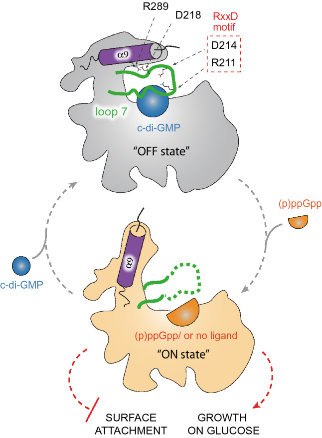
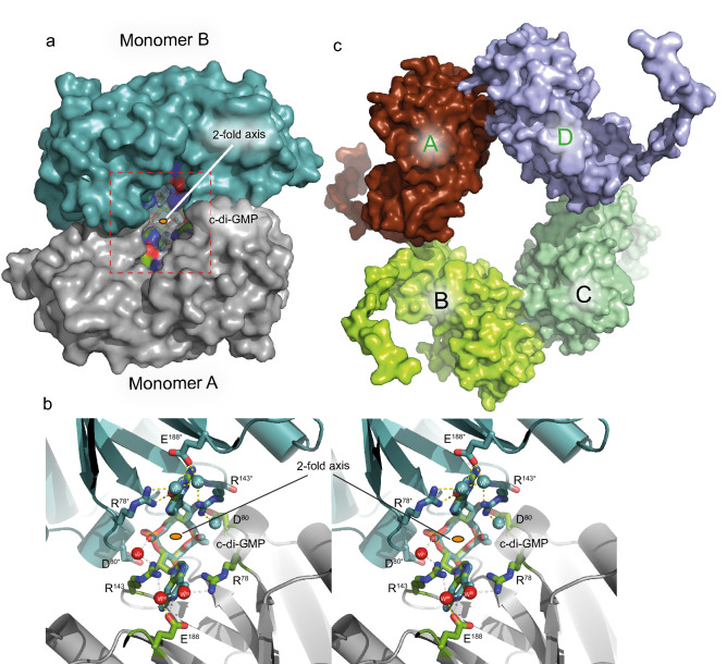
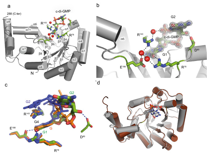
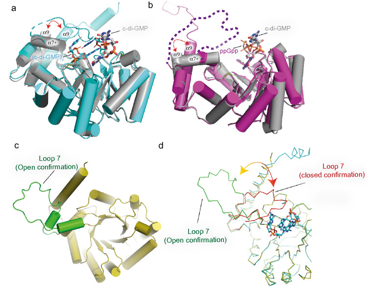
Bacterial second messengers c-di-GMP and (p)ppGpp have broad functional repertoires ranging from growth and cell cycle control to the regulation of biofilm formation and virulence. The recent identification of SmbA, an effector protein from Caulobacter crescentus that is jointly targeted by both signaling molecules, has opened up studies on how these global bacterial networks interact. C-di-GMP and (p)ppGpp compete for the same SmbA binding site, with a dimer of c-di-GMP inducing a conformational change that involves loop 7 of the protein that leads to downstream signaling. Here, we report a crystal structure of a partial loop 7 deletion mutant, SmbA∆loop in complex with c-di-GMP determined at 1.4 Å resolution. SmbA∆loop binds monomeric c-di-GMP indicating that loop 7 is required for c-di-GMP dimerization. Thus the complex probably represents the first step of consecutive c-di-GMP binding to form an intercalated dimer as has been observed in wild-type SmbA. Considering the prevalence of intercalated c-di-GMP molecules observed bound to proteins, the proposed mechanism may be generally applicable to protein-mediated c-di-GMP dimerization. Notably, in the crystal, SmbA∆loop forms a 2-fold symmetric dimer via isologous interactions with the two symmetric halves of c-di-GMP. Structural comparisons of SmbA∆loop with wild-type SmbA in complex with dimeric c-di-GMP or ppGpp support the idea that loop 7 is critical for SmbA function by interacting with downstream partners. Our results also underscore the flexibility of c-di-GMP, to allow binding to the symmetric SmbA∆loop dimer interface. It is envisaged that such isologous interactions of c-di-GMP could be observed in hitherto unrecognized targets.


Conflict of interest statement

The authors declare no competing interests.

Figures

Figure 1
Figure 1
Second messenger mediated regulation of SmbA. Binding of a c-di-GMP dimer (blue sphere) inactivates SmbA (“OFF state”, grey), while its dissociation or displacement by a ppGpp monomer (an orange half-sphere) activates the protein (“ON state”, light orange). Loop 7 is shown in green, the C-terminal α9 helix is represented by a magenta cylinder. Amino acid residues essential for salt bridge formation between α9 helix and loop 7 are indicated. Key residues of the RxxD motif in loop 7 are shown in the red box. The physiological functions of activated SmbA are indicated with red dashed lines (Adopted from Shyp et al.).
Figure 2
Figure 2
Crystal structures of SmbAΔloop with c-di-GMP bound across the crystallographic dyad and of apo SmbAΔloop. (a) The two monomers are depicted as surface (negatively charged atoms in red, positively charged atoms in blue and carbon atoms in green) with monomer A (gray) in standard orientation and monomer B (symmetry mate) in cyan. c-di-GMP in the dimer interface is shown as ball-and-stick model. (b) Stereoview down the twofold axis (indicated as a small orange ellipsoid), showing c-di-GMP forming isologous interactions with the two SmbAΔloop protomers. Relevant residues are shown as color-coded sticks (oxygen, red; nitrogen, blue; carbon, green or cyan and waters as red and cyan spheres) and labeled. Residues and waters of the symmetry mate monomer are marked with an asterisk. Hydrogen bonds between subunits and c-di-GMP are indicated as yellow dotted lines. (c) Crystal packing of apo SmbAΔloop shown in surface representation. The four molecules are arranged in an asymmetric unit form two local dimers (A and D, B and C) with 2-fold symmetry.
Figure 3
Figure 3
Detailled crystal structures of SmbAΔloop in presence and absence of c-di-GMP and and structural comparison with wild-type SmbA ligands. (a) Crystal structure of the SmbAΔloop with the backbone drawn in grey cartoon and monomeric c-di-GMP shown in a stick. Residues in the SmbAΔloop important in interaction with the c-di-GMP molecule are drawn in stick representation. Carbon atoms are shown in green, nitrogen in blue and oxygen in red. (b) 2Fo-Fc omit maps contoured at 1.2 σ of c-di-GMP and full structural details of the interacting residues. H-bonds (length < 3.5 Å) are indicated by gray lines and water molecules in red spheres. (c) View of c-di-GMP (green) as bound to SmbAΔloop, and the proximal c-di-GMP molecule (blue) of dimeric c-di-GMP and ppGpp (orange) as bound to wild-type SmbA. The proximal guanyl of monomeric c-di-GMP (G1), guanyl of ppGpp (G) and G4 of dimeric c-di-GMP overlap closely. While the other guanyl (G2) of the monomeric ligand has moved out considerably, to form isologous interactions with the second SmbAΔloop molecule (not shown). (d) structural superposition of SmbAΔloop/c-di-GMP (gray) with SmbAΔloop (chocolate) yielding a RMSD of 0.49 Å.
Figure 4
Figure 4
Analytical ultracentrifugation (AUC) analysis of SmbAwt and SmbAΔloop. (a) SV-AUC absorbance c(s) distributions of SmbAwt, SmbAwt/(c-di-GMP)2, SmbAΔloop and SmbAΔloop/c-di-GMP. (b) Mass estimation and s and f/fO values of SmbAwt, SmbAwt/(c-di-GMP)2, SmbAΔloop and SmbAΔloop/c-di-GMP.
Figure 5
Figure 5
Structural comparison of SmbAΔloop/c-di-GMP with SmbAwt/(c-di-GMP)2, SmbAwt/ppGpp and Alphafold model of SmbAwt. (a) Superposition of SmbAΔloop/c-di-GMP (gray) with SmbAwt/(c-di-GMP)2 (cyan) with RMSD of 0.4. Relevant secondary structure elements are labeled. Dimeric c-di-GMP (cyan) and monomeric (thick) are shown as ball-and-stick models. (b) Superposition of SmbAΔloop/c-di-GMP (gray) with SmbA/ppGpp (Magenta) with RMSD of 0.5. Relevant secondary-structure elements are labeled. ppGpp (magenta) and monomeric (thick in gray) are shown as ball-and-stick models. The disordered part of loop 7 is marked by broken lines. (c) AlphaFold2 predicted model of SmbAwt (yellow) with loop 7 is show in green color. (d) Superposition of SmbAwt/(c-di-GMP)2 (green) with AlphaFold2 model of SmbAwt (yellow). Loop 7 from SmbAwt/(c-di-GMP)2 and Alphfold model of SmbAwt apo are show in red and green repectively.
Figure 6
Figure 6
Observed c-di-GMP conformations in SmbAloop and its comparison with SmbAwt, PdeL and LapD. (a) and (b) shows the partial open-twisted form of monomeric c-di-GMP in C3'-endo sugar pucker conformation observed in SmbA mutant. Guanine distances are shown in black dotted line. (c) Superimposition of c-di-GMP from SmbAΔloop, SmbAwt and LapDEAL. GMP moiety from both structures shows the same conformation, the C3′-endo sugar pucker; however, there are considerable differences in the G1 and G2 base orientation (indicated by the gray arrow). (d) Superimposition of c-di-GMP from SmbAΔloop with monomeric c-di-GMP as observed when bound to a phosphodiesterase PdeL and degenerated-phosphodiesterase LapD. Distinct sugar pucker of the base at the right (G2) appears responsible for the fully elongated form of c-di-GMP when bound to PdeL or LapD. In contrast, all bases at the left (G1) show the same sugar pucker, i.e. C3′-endo as also observed for SmbAΔloop in this study. (e) Superimposition of crystal structure of c-di-GMP/Mg2+ and dimeric c-di-GMP from SmbAwt. Guanine distances are shown in red and green dotted lines of c-di-GMP/Mg2+ and dimeric c-di-GMP from SmbAwt respectevily.
Figure 7
Figure 7
Sequence alignment and distance of SmbA homologs. (a) Pairwise Needleman-Wunsch global alignment scores of SmbA and CckA reciprocal best BLAST hits (BBH) for species sampled from prosthecate Caulobacterales (PC), non-prosthecate Caulobacterales (NPC), and other bacterial groups (OG). Alignment scores are reported relative to self-alignment of SmbA (Q9A5E6) and CckA (H7C7G9) from Caulobacter crescentus. For the null models, CckA BBH was scored against SmbA and vice versa. The latter BBH was identified using BLASTp against the NCBI-NR database using the BLOSUM45 scoring matrix. (b) A phylogenetic tree of 24 SmbA orthologs inferred using the Maximum Likelihood method based on the JTT model as implemented in MEGA7. Branch lengths indicate the number of substitutions per site. The tree with the highest log likelihood (− 9134.38) is shown, with bootstrap support from 100 replicates indicated at branches. (c) Sequence alignment and logo of SmbA orthologs. The sequence logo was generated using the WebLogo server from the global alignment of SmbA orthologs used to build the distance tree.

References

    1. Hauryliuk V, Atkinson GC, Murakami KS, Tenson T, Gerdes K. Recent functional insights into the role of (p)ppGpp in bacterial physiology. Nat. Rev. Microbiol. 2015;13:298–309. doi: 10.1038/nrmicro3448. - DOI - PMC - PubMed
    1. Jenal U, Reinders A, Lori C. Cyclic di-GMP: Second messenger extraordinaire. Nat. Rev. Microbiol. 2017;15:271–284. doi: 10.1038/nrmicro.2016.190. - DOI - PubMed
    1. Cashel M, Gallant J. Two compounds implicated in the function of the RC gene of Escherichia coli. Nature. 1969;221:838–841. doi: 10.1038/221838a0. - DOI - PubMed
    1. Dalebroux ZD, Swanson MS. ppGpp: Magic beyond RNA polymerase. Nat. Rev. Microbiol. 2012;10:203–212. doi: 10.1038/nrmicro2720. - DOI - PubMed
    1. Kalia D, et al. Nucleotide, c-di-GMP, c-di-AMP, cGMP, cAMP, (p)ppGpp signaling in bacteria and implications in pathogenesis. Chem. Soc. Rev. 2012;42:305–341. doi: 10.1039/C2CS35206K. - DOI - PubMed

Publication types