. 2023 Apr;24(2):159-170.
doi: 10.1007/s10162-023-00891-5. Epub 2023 Feb 21.

Enhanced Expression of Plasminogen Activators and Inhibitor in the Healing of Tympanic Membrane Perforation in Rats

Affiliations

Enhanced Expression of Plasminogen Activators and Inhibitor in the Healing of Tympanic Membrane Perforation in Rats

Maria Makuszewska et al. J Assoc Res Otolaryngol. 2023 Apr.

Abstract

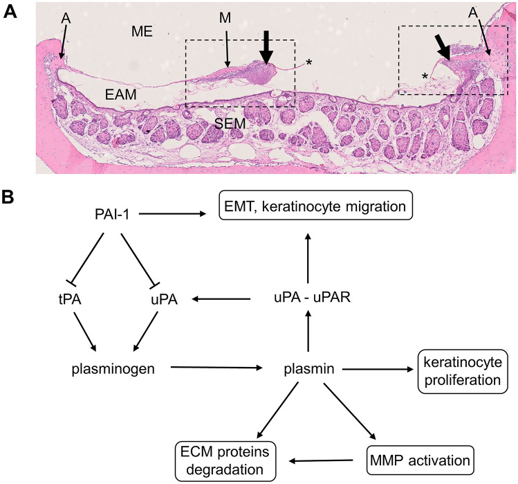
The significance of plasminogen activation during the tympanic membrane (TM) healing is known mainly from studies performed on knock-out mice. In the previous study, we reported activation of genes coding proteins of plasminogen activation and inhibition system in rat's TM perforation healing. The aim of the present study was the evaluation of protein products expressed by these genes and their tissue distribution using Western blotting and immunofluorescent method, respectively, during 10-day observation period after injury. Otomicroscopical and histological evaluation were employed to assess the healing process. The expression of urokinase plasminogen activator (uPA) and its receptor (uPAR) were significantly upregulated in the proliferation phase, with subsequent gradual attenuation during remodeling phase of healing process, when keratinocyte migration was weakening. The expression of plasminogen activator inhibitor type 1 (PAI-1) also showed the highest levels during the proliferation phase. The increase of tissue plasminogen activator (tPA) expression was observed during the whole observation period, with the highest activity during the remodeling phase. Immunofluorescence of these proteins was present mainly in migrating epithelium. Our study found that plasminogen activation (uPA, uPAR, tPA) and inhibitory (PAI-1) molecules form a well-structured regulatory system of the epithelial migration that is critical to the healing of TM after its perforation.

Keywords: Plasminogen activator inhibitor type 1; Rats; Tissue-type plasminogen activator; Tympanic membrane perforation; Urokinase-type plasminogen activator; Urokinase-type plasminogen activator receptor.


Conflict of interest statement

The authors declare no competing interests.

Figures

Fig. 1
Fig. 1
A Horizontal section through pars tensa of healing TM on day 3 after injury. ME, middle ear; EAM, external acoustic meatus; SEM, skin of external acoustic meatus; A, tympanic annulus; M, malleus handle (near umbo); single asterisk indicates edge of the perforation; thick arrows, proliferating squamous epithelium near malleus handle and annulus on the side of the perforation. Rectangles marked with a dashed line demarcate the regions of TM presented on Fig. 2 (right rectangle) and Fig. 3 (left rectangle). B The organization of the plasminogen activation/inhibition system and its influence on processes involved in epidermal healing, which are presented within frames. ECM, extracellular matrix; EMT, epithelial to mesenchymal transition; MMP, metalloproteinases
Fig. 2
Fig. 2
H&E and immunofluorescent staining for uPA (red), uPAR (pink), and PAI-1 (yellow), cell nuclei are stained blue. Magnification × 20 and × 40 on day 10. ME, middle ear; EAM, external acoustic meatus; A, tympanic annulus; single asterisk indicates proliferating epithelium control. Pars tensa of the TM, area around the annulus (H&E). Immunofluorescent staining visible in the thin layer of epithelium covering external surface of TM, mainly in the annular region, and in the epithelium covering neighboring meatus. Staining is also visible in the outer portion of annulus, especially prominent for uPA, and weak for uPAR and PAI-1. Day 1, the area near the annuls with proliferating epithelium, migrating on the surface of TM remnant (H&E). Immunofluorescent staining visible in the proliferating epithelium. The green signal visible on PAI-1 stain presumably comes from sebum autofluorescence. Day 2, the same area as on day 1 with thicker layer of proliferating epithelium migrating on TM remnant (H&E). Immunofluorescent staining visible in the proliferating epithelium. The green signal visible on PAI-1 stain presumably comes from sebum autofluorescence. Day 3, thick layer of proliferating epithelium on the external surface and thinner on the inner surface of TM in the vicinity of annulus (H&E). Immunofluorescent staining visible in the proliferating epithelium on the external surface of TM and in the superficial layer of proliferating epithelium on the inner side. Day 5, the edge of perforation and remnant of fibrous layer of pars tensa surrounded by proliferating squamous epithelium (H&E). Immunofluorescent staining prominent in the layer of proliferating squamous epithelium especially close to the growing front. Weak staining for uPA and uPAR is also present in proliferating fibroblasts. Day 7, the edge of perforation and remnant of fibrous layer of pars tensa surrounded by proliferating fibroblasts and thinner than on day 5 proliferating squamous epithelium on external side of TM. Keratin squames with inflammatory cells on its front (H&E). Immunofluorescent staining visible in the layer of proliferating squamous epithelium and in the thin mucosal layer. Day 10, closed perforation with the layer of squamous epithelium consisting of several layers and loose connective tissue composed of fibroblasts (H&E). Immunofluorescent staining clearly visible in the epithelial layer
Fig. 3
Fig. 3
H&E and immunofluorescent staining for tPA (green), cell nuclei are stained blue. Magnification × 20 and on day 10 × 40. ME, middle ear; EAM, external acoustic meatus; M, malleus handle; single asterisk indicates proliferating epithelium control. Pars tensa of TM, section through malleus handle (H&E). Immunofluorescent staining for tPA is visible in the thin layer of epithelium covering external surface of the TM mainly in the malleus region and in the superficial layers of epithelium covering external acoustic meatus. The green signal visible inside the EAM space and in the sebaceous glands presumably comes from sebum autofluorescence. Day 1, the area near the umbo with a layer of proliferating epithelium (H&E). Immunofluorescent staining for tPA visible in the proliferating epithelium. Day 2, section through the malleus handle with proliferating epithelium covering the surface of malleus handle and surrounding it on the side of perforation (H&E). Immunofluorescent staining for tPA visible in the proliferating epithelium. Day 3, section through the lower part of the malleus handle, the ridge of proliferating epithelial cells near the malleus, migrating on the external surface of TM remnant (H&E). Immunofluorescent staining for tPA visible in the proliferating epithelium. Day 5, section through the malleus handle with proliferating epithelium and thick layer of proliferating fibroblasts adjacent to the perforation (H&E). Immunofluorescent staining for tPA clearly visible in the layer of proliferating squamous epithelium. Day 7, section through the malleus handle with proliferating epithelium (H&E). Immunofluorescent staining for tPA visible in the proliferating epithelium. Day 10, closed perforation with uneven layer of squamous epithelium and loose connective tissue composed of fibroblasts (H&E). Immunofluorescent staining for tPA visible in the epithelium, especially in its superficial layer
Fig. 4
Fig. 4
Expression of uPA, uPAR, tPA, and PAI-1 proteins in consecutive time points after TM perforation evaluated using Western blot technique. Individual values are marked with dots, boxes represent 25–75 percentile; the line inside the box—the median and the whiskers indicate the minimal and maximal values obtained from 4 to 8 animals in each time point. The abundance of the protein was related to its expression in the control group, which was set as 1. *p < 0.05; **p < 0.01; ***p < 0.001; ****p < 0.0001 (ANOVA and post hoc Tukey’s test or Welch’s ANOVA and post hoc Dunnett’s test)

References

    1. Reijnen C, Kuijpers W. The healing pattern of the drum membrane. Acta Otolaryngol Suppl 287:1–74.Blasi F, Sidenius N. (2010) The urokinase receptor: Focused cell surface proteolysis, cell adhesion and signaling. FEBS Lett. 1971;584:1923–1930. doi: 10.1016/j.febslet.2009.12.039. - DOI - PubMed
    1. Stenfors L-E, Carlsöö B, Salén B, Winblad B. Repair of experimental tympanic membrane perforations. Acta Otolaryngol. 1980;90:332–341. doi: 10.3109/00016488009131734. - DOI - PubMed
    1. Johnson AP, Smallman LA, Kent SE. The mechanism of healing of tympanic membrane perforations. Acta Otolaryngol. 1990;109:406–415. doi: 10.3109/00016489009125162. - DOI - PubMed
    1. Wang WQ, Wang ZM, Chi FL. Spontaneous healing of various tympanic membrane perforations in the rat. Acta Otolaryngol. 2004;124:1141–1144. doi: 10.1080/00016480410022921. - DOI - PubMed
    1. Santa Maria PL, Redmond SL, Atlas MD, Ghassemifar R. Histology of the healing tympanic membrane following perforation in rats. Laryngoscope. 2010;120:2061–2070. doi: 10.1002/lary.20998. - DOI - PubMed

Publication types

MeSH terms

Substances

LinkOut - more resources