. 2023 Jul 14;44(27):2483-2494.
doi: 10.1093/eurheartj/ehad086.

Phosphodiesterase 8 governs cAMP/PKA-dependent reduction of L-type calcium current in human atrial fibrillation: a novel arrhythmogenic mechanism

Affiliations

Phosphodiesterase 8 governs cAMP/PKA-dependent reduction of L-type calcium current in human atrial fibrillation: a novel arrhythmogenic mechanism

Nefeli Grammatika Pavlidou et al. Eur Heart J. .

Abstract

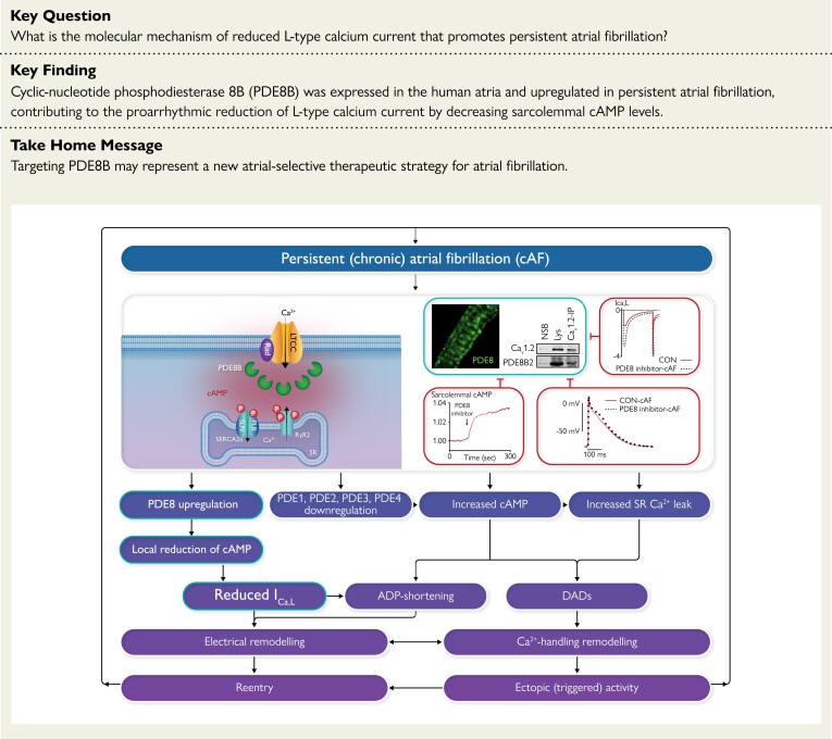
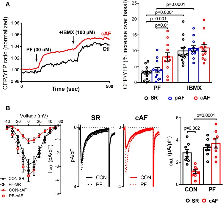
Aims: Atrial fibrillation (AF) is associated with altered cAMP/PKA signaling and an AF-promoting reduction of L-type Ca2+-current (ICa,L), the mechanisms of which are poorly understood. Cyclic-nucleotide phosphodiesterases (PDEs) degrade cAMP and regulate PKA-dependent phosphorylation of key calcium-handling proteins, including the ICa,L-carrying Cav1.2α1C subunit. The aim was to assess whether altered function of PDE type-8 (PDE8) isoforms contributes to the reduction of ICa,L in persistent (chronic) AF (cAF) patients.

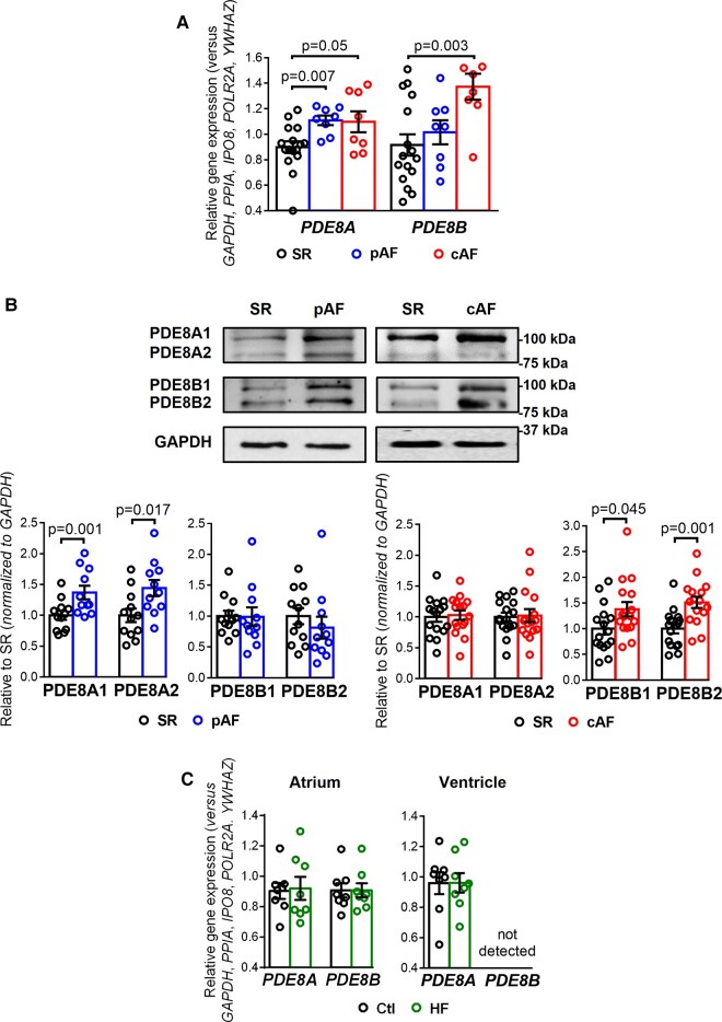
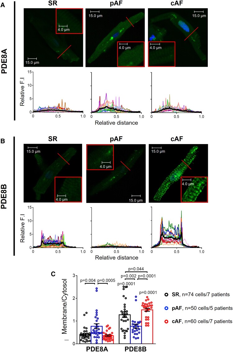
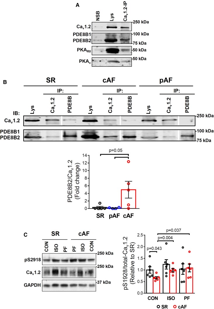
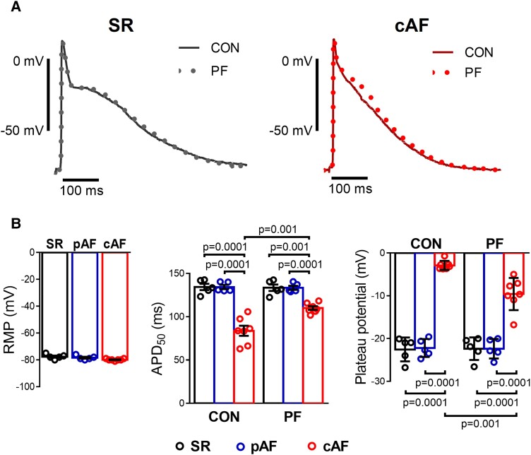
Methods and results: mRNA, protein levels, and localization of PDE8A and PDE8B isoforms were measured by RT-qPCR, western blot, co-immunoprecipitation and immunofluorescence. PDE8 function was assessed by FRET, patch-clamp and sharp-electrode recordings. PDE8A gene and protein levels were higher in paroxysmal AF (pAF) vs. sinus rhythm (SR) patients, whereas PDE8B was upregulated in cAF only. Cytosolic abundance of PDE8A was higher in atrial pAF myocytes, whereas PDE8B tended to be more abundant at the plasmalemma in cAF myocytes. In co-immunoprecipitation, only PDE8B2 showed binding to Cav1.2α1C subunit which was strongly increased in cAF. Accordingly, Cav1.2α1C showed a lower phosphorylation at Ser1928 in association with decreased ICa,L in cAF. Selective PDE8 inhibition increased Ser1928 phosphorylation of Cav1.2α1C, enhanced cAMP at the subsarcolemma and rescued the lower ICa,L in cAF, which was accompanied by a prolongation of action potential duration at 50% of repolarization.

Conclusion: Both PDE8A and PDE8B are expressed in human heart. Upregulation of PDE8B isoforms in cAF reduces ICa,L via direct interaction of PDE8B2 with the Cav1.2α1C subunit. Thus, upregulated PDE8B2 might serve as a novel molecular mechanism of the proarrhythmic reduction of ICa,L in cAF.

Keywords: Atrial fibrillation; Calcium handling; ICa,L; PDE8; Phosphodiesterases; cAMP.


Conflict of interest statement

Conflict of interest The authors have no competing interests. R.B.S. reports grants from European Union Horizon 2020, the German Ministry of Research and Education, ERACOSysMed3 and personal fees from BMS/Pfizer, outside this work.

Figures

Structured Graphical Abstract
The reduction of L-type Ca2+-current in atrial myocytes promotes atrial fibrillation. Phosphodiesterase type 8B binds L-type Ca2+ channels and reduces local cAMP levels and PKA-dependent channel phosphorylation, thereby decreasing L-type Ca2+-current and abbreviating atrial action potential that promotes atrial fibrillation persistence. ADP, adenosine diphosphate; cAF, persistent (chronic) atrial fibrillation; cAMP, cyclic adenosine monophosphate; DAD, delayed afterdepolarization; ICa,L, L-type Ca2+ current; LTCC, L-type Ca2+ channel; PDE, phosphodiesterase; PLB, phospholamban; RyR2, ryanodine receptor type 2; SERCA2a, sarcoplasmic reticulum Ca2 + ATPase type 2a; SLN, sarcolipin; SR, sinus rhythm.
Figure 1
Figure 1
PDE8A and PDE8B expression in human atrium. (A) Plot of the individual and mean ± SEM gene expression ratio of PDE8A and PDE8B normalized to a set of five housekeeping genes (POLR2A, YWHAZ, GAPDH, IPO8, PPIA) in atrial tissue homogenates from 16 sinus rhythm (SR), 8 paroxysmal atrial fibrillation (pAF) and 8 persistent (chronic) atrial fibrillation (cAF) patients. *P < 0.05 vs. sinus rhythm SR based on analysis of variance (ANOVA with a Kruskal–Wallis test). (B) Representative western blot (top) and protein expression quantification (bottom, mean ± SEM) of PDE8A and PDE8B in atrial tissue homogenates from 16 SR, 10 pAF and 16 cAF patients. GAPDH was used as loading control. *P < 0.05 vs. SR based on unpaired Student’s t-test analysis. (C) Plot of the individual and mean ± SEM gene expression ratio of PDE8A and PDE8B normalized to the set of five housekeeping genes in right atrial and left ventricular tissue homogenates from eight healthy control (Ctl) and eight end-stage heart failure (HF) patients. *P < 0.05 vs. Ctl based on ANOVA and Mann–Whitney test.
Figure 2
Figure 2
PDE8A and PDE8B localization in human atrial myocytes. (A) Representative immunocytochemistry confocal images of PDE8A showing its cytosolic distribution in 36 isolated human atrial myocytes (HAMs) from 7 sinus rhythm (SR), 25 HAMs from 5 paroxysmal atrial fibrillation (pAF) and 29 HAMs from 7 persistent (chronic) atrial fibrillation (cAF) patients. Lower panels show transversal line profiles of fluorescence intensity (F.I.) of PDE8A staining for all the myocytes analyzed (thin colored lines) and the average within all cells (thick black lines). (B) Similar representative immunocytochemistry confocal images and fluorescence intensity profiles of PDE8B showing mainly plasma membrane localization of this PDE isoform, in 38 HAMs from 7 SR, 25 HAMs from 5 pAF and 31 HAMs from 7 cAF patients. (C) Average F.I. of PDE8A and PDE8B at the plasma membrane (first and last 10% of the cell width) related to the F.I. in the cytosol (middle), in SR, pAF, and cAF. *P < 0.05 between groups of patients, # P < 0.05 vs. PDE8A, by mixed ANOVA followed by Wald χ2 test and Sidak multiple comparison test. All images were captured under the same conditions in order to obtain comparable fluorescence intensities.
Figure 3
Figure 3
PDE8 co-localizes with LTCC. (A) Co-immunoprecipitation (Co-IP) assay followed by immunoblotting (IB) confirmed that with L-type Ca2+ channel (Cav1.2) binds to PDE8B, PKARII and PKAC in human atria from patients with persistent (chronic) atrial fibrillation (cAF). NSB, non-specific binding. (B) Top, representative Co-IP and IB of Cav1.2 binding to PDE8B in human atrial samples from patients in sinus rhythm (SR), paroxysmal AF (pAF) and cAF. Bottom, mean data of PDE8B binding to Cav1.2 for all groups (n = 6 SR, n = 4 pAF, and 5 cAF patients). Lys, Lysate. Vertical white lines separate non-adjacent lanes on the same blot. (C) Representative western blot (left) and quantification (right, mean ± SEM) of relative steady-state LTCC phosphorylation (pSer1928/total Cav1.2) in atrial tissue homogenates from 6 SR and 6 cAF patients, at baseline and after 5 min stimulation with the selective PDE8 inhibitor PF-04957325 (30 nM) or the β-adrenoceptor agonist isoprenaline (ISO, 100 nM). GAPDH was used as loading control. *P < 0.05 based on ANOVA with a Kruskal–Wallis test.
Figure 4
Figure 4
Functional consequences of selective PDE8 inhibition in human atrial myocytes (HAMs). (A) Left panel, representative experiments showing the time course of the FRET signals indicating cAMP changes in HAMs from sinus rhythm (SR) and in persistent (chronic) atrial fibrillation (cAF) patients exposed to selective PDE8 inhibition with PF-04957325 (30 nM). Right panel, effects of the selective PDE8 inhibitor and the non-selective PDE blocker IBMX (100 mM) on the individual and mean values of cAMP levels expressed as percentage of increase of CFP/YFP over its control value measured in 14 HAMs from 6 SR, 12 HAMs from 6 paroxysmal AF (pAF), and 12 HAMs from 6 cAF patients. The changes in FRET ratio were expressed as a percentage change vs. basal. *P < 0.05 compared with SR, # P < 0.05 compared with PF. (B) Left panel, current-voltage (I-V) relationship for peak inward ICa,L density in HAMs from SR and cAF patients before (CON) and after exposure to PF. Middle panels, representative L-type Ca2+ current (ICa,L) recordings measured at 0 mV in HAMs from SR and cAF patients before (CON) and after exposure to PF. Right panel, average current densities before and after exposure to PF (n = 8 cells/5 patients SR and 8/5 cAF). *P < 0.05 vs. sinus rhythm SR, # P < 0.05 vs. CON. Statistical differences analyzed by mixed ANOVA followed by Wald χ2 test and Sidak multiple comparison test.
Figure 5
Figure 5
Effect of selective PDE8 inhibition on action potential (AP) properties of human atrial trabeculae. (A) Representative traces of AP at baseline (CON) and after 20 min of exposure to the selective PDE8 inhibitor PF-04957325 (PF, 100 nM) recorded in atrial trabeculae from patients in sinus rhythm (SR) and in persistent (chronic) atrial fibrillation (cAF). (B) Left panel, average resting membrane potentials in trabeculae from five SR, five paroxysmal AF (pAF), and seven cAF patients. Middle panel, individual and mean values of the effects of the selective PDE8 inhibitor (PF) on AP duration at 50% of repolarization (APD50) in the three groups of patients. Right panel, plateau potential before (CON) and after exposure to the selective PDE8 inhibitor (PF). *P < 0.05 vs. SR, # P < 0.05 vs. CON based on ANOVA with a Kruskal–Wallis test.

Comment in

References

    1. Larupa Santos J, Rodríguez I, Olesen MS, Hjorth Bentzen B, Schmitt N. Investigating gene-microRNA networks in atrial fibrillation patients with mitral valve regurgitation. PLoS One 2020;15:e0232719. 10.1371/journal.pone.0232719 - DOI - PMC - PubMed
    1. Nattel S, Heijman J, Zhou L, Dobrev D. Molecular basis of atrial fibrillation pathophysiology and therapy: a translational perspective. Circ Res 2020;127:51–72. 10.1161/CIRCRESAHA.120.316363 - DOI - PMC - PubMed
    1. Voigt N, Li N, Wang Q, Wang W, Trafford AW, Abu-Taha I, et al. . Enhanced sarcoplasmic reticulum Ca2 + leak and increased na+-Ca2 + exchanger function underlie delayed afterdepolarizations in patients with chronic atrial fibrillation. Circulation 2012;125:2059–2070. 10.1161/CIRCULATIONAHA.111.067306 - DOI - PMC - PubMed
    1. Llach A, Molina CE, Prat-Vidal C, Fernandes J, Casado V, Ciruela F, et al. . Abnormal calcium handling in atrial fibrillation is linked to up-regulation of adenosine A2A receptors. Eur Heart J 2011;32:721–729. 10.1093/eurheartj/ehq464 - DOI - PubMed
    1. Shanmugam M, Molina CE, Gao S, Severac-Bastide R, Fischmeister R, Babu GJ. Decreased sarcolipin protein expression and enhanced sarco(endo)plasmic reticulum Ca2 + uptake in human atrial fibrillation. Biochem Biophys Res Commun 2011;410:97–101. 10.1016/j.bbrc.2011.05.113 - DOI - PMC - PubMed

Publication types