. 2024 May;18(5):1076-1092.
doi: 10.1002/1878-0261.13390. Epub 2023 Feb 22.

Bovine meat and milk factor protein expression in tumor-free mucosa of colorectal cancer patients coincides with macrophages and might interfere with patient survival

Affiliations

Bovine meat and milk factor protein expression in tumor-free mucosa of colorectal cancer patients coincides with macrophages and might interfere with patient survival

Ekaterina Nikitina et al. Mol Oncol. 2024 May.

Abstract

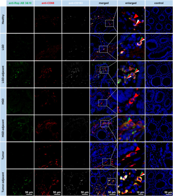
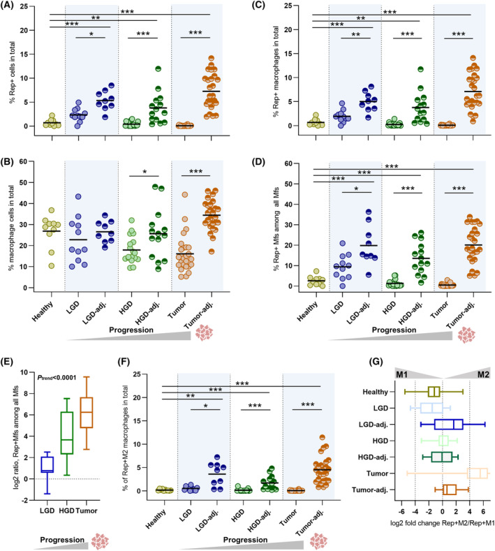
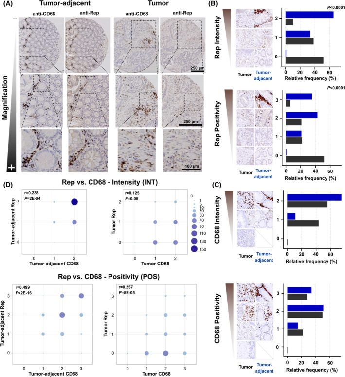
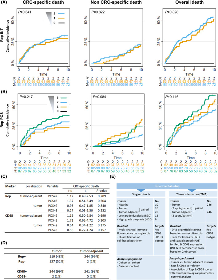
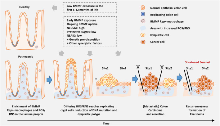
Bovine milk and meat factors (BMMFs) are plasmid-like DNA molecules isolated from bovine milk and serum, as well as the peritumor of colorectal cancer (CRC) patients. BMMFs have been proposed as zoonotic infectious agents and drivers of indirect carcinogenesis of CRC, inducing chronic tissue inflammation, radical formation and increased levels of DNA damage. Data on expression of BMMFs in large clinical cohorts to test an association with co-markers and clinical parameters were not previously available and were therefore assessed in this study. Tissue sections with paired tumor-adjacent mucosa and tumor tissues of CRC patients [individual cohorts and tissue microarrays (TMAs) (n = 246)], low-/high-grade dysplasia (LGD/HGD) and mucosa of healthy donors were used for immunohistochemical quantification of the expression of BMMF replication protein (Rep) and CD68/CD163 (macrophages) by co-immunofluorescence microscopy and immunohistochemical scoring (TMA). Rep was expressed in the tumor-adjacent mucosa of 99% of CRC patients (TMA), was histologically associated with CD68+/CD163+ macrophages and was increased in CRC patients when compared to healthy controls. Tumor tissues showed only low stromal Rep expression. Rep was expressed in LGD and less in HGD but was strongly expressed in LGD/HGD-adjacent tissues. Albeit not reaching statistical significance, incidence curves for CRC-specific death were increased for higher Rep expression (TMA), with high tumor-adjacent Rep expression being linked to the highest incidence of death. BMMF Rep expression might represent a marker and early risk factor for CRC. The correlation between Rep and CD68 expression supports a previous hypothesis that BMMF-specific inflammatory regulations, including macrophages, are involved in the pathogenesis of CRC.

Keywords: bovine meat and milk factors; chronic inflammation; colorectal cancer; infectious indirect carcinogenesis.


Conflict of interest statement

The authors declare no conflict of interest.

Figures

Fig. 1
Fig. 1
Co‐immunodetection of Rep/CD68/CD163 expression. Rep/CD68/CD163 co‐immunofluorescence in FFPE tissue sections prepared from the mucosa of healthy donors, from low‐/high‐grade dysplasia (LGD/HGD) and adjacent tissue and from CRC tumors and tumor‐adjacent tissues (paired). Co‐immunofluorescence microscopy shows cytoplasmic expression of Rep (green, green arrowheads), which is strongest in the tumor‐adjacent mucosa and is congruent with CD68 (red, red arrowheads) and CD163 (white, white arrowheads) expression in interstitial macrophages [scale bar: 50 μm, representative for each full column, DAPI staining of nuclei in blue, fivefold enlargements (scale bar: 10 μm, representative for the full column of enlargements) of indicated tissue parts and control staining performed with PBT diluent in combination with secondary detection antibodies].
Fig. 2
Fig. 2
Quantification of Rep/CD68/CD163‐positive interstitial cells based on co‐immunofluorescence microscopy. Quantification of Rep+ interstitial cells (A) and CD68+ and/or CD163+ macrophages (Mfs) (B) is given in % of all interstitial cells. High levels of Rep+ Mfs (within all interstitial cells) (C) or within the population of Mfs (D) were observed in tissues adjacent to tumors and adjacent to LGD & HGD (for A–D: means illustrated as horizontal lines, full table of calculated P‐values in Table S2, Wilcoxon rank test for unpaired samples, Wilcoxon signed‐rank test for paired samples (dysplastic vs. adjacent) and P‐values adjusted by Holm correction). (E) The log2 ratio of adjacent vs. dysplastic detection of Rep+ Mfs increases from LGD over HGD to CRC (error bars indicating maximum/minimum, Jonckheere–Terpstra Test for trend: P < 0.0001). (F) Among Rep+ Mfs, the M2 phenotype was prevalent in the tissues adjacent to LGD, HGD and tumor (means illustrated as horizontal lines, Wilcoxon rank test for unpaired samples, Wilcoxon signed‐rank test for paired samples (dysplastic vs. adjacent) and P‐values adjusted by Holm correction). (G) This is also reflected by the increased log2‐fold ratio of Rep+ M2 vs. Rep+ M1 macrophages (error bars indicating maximum/minimum). Cohort sizes: healthy donors, n = 10; LGD – low‐grade dysplasia and adjacent tissue, n = 11 and n = 9, respectively; HGD – high‐grade dysplasia and adjacent tissue, n = 18 and n = 14, respectively; and CRC – colorectal cancer tumor and tumor‐adjacent tissues (paired), n = 26 for both. For log2‐fold‐based representations, detection events of exactly 0 were adjusted to half of the minimum of the respective cohort (frequency of corrections: 5× for Rep+ from all nuclei in CRC; 2× for Rep+ MF from all nuclei in HGD, 5× in CRC; and 2× for Rep+ MF within MF in HGD, 5× in CRC). Mf – macrophage, LGD – low‐grade dysplasia and HGD – high‐grade dysplasia. Significance: *P < 0.05, **P < 0.01 and ***P < 0.001.
Fig. 3
Fig. 3
Quantification of Rep and CD68 expression based on CRC TMA. (A) Immunohistochemical Rep and CD68 macrophage staining for representative TMA tissue spots of the (paired) tumor‐adjacent mucosa (left panel) and tumor (right panel) of CRC patients [scale bar: 250 μm for the first two magnifications (upper and center rows) and 100 μm for the highest magnification (lower row)]. Distribution of Rep (B) and CD68 expression (C) in the paired tumor‐adjacent mucosa and tumor of CRC patients stratified in intensity (INT, scores 0–2) and spatial spread (POS, scores 0–3), including representative tissues stainings for each scoring (n = 246 individuals) (test for increased tumor‐adjacent vs. tumor Rep/CD68 POS and INT, respectively, by exact Wilcoxon–Pratt signed‐rank test). (D) Representation of Rep and CD68 INT and POS for tumor‐adjacent mucosa and tumor tissues (correlation of Rep and CD68 POS and INT tested by Spearman's rank correlation coefficient rho).
Fig. 4
Fig. 4
CRC TMA survival analysis and scoring. Individual cumulative incidence curves for intensity (INT, scores 0–2) (A) and spatial spread (POS, scores 0–3) of Rep staining (B) in the tumor‐adjacent mucosa for CRC‐specific, non‐CRC‐specific and overall death (including Gray (log‐rank type) tests for equality of cumulative incidence curves). (C) Hazard ratios (HR) including 95% confidence intervals (CI) and P‐values in cause‐specific multivariate proportional hazard models based on Rep and CD68 POS for the tumor‐adjacent mucosa or tumor accounting for clinical parameters. Cause‐specific multivariate proportional hazard models were adjusted for cancer stage and calculated based on linear age, ignoring BRAF or KRAS mutations due to unavailability and family history. Reference category for each of the CD68/Rep categories is POS “0/1”. (D) Summary of individual tumor or tumor‐adjacent mucosa patient tissues tested positive or negative for Rep (INT and/or POS > 0) or CD68 (INT and/or POS > 0). (E) Overview of the experimental setup for the cell‐based (IF) quantification of the single cohorts including clinical case‐vs.‐control cohorts and pretumor dysplasia (left) and for the CRC TMA cohort (right).
Fig. 5
Fig. 5
Model for BMMF as indirect drivers of CRC and risk factors for limited CRC‐specific survival (figure modified from [11] and [14]). The lack of an early encounter with BMMF, for example by prolonged breastfeeding in the first 6–12 months of life and avoiding bovine products results only in a low or moderate increase in BMMF Rep expression over time. In contrast, early encounter with BMMF causes an immunological condition with less efficient clearance of BMMF in the colonic/rectal mucosa (less resilience toward BMMF) and steadily increasing Rep expression levels. Consistently high BMMF exposure together with Neu5Gc uptake and low levels of protective sugars (such as the human milk oligosaccharides 2′−/3′‐fucosyllactose and lactotetraose) and additional synergistic contributions will result in persisting, high Rep expression in mucosal macrophages and increased chronic inflammation. This will increase the levels of diffusing reactive oxygen and nitrogen species (ROS/RNS) and contribute to DNA mutations in replicating crypt cells and induction of dysplasia and adenoma as precursors of CRC tumors. Even after resection of tumors, high mucosal Rep expression persists resulting in pro‐tumorigenic tissue conditions and ongoing induction of tumors/tumor recurrence and might also shape an environment promoting metastasis and therapy resistance.

Similar articles

Cited by

References

    1. Falida K, Eilebrecht S, Gunst K, zur Hausen H, de Villiers EM. Isolation of two virus‐like circular DNAs from commercially available Milk samples. Genome Announc. 2017;5(17):e00266‐17. - PMC - PubMed
    1. Funk M, Gunst K, Lucansky V, Muller H, zur Hausen H, de Villiers EM. Isolation of protein‐associated circular DNA from healthy cattle serum. Genome Announc. 2014;2(4):e00846‐14. - PMC - PubMed
    1. Gunst K, zur Hausen H, de Villiers EM. Isolation of bacterial plasmid‐related replication‐associated circular DNA from a serum sample of a multiple sclerosis patient. Genome Announc. 2014;2(4):e00847‐14. - PMC - PubMed
    1. Lamberto I, Gunst K, Muller H, zur Hausen H, de Villiers EM. Mycovirus‐like DNA virus sequences from cattle serum and human brain and serum samples from multiple sclerosis patients. Genome Announc. 2014;2(4):e00848‐14. - PMC - PubMed
    1. Whitley C, Gunst K, Muller H, Funk M, zur Hausen H, de Villiers EM. Novel replication‐competent circular DNA molecules from healthy cattle serum and milk and multiple sclerosis‐affected human brain tissue. Genome Announc. 2014;2(4):e00849‐14. - PMC - PubMed

Publication types

MeSH terms