. 2023 Apr 3;222(4):e202008061.
doi: 10.1083/jcb.202008061. Epub 2023 Feb 22.

An interaction between β'-COP and the ArfGAP, Glo3, maintains post-Golgi cargo recycling

Affiliations

An interaction between β'-COP and the ArfGAP, Glo3, maintains post-Golgi cargo recycling

Boyang Xie et al. J Cell Biol. .

Abstract

The essential COPI coat mediates retrieval of transmembrane proteins at the Golgi and endosomes following recruitment by the small GTPase, Arf1. ArfGAP proteins regulate COPI coats, but molecular details for COPI recognition by ArfGAPs remain elusive. Biochemical and biophysical data reveal how β'-COP propeller domains directly engage the yeast ArfGAP, Glo3, with a low micromolar binding affinity. Calorimetry data demonstrate that both β'-COP propeller domains are required to bind Glo3. An acidic patch on β'-COP (D437/D450) interacts with Glo3 lysine residues located within the BoCCS (binding of coatomer, cargo, and SNAREs) region. Targeted point mutations in either Glo3 BoCCS or β'-COP abrogate the interaction in vitro, and loss of the β'-COP/Glo3 interaction drives Ste2 missorting to the vacuole and aberrant Golgi morphology in budding yeast. These data suggest that cells require the β'-COP/Glo3 interaction for cargo recycling via endosomes and the TGN, where β'-COP serves as a molecular platform to coordinate binding to multiple proteins, including Glo3, Arf1, and the COPI F-subcomplex.


Conflict of interest statement

Disclosures: The authors declare no competing interests exist.

Figures
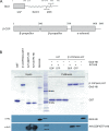
Figure 1.
Figure 1.
β′-COP propeller (WD-repeat) domains directly bind Glo3 and Arf1 in vitro. (A) Schematics of yeast Glo3 and β′-COP proteins. Glo3 contains a GAP domain (residues 1–140), BoCCS region (defined as residues 214–375), and GRM region (residues 375–493). β′-COP contains two WD-repeat (also known as β-propeller) domains followed by an α-solenoid. The N-terminal propeller domain binds dilysine motifs in transmembrane cargo, while the α-solenoid interacts with α-COP. (B) Pulldown experiments using GST-tagged β′-COP propeller domains (residues 1–604 with C-terminal GST tag) as bait and either full-length Arf1-H6 or full-length Glo3-H8 as prey. We tested binding to both GDP-locked (T31N) Arf1 and GTP-locked (Q71L) Arf1. β′-COP interacts directly with both Arf1 constructs and with Glo3 in vitro. The top panel shows a Coomassie-stained SDS-PAGE gel, while the bottom two panels show Western blots against the Glo3 His8 tag (α-His; Abcam NB100-63173) or against yeast Arf1 (α-Arf1, Todd Graham lab, Vanderbilt University).
Figure S1.
Figure S1.
β′-COP, Arf1, and Glo3 form a ternary complex in vitro. (A) Glo3 and Arf1 were identified as potential direct binding partners using GST-tagged β′-COP protein as bait in yeast cell lysates. The table summarizes mass spectrometry results for these two hits. (B) GST-pulldown experiments using GST-tagged β′-COP propeller domains (residues 1–604 with C-terminal GST tag) as bait and full-length Arf1-H6 with and without full-length Glo3-H8 as prey. We tested binding to both nucleotide-bound forms of Arf1: GDP-locked (T31N) or GTP-locked (Q71L). β′-COP can pull down both Arf1 and Glo3 simultaneously. β′-COP does not appear to show a preference for Arf1 nucleotide state; it pulls down Arf1 T31N or Q71L equally well when Arf1 is added at a 5:1 molar ratio.
Figure S2.
Figure S2.
β′-COP WD-repeat domains directly bind the Glo3 BoCCS region in vitro. GST-tagged Glo3 fragments (GAP-GST, residues 1–150; GST-BoCCS, residues 208–383; or GST-GRM, residues 350–493) were used as bait with β′-COP-H6 to determine which portion of Glo3 binds β′-COP. Only the GST-BoCCS fragment exhibited direct binding. Both the BoCCS and GRM fragments are unstable in solution, most likely because they contain long regions predicted to be unstructured. Mass spectrometry data (not shown) confirms the top two bands in the GST-BoCCS input lane correspond to Glo3 BoCCS peptides.
Figure 2.
Figure 2.
Residues within the Glo3 BoCCS region directly bind both β′-COP propellers with low micromolar affinity. (A) Purified recombinant proteins (untagged β′-COP residues 1–604 and Glo3 fragments as labeled) were used in isothermal titration calorimetry (ITC) experiments to quantify binding affinities; representative traces are shown. β′-COP binds a Glo3 fragment located within the BoCCS region. The highest affinity interaction occurs between β′-COP 1-604 and Glo3 residues 230–290, but all fragments exhibit low micromolar KD values (0.8–6 μM) and 1:1 stoichiometry (Table S1). (B) Representative ITC experiments between untagged Glo3 BoCCS fragment (residues 230–300) and N-terminal β′-COP propeller (residues 1–304), C-terminal β′-COP propeller (residues 300–604), or both β′-COP propellers (residues 1–604). Each propeller domain on its own is insufficient to produce measurable binding by calorimetry, which suggests both propellers are required to bind Glo3. Unless otherwise noted, 0.05 mM β′-COP protein was placed in the cell and 0.3 mM Glo3 protein in the syringe (see Materials and methods).
Figure S3.
Figure S3.
The Glo3 BoCCS region interacts directly with β′-COP. (A) An ITC experiment with full BoCCS Glo3 fragment (residues 208–383) shows a low micromolar KD. (B) ITC experiments to ascertain the minimum Glo3 fragment required for binding. Glo3 N-terminal residues 230–240 are required for measurable binding in the calorimeter, while removing residues 220–230 exhibits no measurable effect on binding affinity. n.b. denotes no measurable binding in the calorimeter (KD > 300 μM). (C) Representative SDS-PAGE gel of β′604/Glo3 (residues 230–300) complex following purification; the complex elutes together over gel filtration columns (data not shown). Although some Glo3 fragments are unstable over time, the recombinant fragments used for ITC runs exhibit high levels of purity and stability. (D–F) Representative models generated using AlphaFold2. The well-established interaction between β′-COP and dilysine motifs served as a positive control computational experiment, but AlphaFold failed to predict binding between dilysine motifs and the N-terminal β′-COP propeller (D) or to both propeller domains (E). The known dilysine binding site is marked by black asterisks. (F) One representative model from an AlphaFold experiment with β′-COP 1-604 and Glo3 residues 230–290. Results from modeling experiments are reported in Table S3.
Figure S4.
Figure S4.
Conserved Glo3 lysine residues mediate an electrostatic interaction with β′-COP C-terminal propeller domain. (A) Glo3 partial sequence alignment highlighting key conserved residues between Saccharomyces cerevisiae Glo3 and mammalian ArfGAP2/3 homologs in Mus musculus and Homo sapiens. Glo3 residues 230–300 are labeled. (B) ITC experiment between wild-type β′-COP residues 1–604 and Glo3 residues 230–270 in 100 mM NaCl and 500 mM NaCl. Near physiological salt concentrations, the two proteins interact with a low micromolar KD. The same experiment in high-salt buffer (500 mM NaCl) disrupts the interaction, suggesting electrostatic residues play an important role. In high salt, binding was too weak to determine a KD. (C) Side view from membrane showing the β′-COP D437/D450 acidic patch that binds Glo3.
Figure 3.
Figure 3.
Key Glo3 lysine residues mediate an electrostatic interaction with an acidic patch on β′-COP. (A) Representative ITC experiments between wild-type β′-COP 1-604 and mutant versions of Glo3 (residues 230–290). Two Glo3 mutants substantially reduce binding in the calorimeter: a single point mutation at K233E and the K251E/K252E/K255E triple mutant. Both mutants exhibit 40-fold weaker binding as compared with wild-type Glo3 (KD ∼ 12 μM; Table S2). The Glo3 K233E/K234E/K235E triple mutant exhibits no measurable binding by calorimetry (KD < 300 μM). (B) Representative ITC experiment with wild-type untagged Glo3 (residues 230–290) and mutant β′-COP (D437A/D450A) proteins; the calculated KD for this interaction is 18 μM. The D437/D450 mutant exhibits 60-fold weaker binding to Glo3, suggesting this acidic patch plays a critical role in the interaction. For these experiments, 0.04 mM wild-type or 0.05 mM mutant β′-COP was placed in the cell. (C) Glo3 associates with COPI primarily using its interaction with the β′-COP subunit. Wild-type GST-Glo3 BoCCS pulled down COPI (B- and F-subcomplexes) while disrupting all six lysine residues disrupted Glo3 association with COPI. (D) Disruption of the β′-COP D437/D450 patch reduces the interaction between COPI and Glo3 in yeast cells. Glo3-6xHis-TEV-3xFLAG (Glo3-HTF) efficiently immunoprecipitates COPI from wild-type SEC27 cell lysates, in contrast to the β′-COP D437A/D450A mutant strain.
Figure S5.
Figure S5.
Glo3 BoCCS and dilysine cargo motifs bind β′-COP simultaneously in vitro. (A) Representative ITC experiments with untagged β′-COP 1–604; Glo3 residues 230–290; and dilysine motif (KTKLL) peptide. The presence of Glo3 does not alter the binding affinity of β′-COP to dilysine motifs in vitro. (B) ITC experiments with β′-COP dilysine binding mutants. The R13A/K15A/R59A (RKR mutant) disrupts binding to the dilysine motif C-terminus, while D98A/D117A disrupts binding to lysine residues. The RKR mutant exhibits weaker binding to Glo3, while D98A/D117A binds Glo3 as well as wild-type protein. These data suggest Glo3 and dilysine motifs do not compete for binding β′-COP in vitro. However, disrupting the overall charge distribution on the N-terminal propeller in the RKR mutant suggests it may play some role in Glo3 binding.
Figure S6.
Figure S6.
Yeast growth assays. Yeast growth assays in glo3Δgcs1Δ background strains with GLO3 or glo3 mutants (K233E/K234E/K235E or K251E/K252E/K255E). The glo3∆ gcs1∆ double mutant is inviable but can be sustained with a wild-type copy of GLO3 on a URA3 marked plasmid (pRS416). Both the wild-type GLO3 and the mutant forms were introduced into this background on a LEU2 marked (pRS315) plasmid. On media that selects for both plasmids, all transformed strains grew like a wild-type strain because they contain the wild-type pRS416-GLO3 plasmid. However, upon switching to 5-FOA media, which selects against the pRS416-GLO3 plasmid, the glo3∆gcs1∆ strain harboring the empty pRS415 plasmid failed to grow, as expected.
Figure 4.
Figure 4.
Ste2 is missorted to the vacuole when the β′-COP/Glo3 interaction is disrupted in S. cerevisiae. (A) Fluorescence imaging of Ste2-GFP in glo3Δgcs1Δ strains with GLO3 or glo3 mutants (K233E/K234E/K235E or K251E/K252E/K255E) introduced on a plasmid. Scale bar represents 2 μm. (B) Box plots showing the percentage of Ste2-GFP observed at the plasma membrane in each strain with median marked (black line). Mutating either lysine cluster in yeast cells causes a significant difference in Ste2-GFP sorting compared to the GLO3 strain. Statistical comparisons were pairwise between GLO3 and mutants; data were analyzed using a one-way ANOVA (Prism); and probability values of <0.001 are represented by ***.
Figure S7.
Figure S7.
Golgi/ER COPI cargo trafficking data. (A) The Emp47-myc reporter construct (visualized using α−myc) remains stable over time in both wild-type GLO3 and mutant glo3 strains. In contrast, the reporter is sent to the vacuole and degraded when the dilysine binding site on sec27 is mutated (sec27RKR; R13A/K15A/R59A) or the first sec27 propeller is deleted (sec27Δ2-304). (B) Representative fluorescence images of GFP-Rer1 in glo3Δgcs1Δ strains with GLO3 or glo3 mutants introduced on a plasmid. GFP-Rer1 trafficking does not appear to change when the β′-COP/Glo3 interaction is disrupted. Scale bar represents 2 μm.
Figure S8.
Figure S8.
SNARE localization and Golgi morphology data. (A) Fluorescence imaging of mNG-Snc1 in glo3Δgcs1Δ strains with GLO3 or glo3 mutants (K233E/K234E/K235E or K251E/K252E/K255E) on a plasmid. Scale bar represents 2 μm. (B) Strip plot showing the percentage of abnormal structures (tubules and rings) with mean (black bar) observed in each strain. Mutating the first lysine cluster resulted in cells exhibiting a significant difference from wild type. Significance was determined using a Mann-Whitney test comparing wild-type and mutant strains. (C) Bar graph showing number of mNG-Snc1 ring structures with standard deviation (black lines) in each strain. (D) Fluorescence imaging of mNG-Bet1 in glo3Δgcs1Δ strains with GLO3 or glo3 mutants (K233E/K234E/K235E or K251E/K252E/K255E) on a plasmid. Scale bar represents 2 μm. (E) Strip plot showing percentage of abnormal structures (tubules and rings) with standard deviation observed in each strain. Although we sometimes observe abnormal structures, we do not find a significant difference, as determined by a Mann-Whitney test. (F) Bar graph showing number of mNG-Bet1 ring structures with standard deviation (black lines) in each strain.
Figure 5.
Figure 5.
β′-COP is a molecular platform on Golgi membranes. Model for the interaction between β′-COP (blue ribbons; WD-repeat domains and solenoid shown), Arf1 (pink ribbons with N-terminal amphipathic helix shown as cylinder), γ-COP appendage (green ribbons), and Glo3 (GAP domain as red ribbons and BoCCS region as red line). The dashed line marks the start of Glo3 GRM region; its position and orientation remain unknown, but it is predicted to have a C-terminal amphipathic helix that may insert into the membrane. The dilysine motif in transmembrane cargoes is shown as grey cylinders. The position of γ-COP was generated based on cryo-ET reconstructions of COPI (PDB ID 5NZS). Our data suggest that the first Glo3 lysine cluster (K233/K234/K235) may interact with the D437/D450 patch on the C-terminal β′-COP propeller, and the second cluster (K251/K252/K255) may interact with the N-terminal β′-COP propeller. γ-COP appendage also binds Glo3; γ-COP F836 is implicated in binding, but the Glo3 residues remain unknown.

References

    1. Arakel, E.C., Huranova M., Estrada A.F., Rau E.-M., Spang A., and Schwappach B.. 2019. Dissection of GTPase-activating proteins reveals functional asymmetry in the COPI coat of budding yeast. J. Cell Sci. 132:jcs232124. 10.1242/jcs.232124 - DOI - PMC - PubMed
    1. Arakel, E.C., and Schwappach B.. 2018. Formation of COPI-coated vesicles at a glance. J. Cell Sci. 131:jcs209890. 10.1242/jcs.209890 - DOI - PubMed
    1. Beller, M., Sztalryd C., Southall N., Bell M., Jäckle H., Auld D.S., and Oliver B.. 2008. COPI complex is a regulator of lipid homeostasis. PLoS Biol. 6:e292. 10.1371/journal.pbio.0060292 - DOI - PMC - PubMed
    1. Bhandari, A., Zheng C., Sindan N., Sindan N., Quan R., Xia E., Thapa Y., Tamang D., Wang O., Ye X., and Huang D.. 2019. COPB2 is up-regulated in breast cancer and plays a vital role in the metastasis via N-cadherin and Vimentin. J. Cell. Mol. Med. 23:5235–5245. 10.1111/jcmm.14398 - DOI - PMC - PubMed
    1. Bykov, Y.S., Schaffer M., Dodonova S.O., Albert S., Plitzko J.M., Baumeister W., Engel B.D., and Briggs J.A.. 2017. The structure of the COPI coat determined within the cell. Elife. 6:e32493. 10.7554/eLife.32493 - DOI - PMC - PubMed

Publication types

MeSH terms