Acute thalamic connectivity precedes chronic post-concussive symptoms in mild traumatic brain injury
- PMID: 36811945
- PMCID: PMC10393415
- DOI: 10.1093/brain/awad056
Acute thalamic connectivity precedes chronic post-concussive symptoms in mild traumatic brain injury
Abstract
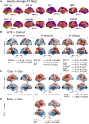
Chronic post-concussive symptoms are common after mild traumatic brain injury (mTBI) and are difficult to predict or treat. Thalamic functional integrity is particularly vulnerable in mTBI and may be related to long-term outcomes but requires further investigation. We compared structural MRI and resting state functional MRI in 108 patients with a Glasgow Coma Scale (GCS) of 13-15 and normal CT, and 76 controls. We examined whether acute changes in thalamic functional connectivity were early markers for persistent symptoms and explored neurochemical associations of our findings using PET data. Of the mTBI cohort, 47% showed incomplete recovery 6 months post-injury. Despite the absence of structural changes, we found acute thalamic hyperconnectivity in mTBI, with specific vulnerabilities of individual thalamic nuclei. Acute fMRI markers differentiated those with chronic post-concussive symptoms, with time- and outcome-dependent relationships in a sub-cohort followed longitudinally. Moreover, emotional and cognitive symptoms were associated with changes in thalamic functional connectivity to known serotonergic and noradrenergic targets, respectively. Our findings suggest that chronic symptoms can have a basis in early thalamic pathophysiology. This may aid identification of patients at risk of chronic post-concussive symptoms following mTBI, provide a basis for development of new therapies and facilitate precision medicine application of these therapies.
Keywords: functional connectivity; mild traumatic brain injury; postconcussive symptoms; resting-state fMRI; thalamus.
© The Author(s) 2023. Published by Oxford University Press on behalf of the Guarantors of Brain.
Conflict of interest statement
D.K.M. reports grant support from the National Institute for Health Research (UK), Medical Research Council (UK), Canadian Institute for Advanced Research and the European Union. He is in receipt of collaborative research grant funding with Lantmannen AB, GlaxoSmithKline Ltd. and Cortirio Ltd., and personal fees from Calico LLC, GlaxoSmithKline Ltd, Lantmannen AB and Integra Neurosciences. All other authors report no competing interests.
Figures


Similar articles
-
Thalamic Functional Connectivity in Mild Traumatic Brain Injury: Longitudinal Associations With Patient-Reported Outcomes and Neuropsychological Tests.Arch Phys Med Rehabil. 2016 Aug;97(8):1254-61. doi: 10.1016/j.apmr.2016.03.013. Epub 2016 Apr 13. Arch Phys Med Rehabil. 2016. PMID: 27085849 Free PMC article.
-
Alterations of connectivity patterns in functional brain networks in patients with mild traumatic brain injury: A longitudinal resting-state functional magnetic resonance imaging study.Neuroradiol J. 2020 Apr;33(2):186-197. doi: 10.1177/1971400920901706. Epub 2020 Jan 28. Neuroradiol J. 2020. PMID: 31992126 Free PMC article.
-
Early Stage Longitudinal Subcortical Volumetric Changes following Mild Traumatic Brain Injury.Brain Inj. 2021 May 12;35(6):725-733. doi: 10.1080/02699052.2021.1906445. Epub 2021 Apr 6. Brain Inj. 2021. PMID: 33822686 Free PMC article.
-
Brain dysfunction underlying prolonged post-concussive syndrome: A systematic review.J Affect Disord. 2020 Feb 1;262:71-76. doi: 10.1016/j.jad.2019.10.058. Epub 2019 Nov 4. J Affect Disord. 2020. PMID: 31710931 Free PMC article.
-
A Systematic Review of Structural and Functional Imaging Correlates of Headache or Pain after Mild Traumatic Brain Injury.J Neurotrauma. 2020 Apr 1;37(7):907-923. doi: 10.1089/neu.2019.6750. Epub 2020 Jan 31. J Neurotrauma. 2020. PMID: 31822205
Cited by
-
Symptom Persistence Relates to Volume and Asymmetry of the Limbic System after Mild Traumatic Brain Injury.J Clin Med. 2024 Aug 30;13(17):5154. doi: 10.3390/jcm13175154. J Clin Med. 2024. PMID: 39274367 Free PMC article.
-
Outcomes and Mechanisms Associated With Selective Thalamic Neuronal Loss in Chronic Traumatic Brain Injury.JAMA Netw Open. 2024 Aug 1;7(8):e2426141. doi: 10.1001/jamanetworkopen.2024.26141. JAMA Netw Open. 2024. PMID: 39106064 Free PMC article.
-
Predicting recovery in patients with mild traumatic brain injury and a normal CT using serum biomarkers and diffusion tensor imaging (CENTER-TBI): an observational cohort study.EClinicalMedicine. 2024 Aug 8;75:102751. doi: 10.1016/j.eclinm.2024.102751. eCollection 2024 Sep. EClinicalMedicine. 2024. PMID: 39720677 Free PMC article.
-
A Scoping Review on the Use of Non-Invasive Brain Stimulation Techniques for Persistent Post-Concussive Symptoms.Biomedicines. 2024 Feb 17;12(2):450. doi: 10.3390/biomedicines12020450. Biomedicines. 2024. PMID: 38398052 Free PMC article.
-
Aberrant dynamic functional network connectivity in patients with diffuse axonal injury.Sci Rep. 2024 Nov 9;14(1):27386. doi: 10.1038/s41598-024-79052-4. Sci Rep. 2024. PMID: 39521859 Free PMC article.
References
-
- Korley FK, Peacock WF, Eckner JT, et al. Clinical gestalt for early prediction of delayed functional and symptomatic recovery from mild traumatic brain injury is inadequate. Acad Emerg Med. 2019;26:1384–1387. - PubMed
-
- Mikolic A, Polinder S, Steyerberg EW, et al. Prediction of global functional outcome and post-concussive symptoms following mild traumatic brain injury: External validation of prognostic models in the CENTER-TBI study. J Neurotrauma. 2020;38:196–209. - PubMed
-
- Maas AIR, Menon DK, Adelson D, et al. The lancet neurology commission traumatic brain injury: Integrated approaches to improve prevention, clinical care, and research executive summary the lancet neurology commission. Lancet Neurol. 2017;16:987–1048. - PubMed

