. 2023 Mar 15;14(6):1080-1094.
doi: 10.1021/acschemneuro.2c00476. Epub 2023 Feb 22.

Elucidation of the GSK3α Structure Informs the Design of Novel, Paralog-Selective Inhibitors

Affiliations

Elucidation of the GSK3α Structure Informs the Design of Novel, Paralog-Selective Inhibitors

Brenda Amaral et al. ACS Chem Neurosci. .

Abstract

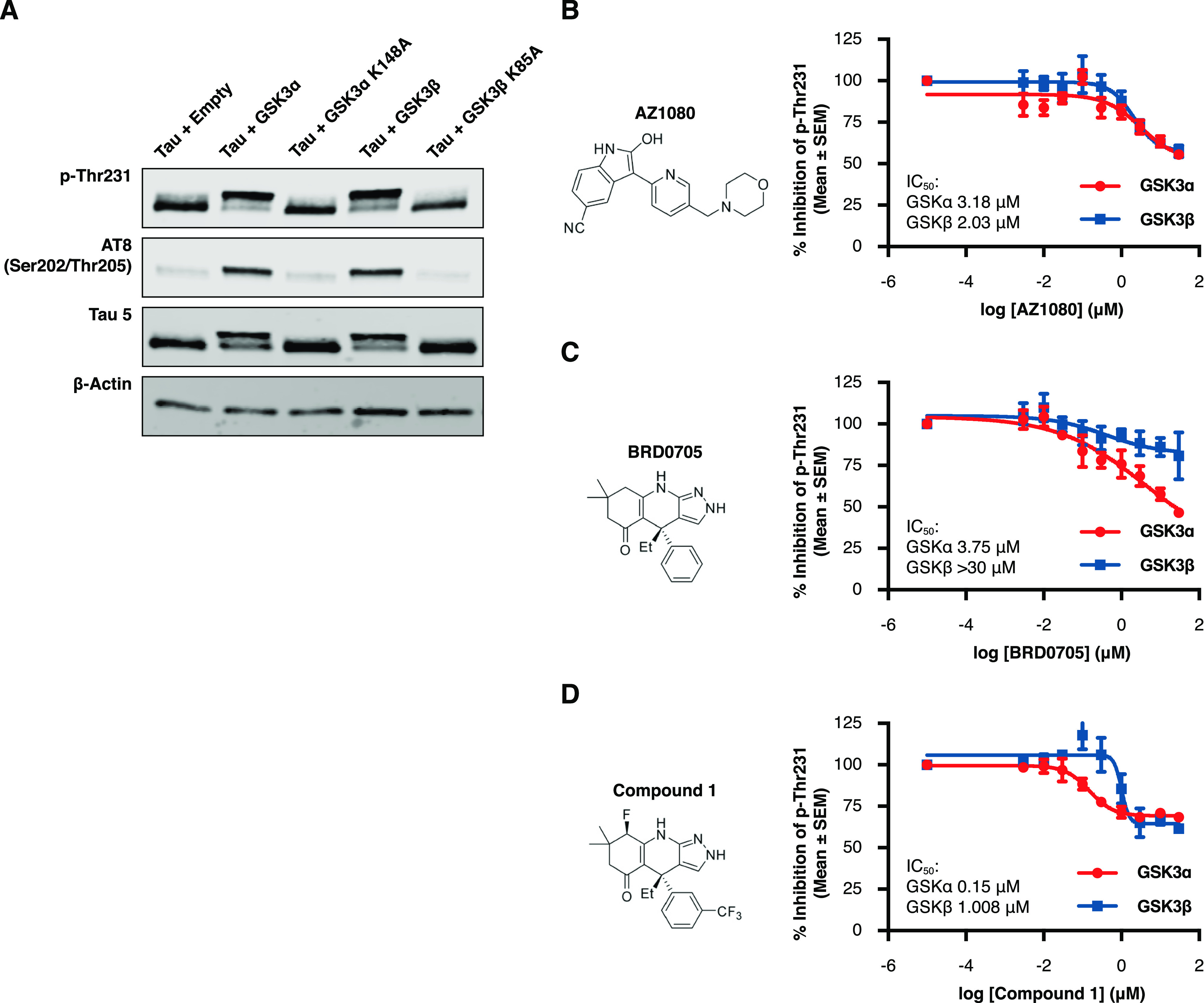
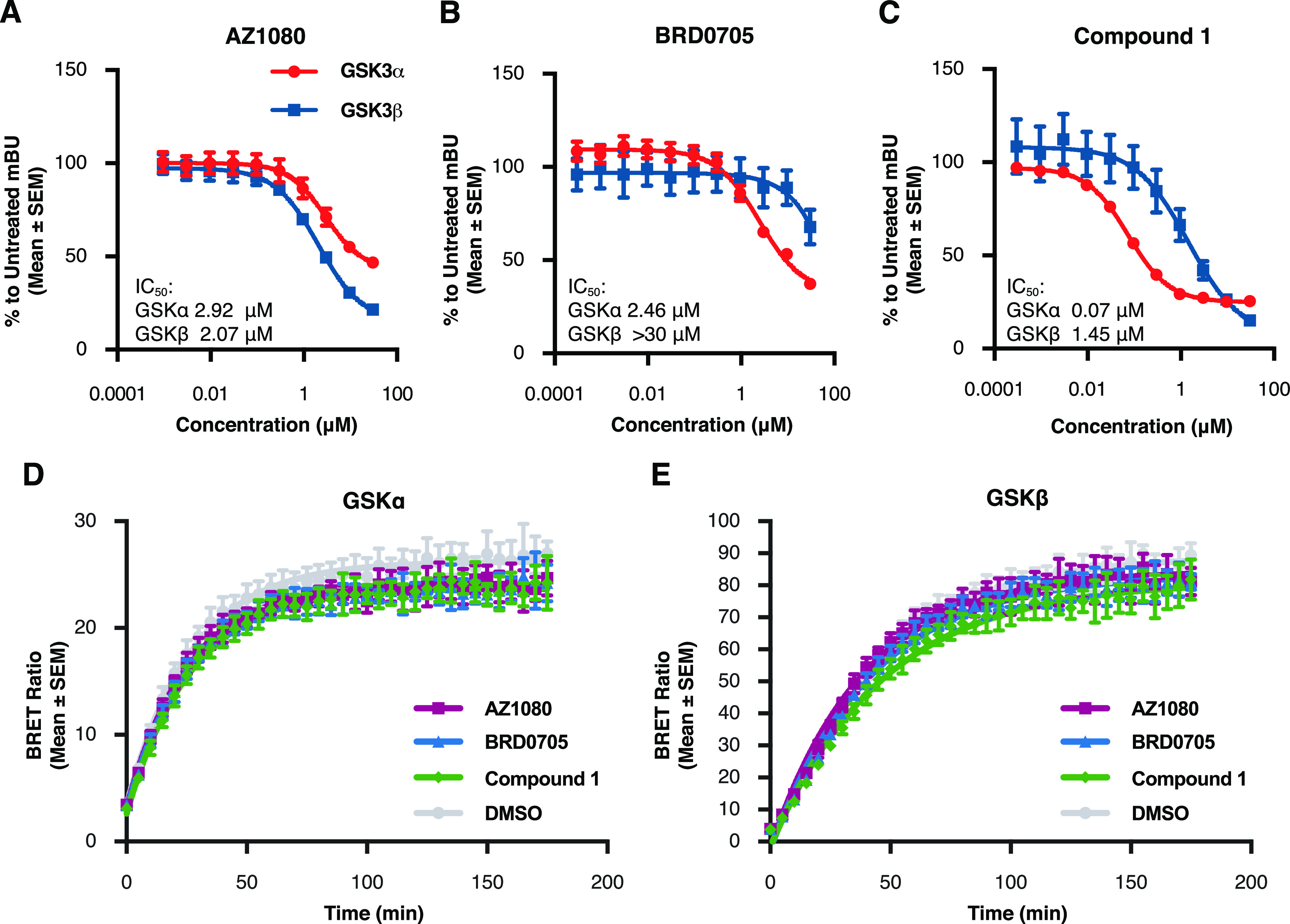
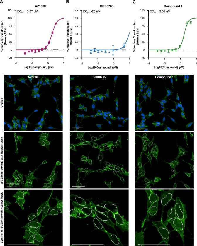
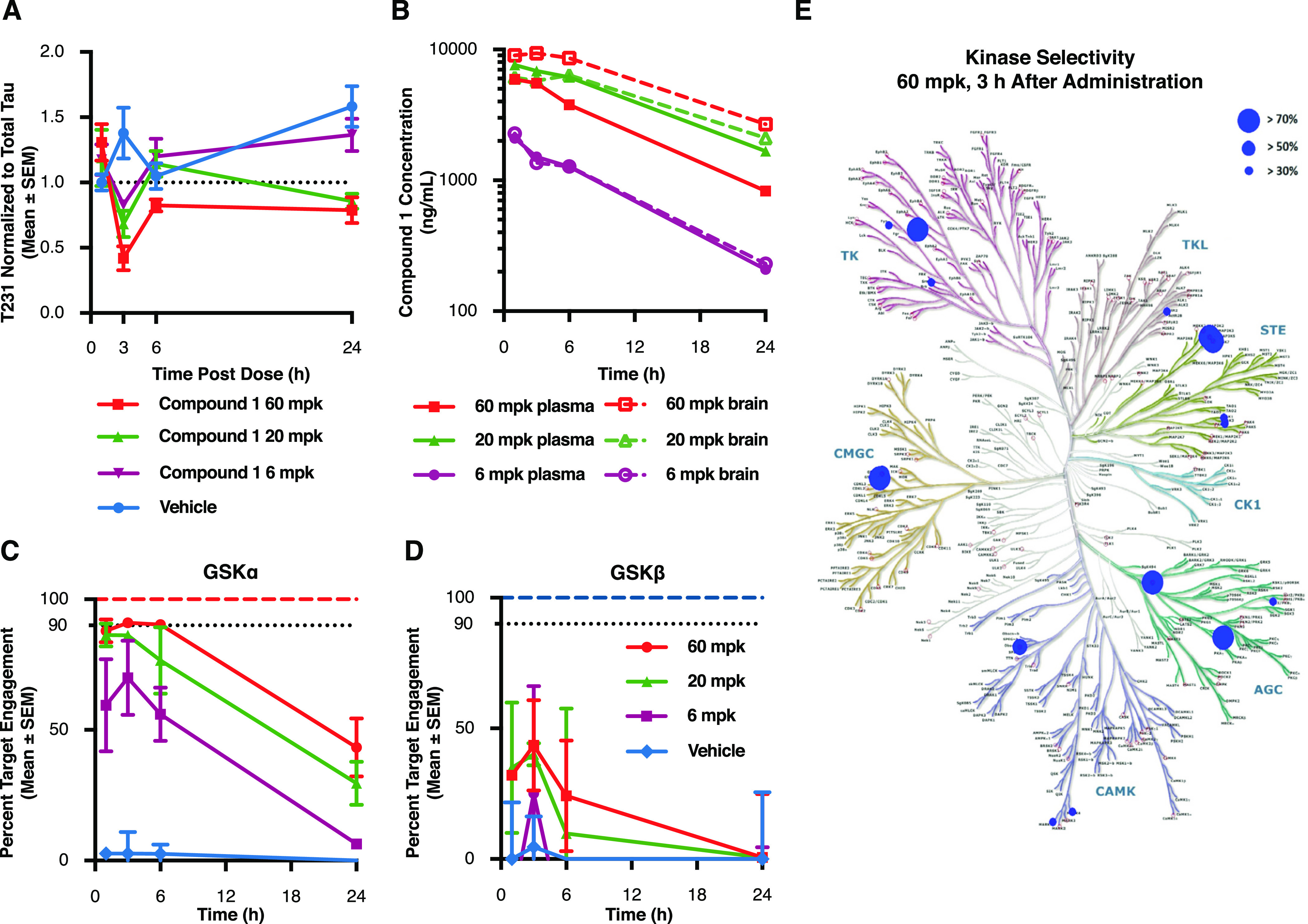
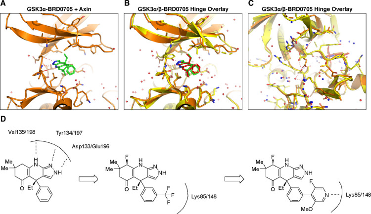
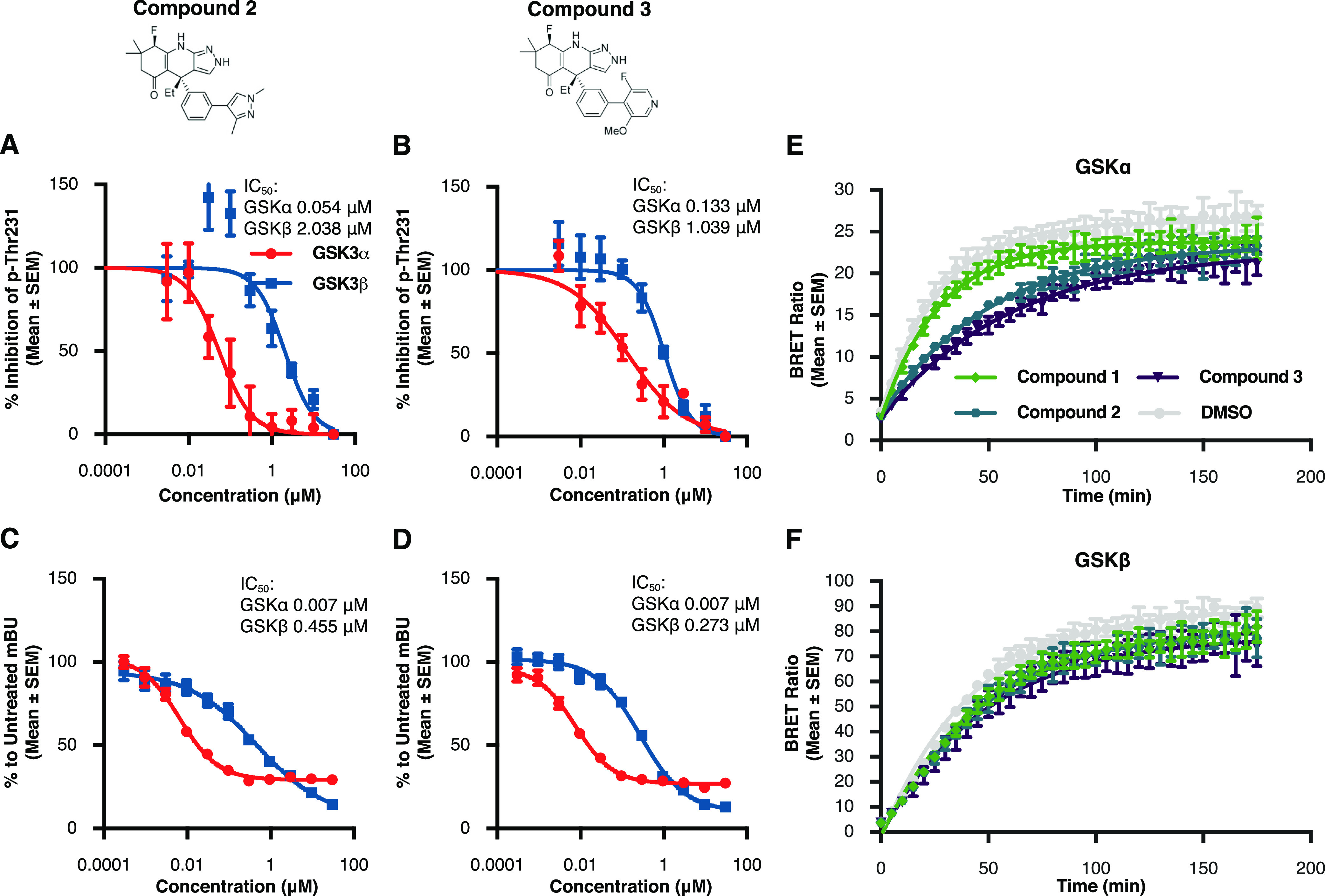
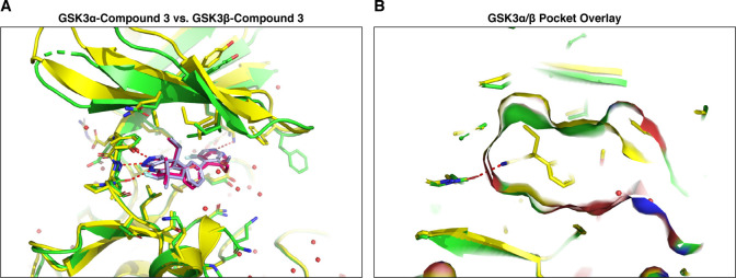
Glycogen synthase kinase 3 (GSK3) remains a therapeutic target of interest for diverse clinical indications. However, one hurdle in the development of small molecule GSK3 inhibitors has been safety concerns related to pan-inhibition of both GSK3 paralogs, leading to activation of the Wnt/β-catenin pathway and potential for aberrant cell proliferation. Development of GSK3α or GSK3β paralog-selective inhibitors that could offer an improved safety profile has been reported but further advancement has been hampered by the lack of structural information for GSK3α. Here we report for the first time the crystal structure for GSK3α, both in apo form and bound to a paralog-selective inhibitor. Taking advantage of this new structural information, we describe the design and in vitro testing of novel compounds with up to ∼37-fold selectivity for GSK3α over GSK3β with favorable drug-like properties. Furthermore, using chemoproteomics, we confirm that acute inhibition of GSK3α can lower tau phosphorylation at disease-relevant sites in vivo, with a high degree of selectivity over GSK3β and other kinases. Altogether, our studies advance prior efforts to develop GSK3 inhibitors by describing GSK3α structure and novel GSK3α inhibitors with improved selectivity, potency, and activity in disease-relevant systems.

Keywords: Alzheimer’s; GSK3α; GSK3β; kinase inhibitor; tau; β-catenin.


Conflict of interest statement

The authors declare no competing financial interest.

Figures

Figure 1
Figure 1
Acute and selective inhibition of GSK3α can reduce tau phosphorylation at disease relevant phospho-sites. HEK293 cells stably expressing human 2N4R tau (HEK-huTau) were transfected with plasmids expressing either human GSK3α, human GSK3β, or kinase dead mutants in which the catalytic lysine was mutated to alanine. 24 h after transfection, cells were lysed. (A) HEK-huTau lysates were run by SDS PAGE to probe for phosphorylation of tau at different epitopes. Both GSK3 isoforms phosphorylate tau at multiple epitopes including the disease enriched epitopes Thr231 and S202/Thr205 (AT8). (B–D) 24 h after transfection, compounds were added at a 10-point dose response (range: 3 nM–30 μM) for 2 h and cell lysates were probed for total tau (Tau5 antibody) and pThr231 using a plate-based assay. The figures represent IC50 curves demonstrating cellular potency and GSK3 isoform selectivity of the non-paralog-selective compound AZ1080 (B), a previously reported GSK3α selective compound BRD0705 (C), and a novel GSK3a selective small molecule, compound 1 (D). Each data point represents the mean ± the standard error of the mean (SEM) from four biological replicates within single run.
Figure 2
Figure 2
GSK3 small molecule inhibitors bind at the ATP binding pocket and have similar residence times. To test for binding at the ATP pocket, HEK293T cells were transiently transfected with either NanoLuc-GSK3α or NanoLuc-GSK3β plasmids and the following day treated for 2 h with the inhibitor compounds in a 10-point dose response curve (range: 3 nM–30 μM) and the NanoBRET Target Engagement Kinase Tracer-8. (A–C) Our results show competitive binding of inhibitors AZ1080 (A), BRD0705 (B), and compound 1 (C) with the kinase tracer indicating displacement at the kinase ATP pocket. Each data point represents the mean ± SEM from four biological replicates within a single run of the assay. mBU: milliBRET Units (please see Methods). To investigate compound residence time, cells were transiently transfected with NanoLuc plasmids and the following day kinase inhibitors were added at a concentration of 10× the previously identified IC50. After 2 h of treatment, media with compound was removed and Tracer-6908 (Promega) was added. The BRET signal was measured every 5 min for a total of 3 h after tracer addition. (D,E) There was no difference in the residence times between any of the inhibitors and either GSK3α (D) or GSK3β (E). Each data point represents the mean ± SEM from eight biological replicates within a single run of the assay.
Figure 3
Figure 3
Combined inhibition of both GSK3 paralogs is necessary for nuclear translocation of β-catenin in SH-SY5Y cells. To examine β-catenin activation, we quantified translocation of this protein to the nucleus in SH-SY5Y cells 6 h after compound addition at a 9-point dose response curve (range: 20 nM–20 μM). (A–C) EC50 curves demonstrating concentration of GSK3 inhibitors AZ1080 (A), BRD0705 (B) and compound 1 (C) necessary to trigger β-catenin translocation to the nucleus. Each data point represents the mean ± SEM from four biological replicates within a single run of the assay. (D) Representative images showing DAPI nuclear stain in blue and β-catenin in green for each GSK3 inhibitor compound at the highest dose tested (20 μM).
Figure 4
Figure 4
Acute GSK3α inhibition reduces tau phosphorylation at disease relevant sites in vivo. P10 rats were injected with compound 1 at 60, 20, and 6 mg/kg and sacrificed at several time points following treatment. (A) Cortical lysates were run in a plate-based assay to quantify levels of phosphorylated Thr231 normalized to total tau. Results demonstrate a significant lowering of T231 phosphorylation beginning 3 h after drug administration when compared to vehicle treated controls (60 mpk p = 0.004; 20 mpk p = 0.0227). Each data point represents the mean ± SEM from three animals. (B) To determine drug exposure, blood and cortical tissue were analyzed at each time point following dosing. Pharmacokinetic data demonstrate dose responsive compound concentrations with a steady Cmax extending to ∼6 h post injection at the highest dose and comparable exposure between blood and brain at each of the doses tested. (C,D) Target engagement at the GSK3 isoforms was measured using competitive chemoproteomics with Sepharose “kinobeads”. Results demonstrate that compound selectivity for the GSK3α paralog was retained in vivo. Each data point represents the mean ± SEM from three animals. (E) Competitive chemoproteomics was also used to monitor other kinases inhibited by compound 1. Our results show that at the highest dose tested (60 mg/kg), there were only 7 out of 251 unique kinases detected (including GSK3α) that had a target engagement of >70%.
Figure 5
Figure 5
Elucidation of novel GSK3α crystal structures; (A) crystal structure of GSK3α with the small molecule inhibitor BRD0705. The compound interacts with the hinge through three hydrogen bonds (indicated with red dashed lines). (B) Overlay of BRD0705 bound with GSK3α and GSK3β shows similar hinge interactions; however, there are sidechain rotamer differences that lead to subtle differences in the binding pocket in Figure 5B vs Figure 5A. (C) Asp vs Glu difference between GSK3α and GSK3β leads to differences in hydrogen bonding interactions behind the binding pocket. (D) Addition of a trifluoromethyl group to the phenyl ring may lead to increased affinity of these compounds for either GSK3 paralog by enhancing hydrogen bonding interactions between the Fluoro group and Lys 85/148.
Figure 6
Figure 6
Structure based design leads to novel, potent GSK3α inhibitors (A,B) results from p-Thr231 cell-based assay illustrate improvements in potency of both compound 2 and compound 3 when compared to previously profiled inhibitors. Each data point represents the mean ± SEM from four biological replicates within a single run of the assay. (C,D) Nanobret assay was used to further verify the potency and selectivity of the novel inhibitors compound 2 and compound 3. Each data point represents the mean ± SEM from four biological replicates within a single run of the assay. (E,F) Both compound 2 and compound 3 show significantly longer residence times for GSK3α in comparison to the parent molecule compound 1. These compounds did not differ in their residence times for GSK3β. Each data point represents the mean ± SEM from eight biological replicates within single run of the assay.
Figure 7
Figure 7
Crystal structure of GSK3α and GSK3β with the novel small molecule inhibitor compound 3 (A) overlay of compound 3 bound to GSK3α (red), and to GSK3β (slate). (B) GSK3α/β pocket surface overlay shows slight differences due to the rotamers of ILE125/62.

Similar articles

Cited by

References

    1. Cole A. R. GSK3 as a Sensor Determining Cell Fate in the Brain. Front. Mol. Neurosci. 2012, 5, 4.10.3389/fnmol.2012.00004. - DOI - PMC - PubMed
    1. Racaud-Sultan C.; Vergnolle N. GSK3β, a Master Kinase in the Regulation of Adult Stem Cell Behavior. Cells 2021, 10, 225.10.3390/cells10020225. - DOI - PMC - PubMed
    1. Henriksen E. J.; Dokken B. B. Role of glycogen synthase kinase-3 in insulin resistance and type 2 diabetes. Curr. Drug Targets 2006, 7, 1435–1441. 10.2174/1389450110607011435. - DOI - PubMed
    1. Bhat R.; Xue Y.; Berg S.; Hellberg S.; Ormö M.; Nilsson Y.; Radesäter A. C.; Jerning E.; Markgren P. O.; Borgegård T.; Nylöf M.; Giménez-Cassina A.; Hernández F.; Lucas J. J.; Díaz-Nido J.; Avila J. Structural insights and biological effects of glycogen synthase kinase 3-specific inhibitor AR-A014418. J. Biol. Chem. 2003, 278, 45937–45945. 10.1074/jbc.M306268200. - DOI - PubMed
    1. Beurel E.; Grieco S. F.; Jope R. S. Glycogen synthase kinase-3 (GSK3): regulation, actions, and diseases. Pharmacol. Ther. 2015, 148, 114–131. 10.1016/j.pharmthera.2014.11.016. - DOI - PMC - PubMed

MeSH terms

Substances