. 2023 Feb 28;120(9):e2221109120.
doi: 10.1073/pnas.2221109120. Epub 2023 Feb 22.

A nucleolar long "non-coding" RNA encodes a novel protein that functions in response to stress

Affiliations

A nucleolar long "non-coding" RNA encodes a novel protein that functions in response to stress

Shuang Feng et al. Proc Natl Acad Sci U S A. .

Abstract

Certain long non-coding RNAs (lncRNAs) are known to contain small open reading frames that can be translated. Here we describe a much larger 25 kDa human protein, "Ribosomal IGS Encoded Protein" (RIEP), that remarkably is encoded by the well-characterized RNA polymerase (RNAP) II-transcribed nucleolar "promoter and pre-rRNA antisense" lncRNA (PAPAS). Strikingly, RIEP, which is conserved throughout primates but not found in other species, predominantly localizes to the nucleolus as well as mitochondria, but both exogenously expressed and endogenous RIEP increase in the nuclear and perinuclear regions upon heat shock (HS). RIEP associates specifically with the rDNA locus, increases levels of the RNA:DNA helicase Senataxin, and functions to sharply reduce DNA damage induced by heat shock. Proteomics analysis identified two mitochondrial proteins, C1QBP and CHCHD2, both known to have mitochondrial and nuclear functions, that we show interact directly, and relocalize following heat shock, with RIEP. Finally, it is especially notable that the rDNA sequences encoding RIEP are multifunctional, giving rise to an RNA that functions both as RIEP messenger RNA (mRNA) and as PAPAS lncRNA, as well as containing the promoter sequences responsible for rRNA synthesis by RNAP I. Our work has thus not only shown that a nucleolar "non-coding" RNA in fact encodes a protein, but also established a novel link between mitochondria and nucleoli that contributes to the cellular stress response.

Keywords: lncRNA; mitochondria; nucleolus; stress.


Conflict of interest statement

The authors declare no competing interest.

Figures
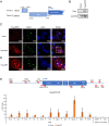
Fig. 1.
Fig. 1.
The non-coding RNA PAPAS encodes a novel nucleolar protein, RIEP. (A) Two putative ORFs in PAPAS, located upstream of 47S pre-rRNA transcription start site, were predicted by ExPASy. Their positions relative to the 47S pre-rRNA transcription start site are indicated. (B) Expression of Flag-RIEP was detected by WB with anti-Flag antibodies in Flag-RIEP but not vector-alone HeLa cells. U2AF65 serves as a loading control. (C) Representative IF images of Flag-RIEP and NPM1 in Flag-RIEP HeLa cells. Flag-RIEP was detected by Flag antibodies (red), nucleophosmin (NPM1) by NPM1 antibodies (green) and DNA by DAPI (blue). (Scale bar, 10 μm.) (D) Selected cells from Fig. 1C, (scale bar, 5 μm.) (E) Chromatin immunoprecipitation of Flag-RIEP at rDNA loci. Schematic at top indicates the rDNA locus and positions of primer pairs used to detect rDNA promoter, upstream core element (UCE), 5′ and 3′ External Transcribed Spacer (ETS), gene body (H8), IGSs (H18, IGS22, H27, IGS28), ORF2 and ORF1 regions, and two RNAP II-transcribed nuclear genes (GAPDH and LIG3). Values from vector-transformed cells (vector) were set at 1.0, and relative values from Flag-RIEP cells (Flag-RIEP) are shown. Data are presented as SE (n = 3).
Fig. 2.
Fig. 2.
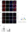
RIEP relocalizes and functions to prevent DNA damage following heat shock. (A) IF representative images showing exogenous Flag-RIEP localization in Flag-RIEP HeLa cells at 37 °C and upon 30 min or 4 h heat shock at 42 °C. Cells were stained for Flag-RIEP (red), NPM1 (green) and DNA with DAPI (blue). (Scale bar, 10 μm.) (B) IF representative images showing less γH2AX in Flag-RIEP expressing cells. Flag-RIEP HeLa were cultured at 37 °C and after 30 min or 4 h heat shock at 42 °C, cells were fixed and analyzed by IF. Flag antibodies (red) and γH2AX antibodies (green) were used and DNA detected with DAPI (blue). (C) Box plot quantification of γH2AX focus intensity per cell as shown in B. Significance was analyzed by Student’s t test. (***) P < 0.001. At least 50 cells were counted under each condition. (D) SETX levels were analyzed by WB in Flag-Vector and Flag-RIEP HeLa cells. GAPDH levels are also shown. Relative SETX levels were quantitated and presented as fold change, shown as mean, SE (n = 3).
Fig. 3.
Fig. 3.
RIEP interacts with mitochondrial proteins CHCHD2 and C1QBP (P32). (A) Immunoprecipitation mass spectrometry analysis of Flag-RIEP from HeLa cells. Top five targets are listed. (B) Immunoprecipitation of Flag-RIEP from vector or Flag-RIEP HeLa cells. Flag-RIEP, CHCHD2, and C1QBP (P32) were detected by WB using appropriate antibodies. (C) IF analysis of HeLa cells transiently expressing vector or Flag-RIEP. Cells were stained for Flag (red), CHCHD2 (green), or with DAPI (blue). Areas highlighted in the “Merge” fields are shown enlarged at the right. (Scale bars, 10 μm.)
Fig. 4.
Fig. 4.
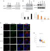
Expression and localization of endogenous RIEP. (A) Detection of endogenous RIEP in vector and Flag-RIEP HeLa cells by WB, using RIEP-1 or RIEP-216 antibodies directed against N- or C-terminal RIEP epitopes, respectively. (B) Detection of endogenous RIEP by WB with RIEP-216 in HeLa cells at 37 °C and following 30 min or 4 h heat shock at 42 °C. Quantification of relative RIEP-216 levels in HeLa cells, normalized to U2AF65 and compared with NT, is shown below. SE is shown. n = 3. (C) Endogenous RIEP levels were determined by WB with RIEP-216 in extracts from vector and Flag-RIEP HeLa cells, treated with an siRNA negative control (NC), two different siRNAs targeting RIEP/PAPAS (siP-78 or siP-100) or a mix of the two siRNAs (siPAPASs) for 72 h. Quantification of relative RIEP levels after RIEP KD as shown in C, normalized to GAPDH and compared with NC, is shown below. SE is shown. n = 3. Significance was analyzed by a Student’s t test. (*P < 0.05, ***P < 0.001). (D) HeLa cells were transfected with an siRNA negative control (NC), two different siRNAs targeting RIEP/PAPAS (siP-78 or siP-100), or a mix of the two siRNAs targeting PAPAS (siPAPASs) for 72 h. Cells were stained for RIEP using RIEP-216. Pol I (RPA 194, purple), RIEP (green), MitoTracker (red), or DAPI (blue) staining are shown. (Scale bar, 10 μm.) (E) Box plot quantification of RIEP foci intensity detected by C terminus RIEP-216 antibody per cell as shown in E.
Fig. 5.
Fig. 5.
RIEP plays a role in the mitochondrial heat shock stress response. HeLa cells were untreated or heat-shocked for 4 h. Cells were stained using RIEP antibodies recognizing the N terminus (RIEP-1) (A) or C terminus (RIEP-216) (B). RIEP (green), MitoTracker (red), or DAPI (blue) staining are shown. (Scale bar, 5 μm.) (C) Illustration of multiple properties of the RIEP-encoding genomic region. One repeat of the human rDNA arrays is diagrammed. The locus contains 47S pre-rRNA coding sequences (TSS indicated by arrow, positions of 5′ and 3′ ETSs, 18S, 5.8S, and 28S rRNAs are shown), the IGS region, and the pre-RNA promoter (UCE and ribosomal gene core element (CORE)). Position of RIEP coding sequence is indicated by the black line under the diagram, and terminators (T) are shown above. (a) 47S pre-rRNA promoter, including transcription factors UBF and SL1. (b) LncRNA PAPAS and interacting chromatin remodeling factors. (c) R loop-forming sequences. (d) RIEP mRNA and protein.

References

    1. Iyer M. K., et al. , The landscape of long noncoding RNAs in the human transcriptome. Nat. Genet 47, 199–208 (2015). - PMC - PubMed
    1. Ingolia N. T., Lareau L. F., Weissman J. S., Ribosome profiling of mouse embryonic stem cells reveals the complexity and dynamics of mammalian proteomes. Cell 147, 789–802 (2011). - PMC - PubMed
    1. Matsumoto A., et al. , mTORC1 and muscle regeneration are regulated by the LINC00961-encoded SPAR polypeptide. Nature 541, 228–232 (2017). - PubMed
    1. Anderson D. M., et al. , A micropeptide encoded by a putative long noncoding RNA regulates muscle performance. Cell 160, 595–606 (2015). - PMC - PubMed
    1. Statello L., Guo C. J., Chen L. L., Huarte M., Gene regulation by long non-coding RNAs and its biological functions. Nat. Rev. Mol. Cell Biol. 22, 96–118 (2021). - PMC - PubMed

Publication types

MeSH terms

LinkOut - more resources