. 2023 Feb 22;9(8):eabq0619.
doi: 10.1126/sciadv.abq0619. Epub 2023 Feb 22.

Unmasking of the von Willebrand A-domain surface adhesin CglB at bacterial focal adhesions mediates myxobacterial gliding motility

Affiliations

Unmasking of the von Willebrand A-domain surface adhesin CglB at bacterial focal adhesions mediates myxobacterial gliding motility

Salim T Islam et al. Sci Adv. .

Abstract

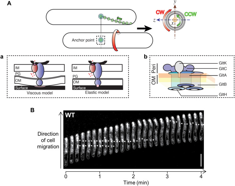
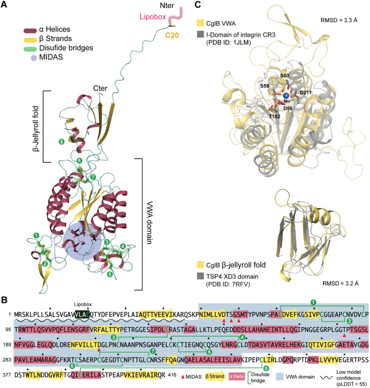
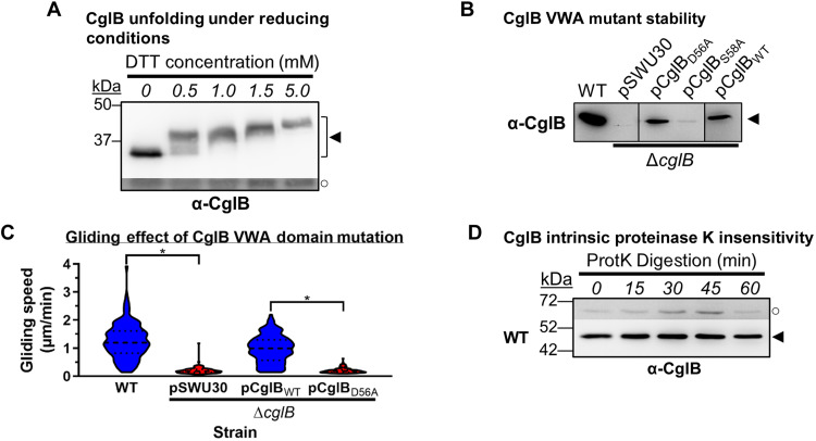
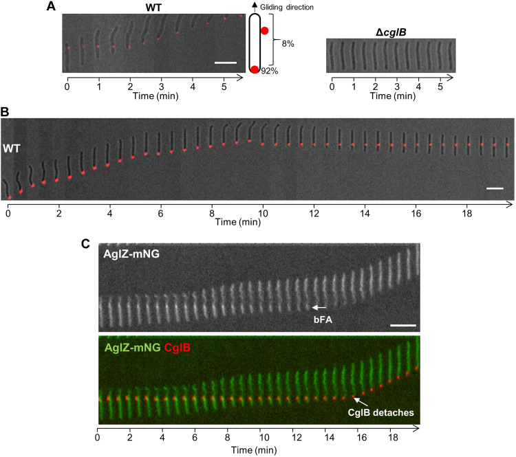
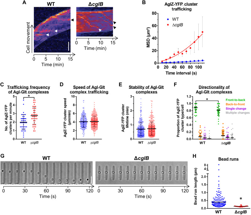
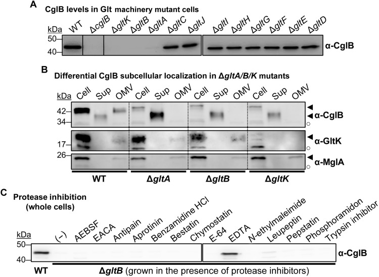
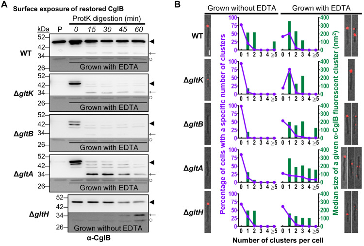
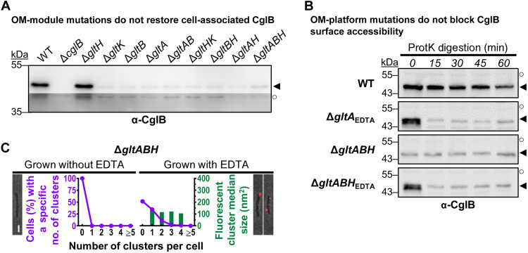
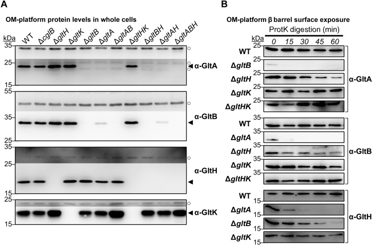
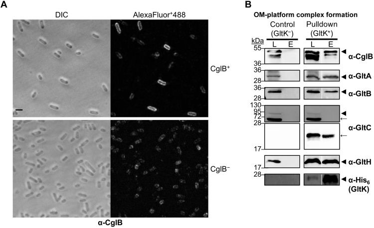
The predatory deltaproteobacterium Myxococcus xanthus uses a helically-trafficked motor at bacterial focal-adhesion (bFA) sites to power gliding motility. Using total internal reflection fluorescence and force microscopies, we identify the von Willebrand A domain-containing outer-membrane (OM) lipoprotein CglB as an essential substratum-coupling adhesin of the gliding transducer (Glt) machinery at bFAs. Biochemical and genetic analyses reveal that CglB localizes to the cell surface independently of the Glt apparatus; once there, it is recruited by the OM module of the gliding machinery, a heteroligomeric complex containing the integral OM β barrels GltA, GltB, and GltH, as well as the OM protein GltC and OM lipoprotein GltK. This Glt OM platform mediates the cell-surface accessibility and retention of CglB by the Glt apparatus. Together, these data suggest that the gliding complex promotes regulated surface exposure of CglB at bFAs, thus explaining the manner by which contractile forces exerted by inner-membrane motors are transduced across the cell envelope to the substratum.


Figures

Fig. 1.
Fig. 1.. Concept of bFA-mediated gliding motility in M. xanthus.
(A) Following assembly at the leading pole, motility complexes move toward the lagging cell pole in a counterclockwise (CCW) rotational trajectory. Clockwise (CW) and CCW directionalities are defined by observing the cell cylinder from the leading pole (yz plane). When the complexes interact with the substratum, bacterial focal adhesions (bFAs) form and propel rotational cell movements. (a) In the “viscous interaction” model for bFA formation, the periplasmic complex accumulates at bFAs and pushes against the elastic peptidoglycan (PG) to create cell-envelope deformations at bFAs, creating viscous substratum interactions. Outer-membrane (OM) complex function is not accounted for in this model. In the “elastic” model, the periplasmic complex transiently interacts (through the PG) with the OM complex, which itself interacts with the substratum via an unknown adhesive molecule (pink circle). IM, inner membrane. Blue, trans-envelope Glt complex; dark red, IM AglRQS H+ motor; dots, protons; black curve, MreB. (b) OM localization of the proposed GltA/B/H/C/K OM platform is based on structural bioinformatic and fractionation analyses presented here and elsewhere (7, 17, 21). The integral association of GltABH is based on bioinformatic and proteinase K accessibility assays herein and elsewhere (17). GltA–GltB, GltA–GltC, and GltB–GltC interactions were demonstrated by pulldown assays (17). Connection with GltH is indicated from results reported in this study. The OM periplasmic (orange) and outer (yellow) leaflets are indicated. Peri, periplasm. (B) Position of a bFA (arrowheads) revealed via fluorescence microscopy of wild-type (WT) M. xanthus expressing AglZ-YFP (yellow fluorescent protein). Scale bar, 5 μm.
Fig. 2.
Fig. 2.. CglB is a cell-surface protein with a potential integrin αI-domain-like VWA fold.
(A) AlphaFold model of CglB. CglB is predicted to contain a VWA domain and a smaller domain adopting a β-jellyroll fold. Within the CglB VWA domain, conserved residues previously described in VWA domains to coordinate divalent cations and constituting the metal ion-dependent adhesion site (MIDAS) are also present (blue circle). CglB also contains a lipobox motif with a conserved cysteine (C20). The other 16 cysteines are likely involved in disulfide bridges that stabilize the CglB structure. (B) Amino acid sequence of CglB. Secondary structures are reported as well as the cysteines potentially forming disulfide bonds (green lines). The limits of the VWA domain are also reported in gray, and the MIDAS residues are highlighted with red triangles. (C) The Dali server was used to scan the CglB structural model against the Protein Data Bank (PDB). Top: Structural alignment of CglB VWA model (yellow) to the αI domain of integrin CR3 (gray, PDB ID: 1JLM) (80). Predicted CglB MIDAS residues superimpose with the MIDAS residues (coordinating Mn2+) of the integrin CR3. Bottom: Structural alignment of CglB smaller domain adopting a β-jellyroll fold (yellow) to the XD3 domain of the bacteriophage tailspike protein 4 (TSP4 in gray, PDB ID: 7RFV) (81). RMSD, root-mean-square deviation; pLDDT, predicted local distance difference test.
Fig. 3.
Fig. 3.. The VWA domain is important for CglB stability and function.
(A) α-CglB Western blot of WT cell lysates treated with increasing dithiothreitol (DTT) concentrations to break disulfide bonds. The lower/darker zone on the blot corresponds to the same blot image section shown with higher contrast to highlight lower-intensity bands. Legend: ◄, full-length CglB; ○, loading control (nonspecific band labeled by α-CglB polyclonal antibody). (B) α-CglB Western immunoblot of CglB MIDAS motif mutant lysates. Nonadjacent lanes from the same blot are separated by vertical black lines. (C) Violin plots of single-cell gliding speeds on hard (1.5%) agar for M. xanthus DZ2 ΔcglB (n = 120 cells) complemented with CglBWT or CglBD56A. The median (dashed line) as well as lower and upper quartiles (dotted lines) are indicated. Asterisks denote datasets displaying statistically significant dataset differences (P < 0.0001) compared to strains harboring either the pSWU30 empty-vector control or the pCglBD56A VWA domain mutant CglB complementation construct, as determined via two-tailed Mann-Whitney U tests. (D) Protein samples from WT cells resuspended in tris-phosphate-magnesium (TPM) buffer and digested with exogenous proteinase K. Aliquots of the digestion mixture were removed at 15-min intervals and trichloroacetic acid (TCA)-precipitated to stop digestion. The higher, darker zone on the blot corresponds to a section of the same blot image for which the contrast has been increased to highlight lower-intensity protein bands. The lack of CglB degradation was not due to lack of proteinase K activity (see below). Legend: ◄, full-length CglB; ○, loading control (nonspecific protein band labeled by α-CglB antibody).
Fig. 4.
Fig. 4.. CglB is a cell-surface protein that localizes to bFA sites.
(A) Montage of live WT and ΔcglB cells immunolabeled with α-CglB 1° antibody, followed by goat α-rabbit 2° antibody conjugated to Alexa Fluor 647 on agar pads at 32°C. Images were acquired at 30-s intervals. Scale bar, 5 μm. (B) Montage of a live immunolabeled WT cell [labeled as in (A)] in which a dragged fluorescent cluster becomes immobilized relative to the substratum upon reversal of gliding direction. Scale bar, 5 μm. (C) Montage of a live immunolabeled cell expressing AglZ-mNeonGreen (82) in which an α-CglB antibody colocalizes with a fixed AglZ-mNeonGreen cluster at a bFA site. Note that the CglB cluster detaches from the surface when it reaches the lagging cell pole. Scale bar, 5 μm.
Fig. 5.
Fig. 5.. CglB is essential for gliding-complex substratum adhesion.
(A) Temporal AglZ-YFP cluster position (dashed lines) kymographs (cells on agar pads). Scale bar, 2 μm. White arrowheads: Clusters followed for entire lifetimes. Black arrowheads: Clusters followed for incomplete lifetimes. (B) Mean square displacement (MSD) of AglZ-YFP cluster position tracking in WT (n = 48 clusters) and ΔcglB (n = 23 clusters) cells. Mean MSD (± SEM) at each time interval is displayed, with a second-order polynomial fit to each dataset. For (C) to (F), experiments were performed via TIRFM on chitosan-coated glass in polydimethylsiloxane microfluidic chambers; mean values are indicated by a black line ± SEM. Distributions of the WT–ΔcglB datasets were compared via unpaired two-tailed Mann-Whitney U tests; those with significant differences (P < 0.05) are indicated (*). (C) AglZ-YFP complex trafficking frequency: WT (n = 44 cells), ΔcglB (n = 41 cells). (D) Agl-Glt complex trafficking speed: WT (n = 260 clusters), ΔcglB (n = 371 clusters). (E) Trafficking Agl–Glt complex stability: WT (n = 333 clusters), ΔcglB (n = 409 clusters). (F) Directionality of trafficked Agl–Glt complexes: WT (n = 44 cells), ΔcglB (n = 41 cells). Front and back are defined as cell poles with high/low AglZ-YFP fluorescence intensity, respectively. (G) Trafficking phenotypes of surface-deposited polystyrene beads. Scale bar, 3 μm. (H) Lengths of tracked bead runs >0.1 μm. Images from 10-s intervals were analyzed. The distributions of the two datasets are significantly different (*) as determined via unpaired two-tailed Mann-Whitney U test (P < 0.05).
Fig. 6.
Fig. 6.. Glt OM-platform constituents mediate cellular retention of CglB.
(A) Whole-cell extracts from different Δglt mutants. Nonadjacent lanes on the same blot are separated by vertical black lines. White space separates two distinct blots processed at the same time. (B) Fractionated samples containing whole cells (Cell), supernatants (Sup), and OM vesicles (OMV) from various genetic backgrounds. Detection of the gliding motility OM lipoprotein GltK was added as a control, with the protein only detected in Cell and OMV samples, showing that the various mutations do not affect OM integrity, with the supernatant localization in this instance being specific to CglB. MglA is a cytoplasmic protein added as a control to show that cell lysis is negligible and does not account for the presence of CglB in supernatants. Legend: ◄, full-length protein; ○, loading control (nonspecific protein band labeled by the respective polyclonal antibody). (C) Whole-cell extracts from ΔgltB cells grown in the presence of different protease inhibitors. White space separates two distinct blots from the same experiment. AEBSF, 4-(2-aminoethyl benzenesulfonyl fluoride HCl; EACA, ε-aminocaproic acid.
Fig. 7.
Fig. 7.. CglB surface exposure is mediated by the Glt OM platform.
(A) Protein samples from intact cells digested with exogenous proteinase K. Digestion mixture aliquots were removed at 15-min intervals and TCA-precipitated to stop digestion. “P”: Lanes containing the untreated parent strain grown without EDTA. Lower/darker zones on each blot correspond to sections of the same blot image for which the contrast has been increased to highlight lower-intensity protein bands. The samples and blot for ΔgltH were obtained at the same time as that for WT (Fig. 3D), indicating that the proteinase K was active during treatment of the latter. Legend: ◄, full-length CglB; ←, CglB degradation band; ○, loading control (nonspecific band labeled by α-CglB antibody). (B) Fluorescence micrographs of live immunolabeled WT, ΔgltK/B/A/H cells grown with(out) EDTA (labeled with α-CglB 1° antibody, followed by goat α-rabbit 2° antibody conjugated to Alexa Fluor 647) on agar pads at 32°C. Representative images are provided for cluster-labeling patterns observed on ~20% or more of analyzed cells for a given strain/treatment. Scale bar, 1 μm. For each strain grown with(out) EDTA, the number of fluorescent clusters detected per cell was counted (x axis) and compared against the proportion of cells with such a labeling phenotype (purple left-side y axis). The size of each cluster was also measured, with the median area (dark green right-side y axis) given for each labeling phenotype. The number of cells analyzed for each treatment is as follows (±EDTA): WT 304/347, ΔgltK 306/306, ΔgltB 437/199, ΔgltA 424/251, and ΔgltH 505/471.
Fig. 8.
Fig. 8.. CglB secretion to the cell surface is not mediated by the Glt OM platform.
(A) α-CglB Western immunoblots for whole-cell extracts from different combinations of Δglt OM-module mutations in the same strain. Legend: ◄, full-length CglB; ○, loading control (nonspecific protein band labeled by α-CglB polyclonal antibody). (B) α-CglB Western immunoblots for protein samples from cells resuspended in TPM buffer and digested with exogenous proteinase K. Aliquots of the digestion mixture were removed at 15-min intervals and TCA-precipitated to stop digestion. Legend: ◄, full-length CglB; ○, loading control (nonspecific protein band labeled by α-CglB polyclonal antibody). (C) Fluorescence micrographs of live immunolabeled ΔgltABH cells grown with(out) EDTA (labeled with α-CglB 1° antibody, followed by goat α-rabbit 2° antibody conjugated to Alexa Fluor 647) on agar pads at 32°C. Representative images are provided for cluster-labeling patterns observed on ~20% or more of analyzed cells for a given treatment. Scale bar, 1 μm. For cultures grown with(out) EDTA, the number of fluorescent clusters detected per cell was counted (x axis) and compared against the proportion of cells with such a labeling phenotype (purple left-side y axis). The size of each cluster was also measured, with the median area (dark green right-side y axis) given for each labeling phenotype. The number of cells analyzed for each treatment is as follows (±EDTA): ΔgltABH 370/344.
Fig. 9.
Fig. 9.. Glt OM-platform constituents exhibit interdependencies.
(A) Western immunoblots of GltA, GltB, GltH, and GltK in various single-, double-, and triple-mutant combinations of OM-platform constituents. Legend: ◄, full-length protein; ○, loading control (nonspecific protein band labeled by the respective α-GltA/α-GltB/α-GltH/α-GltK polyclonal antibody). (B) Western immunoblots for Glt OM-platform β-barrel constituent susceptibility to digestion by proteinase K in Glt OM-module mutant strains. Digestion aliquots were removed at 15-min intervals and TCA-precipitated to stop digestion.
Fig. 10.
Fig. 10.. CglB directly interacts with the Glt OM-platform heteroligomeric complex.
(A) Fluorescence micrographs of E. coli BL21(DE3) cells immunolabeled with α-CglB 1° antibody, followed by goat α-rabbit 2° antibody conjugated to Alexa Fluor Plus 488 (AlexaFluor+488). Cells had been transformed with the following plasmid combinations: “pCDF-Duet-GltK6H+GltBACS & pET-Duet-GltH-CglB” (for coexpression of GltA, GltB, GltC-StrepII, GltK-His6, GltH, and CglB) or “pCDF-Duet and pET-Duet” (as empty-vector controls). Cells were induced overnight with 1.0 mM IPTG and then fixed with paraformaldehyde before immunolabeling. Scale bar, 2 μm. DIC, differential interference contrast. (B) Western immunoblotting of purified OM-platform proteins from the pulldown assay (right side) or negative control (left side) using α-CglB, α-GltA, α-GltB, α-GltH, α-His (GltK), and α-GltC 1º antibodies. Calculated molecular weights for monomeric forms of each protein construct (lacking signal peptide): CglB (42.3 kDa), GltA (25.4 kDa), GltB (27.5 kDa), GltC-StrepII (74.4 kDa), GltH (20.0 kDa), and GltK-His6 (17.5 kDa). Nonadjacent lanes from the same blot are separated by white spaces. Lane legend: L, column loading fraction; E, column elution fraction. Blot legend: ◄, full-length protein; ←, degradation product of the protein of interest.

Similar articles

Cited by

References

    1. Z. Sun, S. S. Guo, R. Fässler, Integrin-mediated mechanotransduction. J. Cell Biol. 215, 445–456 (2016). - PMC - PubMed
    1. P. Kanchanawong, G. Shtengel, A. M. Pasapera, E. B. Ramko, M. W. Davidson, H. F. Hess, C. M. Waterman, Nanoscale architecture of integrin-based cell adhesions. Nature 468, 580–584 (2010). - PMC - PubMed
    1. S. T. Islam, I. Vergara Alvarez, F. Saïdi, A. Guiseppi, E. Vinogradov, G. Sharma, L. Espinosa, C. Morrone, G. Brasseur, J. F. Guillemot, A. Benarouche, J. L. Bridot, G. Ravicoularamin, A. Cagna, C. Gauthier, M. Singer, H. P. Fierobe, T. Mignot, E. M. F. Mauriello, Modulation of bacterial multicellularity via spatio-specific polysaccharide secretion. PLOS Biol. 18, e3000728 (2020). - PMC - PubMed
    1. F. Saïdi, N. Y. Jolivet, D. J. Lemon, A. Nakamura, A. M. Belgrave, A. G. Garza, F. J. Veyrier, S. T. Islam, Bacterial glycocalyx integrity drives multicellular swarm biofilm dynamism. Mol. Microbiol. 116, 1151–1172 (2021). - PubMed
    1. F. Saïdi, U. Mahanta, A. Panda, A. A. Kezzo, N. Y. Jolivet, R. Bitazar, G. John, M. Martinez, A. Mellouk, C. Calmettes, Y. W. Chang, G. Sharma, S. T. Islam, Bacterial outer membrane polysaccharide export (OPX) proteins occupy three structural classes with selective β-barrel porin requirements for polymer secretion. Microbiol. Spectr. 10, e01290-22 (2022). - PMC - PubMed