

Ring-stacked capsids of white spot syndrome virus and structural transitions with genome ejection
- PMID: 36812312
- PMCID: PMC9946344
- DOI: 10.1126/sciadv.add2796
Ring-stacked capsids of white spot syndrome virus and structural transitions with genome ejection
Abstract
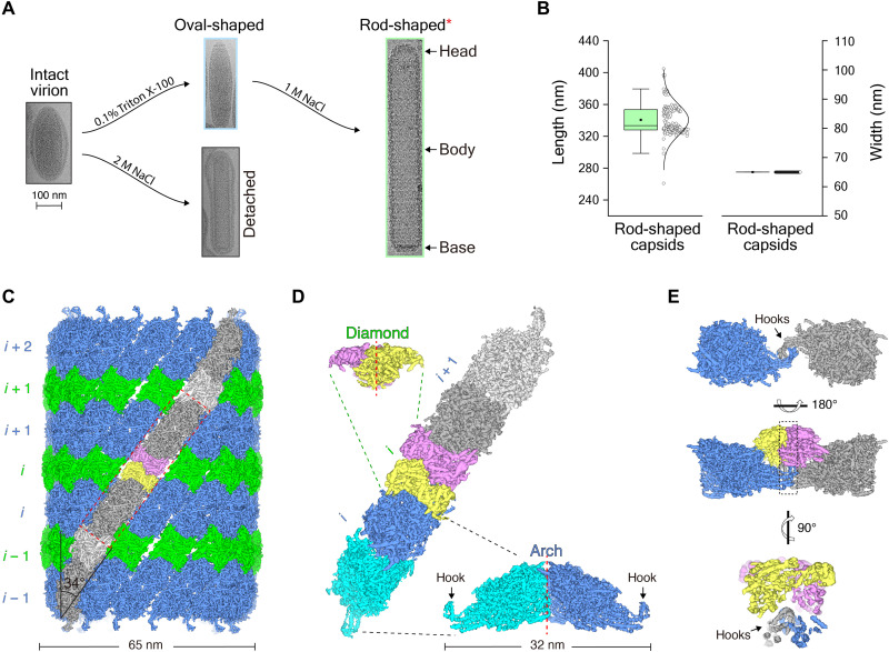
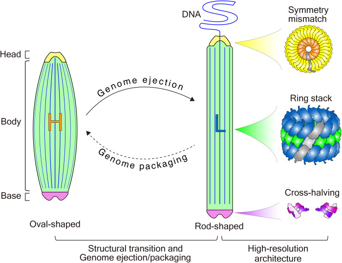
White spot syndrome virus (WSSV) is one of the largest DNA viruses and the major pathogen responsible for white spot syndrome in crustaceans. The WSSV capsid is critical for genome encapsulation and ejection and exhibits the rod-shaped and oval-shaped structures during the viral life cycle. However, the detailed architecture of the capsid and the structural transition mechanism remain unclear. Here, using cryo-electron microscopy (cryo-EM), we obtained a cryo-EM model of the rod-shaped WSSV capsid and were able to characterize its ring-stacked assembly mechanism. Furthermore, we identified an oval-shaped WSSV capsid from intact WSSV virions and analyzed the structural transition mechanism from the oval-shaped to rod-shaped capsids induced by high salinity. These transitions, which decrease internal capsid pressure, always accompany DNA release and mostly eliminate the infection of the host cells. Our results demonstrate an unusual assembly mechanism of the WSSV capsid and offer structural insights into the pressure-driven genome release.
Figures
Similar articles
-
Multiple Nucleocapsid Structural Forms of Shrimp White Spot Syndrome Virus Suggests a Novel Viral Morphogenetic Pathway.Int J Mol Sci. 2023 Apr 19;24(8):7525. doi: 10.3390/ijms24087525. Int J Mol Sci. 2023. PMID: 37108688 Free PMC article.
-
White Spot Syndrome Virus Benefits from Endosomal Trafficking, Substantially Facilitated by a Valosin-Containing Protein, To Escape Autophagic Elimination and Propagate in the Crustacean Cherax quadricarinatus.J Virol. 2020 Nov 23;94(24):e01570-20. doi: 10.1128/JVI.01570-20. Print 2020 Nov 23. J Virol. 2020. PMID: 32967962 Free PMC article.
-
Cryo-electron Microscopy Study of the Genome Release of the Dicistrovirus Israeli Acute Bee Paralysis Virus.J Virol. 2017 Jan 31;91(4):e02060-16. doi: 10.1128/JVI.02060-16. Print 2017 Feb 15. J Virol. 2017. PMID: 27928006 Free PMC article.
-
Breaking Symmetry in Viral Icosahedral Capsids as Seen through the Lenses of X-ray Crystallography and Cryo-Electron Microscopy.Viruses. 2018 Feb 7;10(2):67. doi: 10.3390/v10020067. Viruses. 2018. PMID: 29414851 Free PMC article. Review.
-
Beyond structures of highly symmetric purified viral capsids by cryo-EM.Curr Opin Struct Biol. 2018 Oct;52:25-31. doi: 10.1016/j.sbi.2018.07.011. Epub 2018 Aug 7. Curr Opin Struct Biol. 2018. PMID: 30096461 Review.
Cited by
-
Structure of AcMNPV nucleocapsid reveals DNA portal organization and packaging apparatus of circular dsDNA baculovirus.Nat Commun. 2025 May 24;16(1):4844. doi: 10.1038/s41467-025-60152-2. Nat Commun. 2025. PMID: 40413174 Free PMC article.
-
Multiple Nucleocapsid Structural Forms of Shrimp White Spot Syndrome Virus Suggests a Novel Viral Morphogenetic Pathway.Int J Mol Sci. 2023 Apr 19;24(8):7525. doi: 10.3390/ijms24087525. Int J Mol Sci. 2023. PMID: 37108688 Free PMC article.
-
Temperature-dependent ejection evolution arising from active and passive effects in DNA viruses.Biophys J. 2024 Oct 1;123(19):3317-3330. doi: 10.1016/j.bpj.2024.07.037. Epub 2024 Jul 31. Biophys J. 2024. PMID: 39091028
-
Novel Artificial Intelligence-Based Approaches for Ab Initio Structure Determination and Atomic Model Building for Cryo-Electron Microscopy.Micromachines (Basel). 2023 Aug 27;14(9):1674. doi: 10.3390/mi14091674. Micromachines (Basel). 2023. PMID: 37763837 Free PMC article. Review.
-
The nucleocapsid architecture and structural atlas of the prototype baculovirus define the hallmarks of a new viral realm.Sci Adv. 2024 Dec 20;10(51):eado2631. doi: 10.1126/sciadv.ado2631. Epub 2024 Dec 18. Sci Adv. 2024. PMID: 39693434 Free PMC article.
References
-
- C. Li, S. Weng, J. He, WSSV-host interaction: Host response and immune evasion. Fish Shellfish Immunol. 84, 558–571 (2019). - PubMed
-
- H.-C. Wang, I. Hirono, M. B. Bacano Maningas, K. Somboonwiwat, G. Stentiford; Ictv Report Consortium , ICTV virus taxonomy profile: Nimaviridae. J. Gen. Virol. 100, 1053–1054 (2019). - PubMed
-
- J. H. Leu, F. Yang, X. Zhang, X. Xu, G. H. Kou, C. F. Lo, Whispovirus. Curr. Top. Microbiol. Immunol. 328, 197–227 (2009). - PubMed
-
- S. K. Chandrika, S. T. Puthiyedathu, Challenges and prospects of viral envelope protein VP28-based control strategies to combat white spot syndrome virus in penaeid shrimps: A review. Rev. Aquac. 13, 734–743 (2021).
MeSH terms
Substances
LinkOut - more resources
Full Text Sources
Other Literature Sources
