. 2023 Feb 22;9(8):eade7923.
doi: 10.1126/sciadv.ade7923. Epub 2023 Feb 22.

High cell density and high-resolution 3D bioprinting for fabricating vascularized tissues

Affiliations

High cell density and high-resolution 3D bioprinting for fabricating vascularized tissues

Shangting You et al. Sci Adv. .

Abstract

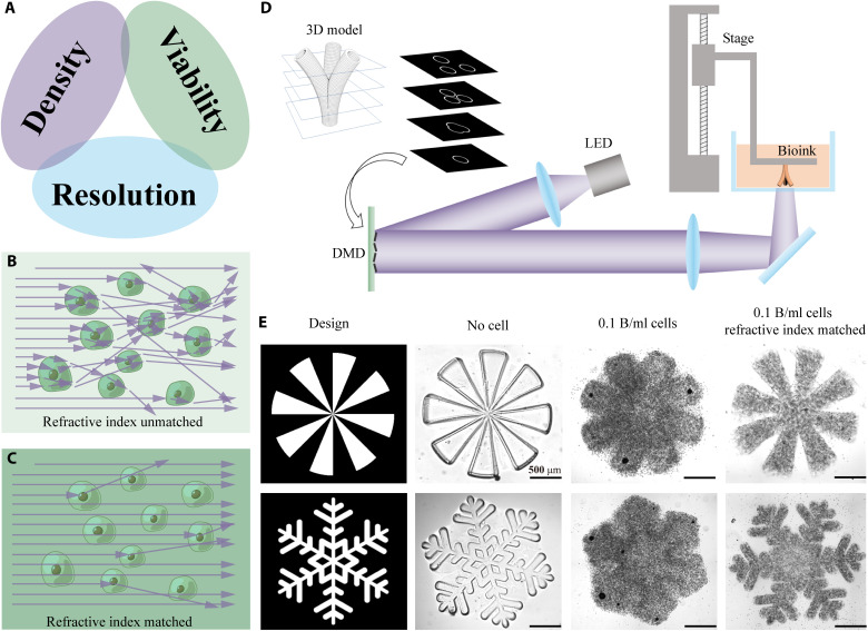
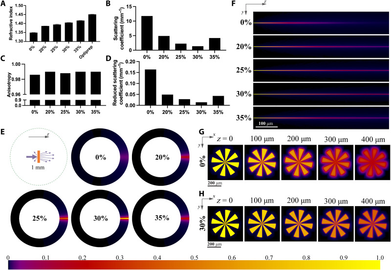
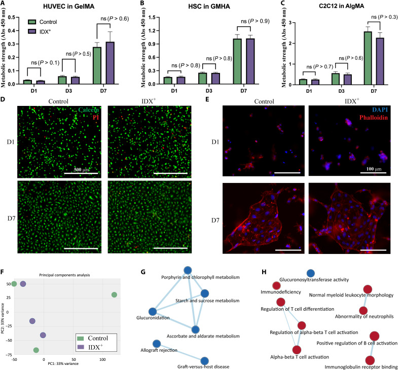
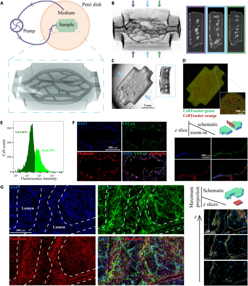
Three-dimensional (3D) bioprinting techniques have emerged as the most popular methods to fabricate 3D-engineered tissues; however, there are challenges in simultaneously satisfying the requirements of high cell density (HCD), high cell viability, and fine fabrication resolution. In particular, bioprinting resolution of digital light processing-based 3D bioprinting suffers with increasing bioink cell density due to light scattering. We developed a novel approach to mitigate this scattering-induced deterioration of bioprinting resolution. The inclusion of iodixanol in the bioink enables a 10-fold reduction in light scattering and a substantial improvement in fabrication resolution for bioinks with an HCD. Fifty-micrometer fabrication resolution was achieved for a bioink with 0.1 billion per milliliter cell density. To showcase the potential application in tissue/organ 3D bioprinting, HCD thick tissues with fine vascular networks were fabricated. The tissues were viable in a perfusion culture system, with endothelialization and angiogenesis observed after 14 days of culture.


Figures

Fig. 1.
Fig. 1.. Achieving high fabrication resolution in HCD bioinks.
(A) The “impossible trinity” in 3D bioprinting: HCD, high cell viability, and high fabrication resolution cannot be satisfied simultaneously. (B) A schematic showing light propagation in a refractive index–unmatched bioink. (C) A schematic showing light propagation in a refractive index–matched bioink, where light scattering is substanrually reduced. (D) A schematic showing how DLP-based 3D bioprinter works. LED, light-emitting diode; DMD, digital micromirror device. (E) Printing resolution comparison among three different bioink compositions: bioink without cells, bioink with 0.1 billion cells/ml, and refractive index–matched bioink with 0.1 billion cells/ml. Scale bars, 500 μm.
Fig. 2.
Fig. 2.. Optical properties and light energy distribution.
(A) Refractive index of 5% GelMA bioink with various IDX concentrations at 405 nm and the refractive index of Optiprep solution (60% IDX). (B to D) Scattering coefficient, anisotropy, and reduced scattering coefficient of 40 million cells/ml cell–encapsulated bioink with various IDX concentrations. (E) Angular intensity distribution of the light scattered by cell-encapsulated bioink with various IDX concentrations. Light propagates in the z direction. (F) Spatial distribution (in YZ cross section) of light in the cell-encapsulated bioink with various IDX concentrations. (G) Projected pattern (in XY cross section) at different z depths of the cell-encapsulated bioink with 0% IDX. (H) Projected pattern (in XY cross section) at different z depths of the cell-encapsulated bioink with 30% IDX. Color bar denotes the relative light intensity in (E) to (H).
Fig. 3.
Fig. 3.. Biocompatibility analysis.
(A to C) Comparison of metabolic strength of the printed tissues using bioinks with or without IDX. (D) Live (green) and dead (red) cells of the printed tissues using bioinks with or without IDX. (E) Comparison of the images of cytoskeleton of the printed tissues using bioinks with or without IDX. (F) Principal components analysis results of HUVEC slabs using bioinks with or without IDX (n = 3). (G) Network analysis of enriched gene sets in the Molecular Signature Database curated collection. (H) Network analysis of enriched gene sets in the ontology collection. Red nodes, up-regulated gene sets; blue nodes, down-regulated gene sets. Abs, absorbance. ns, not significant.
Fig. 4.
Fig. 4.. 3D printing of vascularized perfusable thick tissues.
(A) Schematic of the perfusion culture system and the 3D render of the printed tissue. (B) μCT images of the printed samples (perspective view and cross sections). (C) Bright-field images of the printed samples (top view and cross section). (D) Fluorescence images of the printed samples. (E) Cell viability in the thick tissue after 14 days of perfusion culture. (F) Immunofluorescence images of a cryosection in a plane perpendicular to the printed channels. (G) Immunofluorescence images of a horizontally cleaved chunk. The z slices are in horizontal planes, and the maximum projection images are stacks of all z slices. The white dashed lines denote the position of the 3D-printed lumen.

References

    1. J. M. Unagolla, A. C. Jayasuriya, Hydrogel-based 3D bioprinting: A comprehensive review on cell-laden hydrogels, bioink formulations, and future perspectives. Appl. Mater. Today 18, 100479 (2020). - PMC - PubMed
    1. C. Yu, J. Schimelman, P. Wang, K. L. Miller, X. Ma, S. You, J. Guan, B. Sun, W. Zhu, S. Chen, Photopolymerizable biomaterials and light-based 3D printing strategies for biomedical applications. Chem. Rev. 120, 10695–10743 (2020). - PMC - PubMed
    1. S. V. Murphy, A. Atala, 3D bioprinting of tissues and organs. Nat. Biotechnol. 32, 773–785 (2014). - PubMed
    1. W. Zhu, K. R. Tringale, S. A. Woller, S. You, S. Johnson, H. Shen, J. Schimelman, M. Whitney, J. Steinauer, W. Xu, T. L. Yaksh, Q. T. Nguyen, S. Chen, Rapid continuous 3D printing of customizable peripheral nerve guidance conduits. Mater. Today 21, 951–959 (2018). - PMC - PubMed
    1. Z. Zhong, J. Wang, J. Tian, X. Deng, A. Balayan, Y. Sun, Y. Xiang, J. Guan, J. Schimelman, H. Hwang, S. You, X. Wu, C. Ma, X. Shi, E. Yao, S. X. Deng, S. Chen, Rapid 3D bioprinting of a multicellular model recapitulating pterygium microenvironment. Biomaterials 282, 121391 (2022). - PMC - PubMed