. 2023 Apr;299(4):103050.
doi: 10.1016/j.jbc.2023.103050. Epub 2023 Feb 21.

The 5-lipoxygenase/cyclooxygenase-2 cross-over metabolite, hemiketal E2, enhances VEGFR2 activation and promotes angiogenesis

Affiliations

The 5-lipoxygenase/cyclooxygenase-2 cross-over metabolite, hemiketal E2, enhances VEGFR2 activation and promotes angiogenesis

Fumie Nakashima et al. J Biol Chem. 2023 Apr.

Abstract

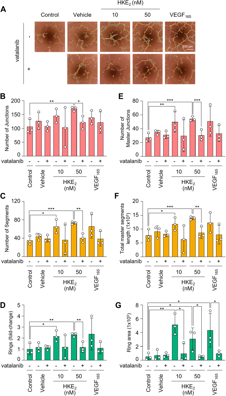
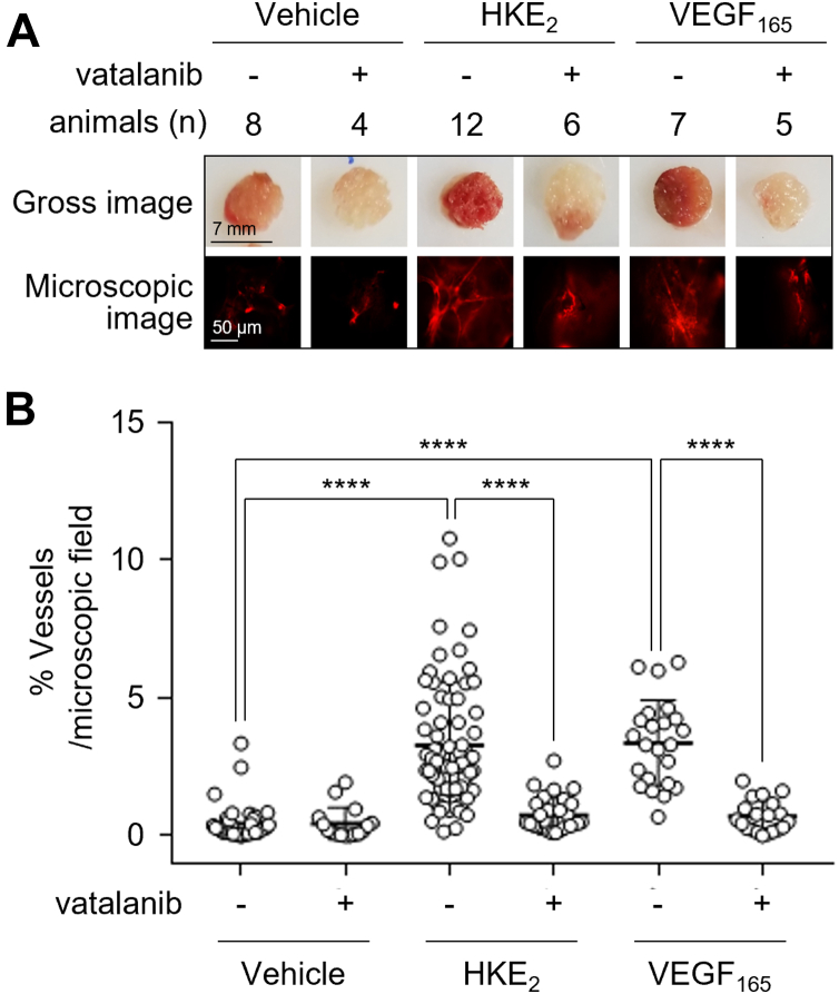
Consecutive oxygenation of arachidonic acid by 5-lipoxygenase and cyclooxygenase-2 yields the hemiketal eicosanoids, HKE2 and HKD2. Hemiketals stimulate angiogenesis by inducing endothelial cell tubulogenesis in culture; however, how this process is regulated has not been determined. Here, we identify vascular endothelial growth factor receptor 2 (VEGFR2) as a mediator of HKE2-induced angiogenesis in vitro and in vivo. We found that HKE2 treatment of human umbilical vein endothelial cells dose-dependently increased the phosphorylation of VEGFR2 and the downstream kinases ERK and Akt that mediated endothelial cell tubulogenesis. In vivo, HKE2 induced the growth of blood vessels into polyacetal sponges implanted in mice. HKE2-mediated effects in vitro and in vivo were blocked by the VEGFR2 inhibitor vatalanib, indicating that the pro-angiogenic effect of HKE2 was mediated by VEGFR2. HKE2 covalently bound and inhibited PTP1B, a protein tyrosine phosphatase that dephosphorylates VEGFR2, thereby providing a possible molecular mechanism for how HKE2 induced pro-angiogenic signaling. In summary, our studies indicate that biosynthetic cross-over of the 5-lipoxygenase and cyclooxygenase-2 pathways gives rise to a potent lipid autacoid that regulates endothelial cell function in vitro and in vivo. These findings suggest that common drugs targeting the arachidonic acid pathway could prove useful in antiangiogenic therapy.

Keywords: 5-lipoxygenase; arachidonic acid; cyclooxygenase-2; eicosanoid; endothelial cell; kinase signaling; sponge assay.


Conflict of interest statement

Conflict of interest The authors declare that they have no conflicts of interest with the contents of this article.

Figures

Figure 1
Figure 1
The 5-LOX/COX-2 cross-over biosynthetic pathway to hemiketal eicosanoids HKE2and HKD2in comparison to prostaglandin formation. The top reactions show prostaglandin biosynthesis by COX-1 and COX-2. The prostaglandin endoperoxide PGH2 formed in the COX reaction undergoes rearrangement to PGE2 and PGD2. The reactions below show the cross-over pathway initiated by 5-LOX formation of 5S-hydroxyeicosatetraenoic acid (5S-HETE) from arachidonic acid followed by COX-2–catalyzed formation of a diendoperoxide that rearranges to HKE2 and HKD2. 5-LOX, 5-lipoxygenase; COX-2, cyclooxygenase-2.
Figure 2
Figure 2
Protein phosphorylation in endothelial cells treated with HKE2. HUVEC were treated with (A) 500 nM of HKE2 for 0 to 120 min or (B) 0 to 500 nM of HKE2 for 30 min, and cellular protein was analyzed by SDS-PAGE and immunoblotting using antibodies against phospho-tyrosine (α-pTyr) and β-actin. Red triangles indicate protein bands that showed increased phosphorylation in response to treatment with HKE2 compared to vehicle control. C, a human phospho-Receptor Tyrosine Kinase (RTK) array was used to analyze HKE2-induced phosphorylation of 49 RTKs in HUVEC lysates. X-ray film images of the membranes treated with vehicle and 50, 100, and 500 nM HKE2 are shown. Protein spots that showed differences in intensity between HKE2 and vehicle control are boxed. D, quantification of the RTK array results using ImageJ software. The five RTK boxed in panel (C) are marked with a black triangle. The mean of two independent repeats of the RTK array using 0 to 500 nM HKE2 is shown. HUVEC, human umbilical vein endothelial cell; RTK, receptor tyrosine kinase.
Figure 3
Figure 3
Phosphorylation of VEGFR2 and downstream kinases in endothelial cells treated with HKE2.A, Western blots of phosphorylated and total VEGFR2 in cell lysates of HUVEC treated with 0 to 500 nM HKE2 for 30 min. The graph shows the ratio of phosphorylated to total VEGFR2 from n = 3 independent repeats. Data are presented as means ± S.D. (∗) indicates significant differences (∗p < 0.05, ∗∗p < 0.01) between vehicle (0 nM HKE2) and HKE2-treated cells. B, cells were treated with the VEGFR2 inhibitor vatalanib 30 min prior to HKE2 treatment, and phosphorylation of VEGFR2 was analyzed by Western blotting. C and D, HUVEC were incubated for 1 h with or without vatalanib and then treated with vehicle, VEGF165 (50 ng/ml) or the indicated concentration of HKE2. Cell lysates were prepared and subjected to Western blotting for phosphorylated and total ERK and Akt, respectively, and for β-actin. Bar graphs show the ratio of phosphorylated over total proteins obtained from n = 3 independent repeats. Data are presented as means ± S.D. (∗) indicates significant differences (∗p < 0.05, ∗∗p < 0.01). HUVEC, human umbilical vein endothelial cell; VEGFR, vascular endothelial growth factor receptor.
Figure 4
Figure 4
HKE2induces formation of capillary-like structures by endothelial cells.A, HUVEC were placed in serum-free Medium 200PRF onto Matrigel coated 96-well plates in the absence or presence of the VEGFR2 inhibitor, vatalanib. After 2 h incubation, HKE2 or VEGF165 (50 ng/ml) was added to the wells. Representative images of tube-like structures were taken 6 h after HKE2 treatment. BG, capillary network formation was quantified using ImageJ software with the Angiogenesis Analyzer plugin counting the number of (B) junctions, (C) segments, (D) rings, (E) master junctions, (F) length of total master segments, and (G) ring area from two independent experiments. Data are presented as means ± S.D. (∗) indicates significant differences (∗p < 0.05, ∗∗p < 0.01, ∗∗∗p < 0.005). HUVEC, human umbilical vein endothelial cell; VEGFR, vascular endothelial growth factor receptor.
Figure 5
Figure 5
Kinase inhibition in HKE2-induced tubulogenesis of HUVEC. HUVEC were plated in 96-well plates containing reduced growth factor Matrigel and were treated with vehicle (DMSO), the MAPK/ERK inhibitor U0126 (10 μM), or the PI3 kinase inhibitor LY294002 (1 μM). After 30 min, cells were treated with ethanol (vehicle) or HKE2 (500 nM). After 6 h, capillary network formation was visualized and quantified by ImageJ software with the Angiogenesis Analyzer plugin counting the number of (A) junctions, (B) segments, and (C) rings. Data are representative of three independent experiments. Data are presented as means ± S.D. (∗) indicates significant differences (∗p < 0.05, ∗∗p < 0.01). HUVEC, human umbilical vein endothelial cell.
Figure 6
Figure 6
In vivo angiogenesis induced by HKE2.A, gross and microscopic images of polyvinyl acetal CF-50 sponges implanted under the dorsal skin of BALB∖c female mice (12 weeks of age, 20 g). The sponges were injected every other day for 2 weeks with 50 μl of vehicle (coconut medium-chain triglyceride (MCT) oil, n = 8 animals), HKE2 (10 μM in MCT oil, n = 12), or VEGF165 (5 μg/ml in PBS, n = 7). In addition, animals received vatalanib (50 mg/kg) or vehicle (DMSO/PEG 400/0.9% sodium chloride: 5/47.5/47.5) by gavage daily (vehicle + inhibitor, n = 4; HKE2 + inhibitor, n = 6; VEGF165 + inhibitor, n = 5) starting from same day of the first injection of HKE2. Fifteen minutes prior to sacrifice, mice were injected intravenously with 50 μl of rhodamine-dextran (2% in PBS). Sponges were then removed, sectioned, and placed under a fluorescence microscope to visualize vascularization. B, vascularity within sponges was quantified by calculating the area occupied by rhodamine-positive structures per microscopic field. The horizontal bar indicates the mean ± S.D. calculated for 5 to 6 images/sponge per treatment. Differences were analyzed by ANOVA followed by Dunnett’s multiple comparison analysis. (∗) indicates significant differences (∗∗∗∗p < 0.0001).
Figure 7
Figure 7
Adduction and inhibition of PTP1B by HKE2.A, recombinant PTP1B (0.5 μM) was treated with HKE2 or N-ethyl maleimide (NEM) at 5 or 45 μM, respectively, followed by the detection of free remaining cysteine residues with biotin-maleimide and analysis by SDS-PAGE. The image shows Western blot detection using HRP-conjugated avidin (top) or an antibody against PTP1B (bottom). B, the graph shows the ratio of unmodified PTP1B (top panel in A) to total PTP1B (bottom panel in A) from n = 3 independent repeats. Data are presented as means ± S.D. (∗) indicates significant differences (∗∗∗∗p < 0.0001) between control and HKE2 or NEM-treated samples. C, recombinant PTP1B (0.5 μM) was treated with HKE2 or N-ethyl maleimide (NEM) at 5 or 45 μM, respectively, followed by the detection of free remaining cysteine residues with PEG-PC-Maleimide and analysis by SDS-PAGE with Coomassie staining. D, the graph shows the relative abundance of shifted bands (band 1–6) and the original PTP1B band for each treatment from n = 3 independent repeats. E, phosphatase activity of PTP1B (1 μM) using pNPP as a substrate from n = 3 independent repeats. Data are presented as means ± S.D. (∗) indicates significant differences (∗∗∗∗p < 0.0001) between control and HKE2 or NEM-treated samples. F, illustration of the Michael-type addition of HKE2 to a reactive cysteine residue of PTP1B. NEM, N-ethyl-maleimide; PTP1B, protein tyrosine phosphatase 1B.

References

    1. Funk C.D. Prostaglandins and leukotrienes: advances in eicosanoid biology. Science. 2001;294:1871–1875. - PubMed
    1. Murphy R.C., Gijon M.A. Biosynthesis and metabolism of leukotrienes. Biochem. J. 2007;405:379–395. - PubMed
    1. Smith W.L., Urade Y., Jakobsson P.J. Enzymes of the cyclooxygenase pathways of prostanoid biosynthesis. Chem. Rev. 2011;111:5821–5865. - PMC - PubMed
    1. Griesser M., Suzuki T., Tejera N., Mont S., Boeglin W.E., Pozzi A., et al. Biosynthesis of hemiketal eicosanoids by cross-over of the 5-lipoxygenase and cyclooxygenase-2 pathways. Proc. Natl. Acad. Sci. U. S. A. 2011;108:6945–6950. - PMC - PubMed
    1. Nakashima F., Suzuki T., Gordon O.N., Golding D., Okuno T., Gimenez-Bastida J.A., et al. Biosynthetic crossover of 5-lipoxygenase and cyclooxygenase-2 yields 5-hydroxy-PGE2 and 5-hydroxy-PGD2. JACS Au. 2021;1:1380–1388. - PMC - PubMed

Publication types

MeSH terms

Substances