. 2023 Mar;615(7950):111-116.
doi: 10.1038/s41586-023-05735-z. Epub 2023 Feb 22.

Essential elements of radical pair magnetosensitivity in Drosophila

Affiliations

Essential elements of radical pair magnetosensitivity in Drosophila

Adam A Bradlaugh et al. Nature. 2023 Mar.

Erratum in

Abstract

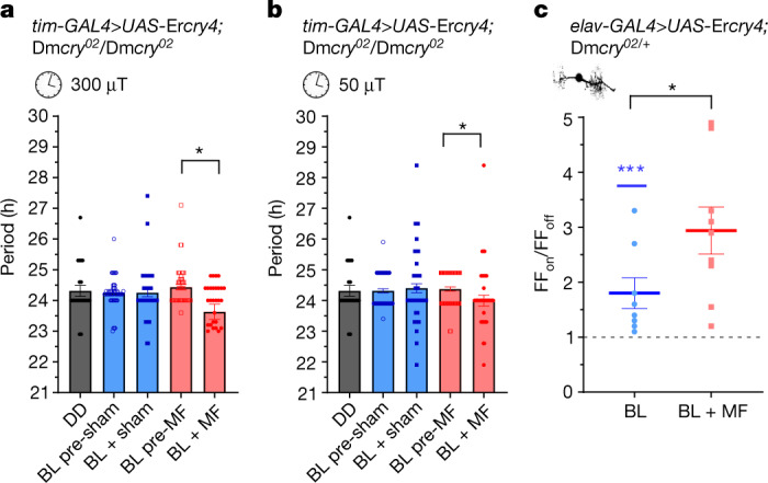
Many animals use Earth's magnetic field (also known as the geomagnetic field) for navigation1. The favoured mechanism for magnetosensitivity involves a blue-light-activated electron-transfer reaction between flavin adenine dinucleotide (FAD) and a chain of tryptophan residues within the photoreceptor protein CRYPTOCHROME (CRY). The spin-state of the resultant radical pair, and therefore the concentration of CRY in its active state, is influenced by the geomagnetic field2. However, the canonical CRY-centric radical-pair mechanism does not explain many physiological and behavioural observations2-8. Here, using electrophysiology and behavioural analyses, we assay magnetic-field responses at the single-neuron and organismal levels. We show that the 52 C-terminal amino acid residues of Drosophila melanogaster CRY, lacking the canonical FAD-binding domain and tryptophan chain, are sufficient to facilitate magnetoreception. We also show that increasing intracellular FAD potentiates both blue-light-induced and magnetic-field-dependent effects on the activity mediated by the C terminus. High levels of FAD alone are sufficient to cause blue-light neuronal sensitivity and, notably, the potentiation of this response in the co-presence of a magnetic field. These results reveal the essential components of a primary magnetoreceptor in flies, providing strong evidence that non-canonical (that is, non-CRY-dependent) radical pairs can elicit magnetic-field responses in cells.


Conflict of interest statement

The authors declare no competing interests.

Figures

Fig. 1
Fig. 1. Luc–CT is sufficient to support magnetosensitivity.
a, The electrophysiology set-up (permanent magnets are shown in red) (left). Top right, BL-exposure of aCC neurons expressing Dmcry increases action potential firing. Bottom right, the co-presence of MF (100 mT) potentiates the effect. The traces are from different preparations. APs, action potentials. b, The relative firing frequency (FF) of aCC neurons expressing Dmcry. BL increases firing 1.69-fold (t9 = 7.72, P ≤ 0.0001, n = 10, FFon/FFoff) compared with in the dark (dashed line). External MF (BL + MF, 100 mT) potentiates the effect to 2.41-fold (BL versus BL + MF, t18 = 3.2, P = 0.005, n = 10; Extended Data Fig. 1a). c, tim-GAL4>UAS-Luc-CT;Dmcry02/Dmcry02 shows period shortening under MF (left) (sham/MF × before/after exposure interaction (F1,377 = 7.6, P = 0.006, three-way ANOVA). n = 28 (DD), n = 108 (BL pre-sham), n = 93 (BL + sham), n = 104 (BL pre-MF), n = 90 (BL + MF). MF-exposed flies show a significantly shorter period. Four repeats showed the same period shortening under an MF (Extended Data Table 1). Right, Luc–CT supports BL-induced firing (1.4-fold, t9 = 4.01, P = 0.003, n = 10; Extended Data Fig. 1b) potentiated twofold after BL + MF treatment (BL versus BL + MF, t18 = 3.71, P = 0.002, n = 10). d, Luc–CT(W536F) revealed significant period shortening after exposure to an MF (left) (significant before/after exposure × MF/sham interaction, F1,198 = 5.1, P = 0.025, two-way ANOVA). n = 29 (DD), n = 47 (BL pre-sham), n = 51 (BL + sham), n = 52 (BL pre-MF), n = 52 (BL + MF). Post hoc tests are shown in Extended Data Table 1. Right, aCC neurons expressing Luc–CT(W536F) show a twofold change in BL-induced firing (t19 = 6.06, P ≤ 0.0001, n = 20). The response to BL + MF was variable, but greater than BL alone (2.69-fold, two-way ANOVA, replicates as factor, F1,16 = 5.09, P = 0.03, n = 20; Extended Data Fig. 1c). Controls are reported in Extended Data Figs. 3 and 4 and Extended Data Table 3. For FFon/FFoff data, the blue asterisks represent significance comparing before versus during BL exposure (same cells, paired two-tailed t-tests) and the black asterisks represent comparisons of BL versus BL + MF (different cells, unpaired two-tailed t-tests). The reported n value for each electrophysiological recording is derived from independent cells from biologically independent animals. The reported n values for each circadian period derives from biologically independent animals. Data are mean ± s.e.m. *P ≤ 0.05, **P ≤ 0.01, ***P ≤ 0.001.
Fig. 2
Fig. 2. Free FAD potentiates the effect of Luc–CT and, at high concentration, supports magnetosensitivity alone.
a, Exposing aCC neurons expressing Luc–CT to FAD (through the recording pipette) increases the response to BL (R2 = 0.71, F1,4 = 10.1, P = 0.03, linear regression). The co-presence of an MF (100 mT) potentiates the response (F1,9 = 9.06, P = 0.015, analysis of covariance (ANCOVA) model). n = 5 except for BL 30 µM and 50 µM FAD, for which n = 6. b, The addition of FAD (50 µM) supports BL sensitivity (Extended Data Fig. 5a), but not magnetosensitivity in the absence of Luc–CT (t18 = 0.521, P = 0.609, n = 10). c, The addition of riboflavin (50 µM) to aCC neurons expressing Luc–CT, supports the response to BL, but not MF potentiation (t18 = 0.12, P = 0.91). n = 10. d, Increased FAD (200 µM) in the Dmcry02-null background supports a BL-induced change in firing (1.27-fold, t19 = 4.29, P = 0.0004, n = 20; Extended Data Fig. 5c). The co-presence of an MF (n = 19) significantly potentiates this effect (1.84-fold, two-way ANOVA, F1,55 = 3.51, P = 0.066, Newman–Keuls post hoc P = 0.003). Riboflavin (200 µM) shows a similar BL effect (1.17-fold, t9 = 2.33, P = 0.045, n = 10; Extended Data Fig. 5c) but no MF potentiation (1.31-fold, Newman–Keuls post hoc, P = 0.67, n = 10). Raw data are reported in Extended Data Fig. 5. The blue asterisks represent significance values before versus during BL exposure (same cells, paired two-tailed t-tests) and the black asterisks represent comparisons of the BL versus BL + MF condition (different cells, unpaired two-tailed t-tests). NS, not significant. The reported n value for each electrophysiological recording is derived from independent cells from biologically independent animals. Data are mean ± s.e.m. NS, P ≥ 0.06.
Fig. 3
Fig. 3. Integrity of the CTT is required for it to facilitate magnetosensitivity.
a, Schematic of the domain structure of full-length DmCRY, including the CT (amino acids 491–542) and CTT (amino acids 521–542). The four Trp residues, presumed to be essential for the canonical RPM, are indicated by red asterisks. A putative PDZ-binding site (EEEV 528–531, shown in red) was mutated (Val531) DmCRY(V531K). The Trp residue (Trp536) mutated in Luc–CT(W536F) is shown in green. b, DmcryV531K expressed in clock neurons (tim-GAL4) does not support magnetosensitivity in the circadian period-shortening assay. A two-way ANOVA revealed no significant main effects or interaction effects (interaction, F1,52 = 0.09, P = 0.77). n = 53 (DD), n = 43 (BL pre-sham), n = 41 (BL + sham), n = 41 (BL pre-MF), n = 38 (BL + MF) (Extended Data Table 4a). c, Expression of DmcryV531K in aCC neurons is sufficient to support BL sensitivity (t9 = 2.934, P = 0.017, n = 10; Extended Data Fig. 7a) but not potentiation in the BL + MF condition (100 mT, t18 = 0.299, P = 0.768, n = 10). d, Expression of DmcryM, a truncated CRY variant lacking the terminal 19 amino acids, including the PDZ-binding motif (528–531), does not support sensitivity to a 300 µT MF (3 Hz, two-way ANOVA, F1,122 = 0.021, P = 0.89). n = 26 (DD), n = 31 (BL pre-sham), n = 31 (BL + sham), n = 32 (BL pre-MF), n = 32 (BL + MF) (Extended Data Table 4b). The blue asterisks represent significance values before versus during BL exposure (same cells, two-tailed paired t-tests) and the black asterisks represent comparisons of the BL versus BL + MF condition (different cells, unpaired two-tailed t-tests). The reported n value for each electrophysiological recording is derived from independent cells from biologically independent animals. The reported n values for each circadian period derives from biologically independent animals. Data are mean ± s.e.m.
Fig. 4
Fig. 4. ErCRY4 is sufficient to support MF sensitivity in Drosophila.
a, Expression of Ercry4 in Drosophila clock neurons (through tim-GAL4) results in significant period shortening in the presence of a 300 µT MF (3 Hz, two-way ANOVA interaction, F1,238 = 4.4, P = 0.036). n = 22 (DD), n = 49 (BL pre-sham), n = 41 (BL + sham), n = 49 (BL pre-MF), n = 30 (BL + MF) (Extended Data Table 5a–c). b, Period shortening is also present in a 50 µT MF (3 Hz, two-way ANOVA interaction, F1,237 = 3.97, P = 0.047). n = 22 (DD), n = 64 (BL pre-sham), n = 59 (BL + sham), n = 64 (BL pre-MF), n = 54 (BL + MF) (Extended Data Table 5d,e). c, Relative firing-frequency recordings of aCC motorneurons expressing Ercry4 for the BL versus BL + MF condition. BL exposure increases action potential firing (1.8-fold, t7 = 3.6, P = 0.0088, n = 8; Extended Data Fig. 7c), an effect that is potentiated by the co-presence of an MF (100 mT, 2.94-fold, BL versus BL + MF, t15 = 2.17, P = 0.046, n = 9). The blue asterisks represent significance values for before versus during BL exposure (same cells, paired two-tailed t-tests) and the black asterisks represent comparisons of BL versus BL + MF (different cells, unpaired two-tailed t-tests). The reported n value for each electrophysiological recording is derived from independent cells from biologically independent animals. The reported n value for each circadian period derives from biologically independent animals. Data are mean ± s.e.m.
Extended Data Fig. 1
Extended Data Fig. 1. Supporting electrophysiological data for main text Fig. 1.
(a). Raw action potential (AP) counts for aCC neurons expressing DmCRY, BL: n = 10, BL+MF: n = 10 (b). Luc-CT, BL: n = 10, BL+MF: n = 10 or (c). Luc-CTW536F, BL: n = 20, BL+MF: n = 20, recorded in the 15 s before and 15 s during exposure to BL or BL±MF. These data were used to derive firing-fold (FF) change reported in main text Fig. 1. Paired t-tests – two tailed – were used to compare AP counts before vs. during for cells exposed to BL (left hand graph) or to BL±MF exposure (right hand graph). MF-potentiation between the two groups was tested by unpaired t-test – two tailed (different cells). The figure number at the top of each panel represents the main text figure supported. The reported n for each electrophysiological recording is derived from independent cells from biologically independent animals. ns p ≥ 0.06, *p ≤ 0.05, **p ≤ 0.01, ***p ≤ 0.001. See main text Fig. 1 for FFon/FFoff ratio comparisons.
Extended Data Fig. 2
Extended Data Fig. 2. Supporting circadian data for main text Fig. 1.
(a). Showing a relative period shortening following expression of Luc-CT under MF (EMF- bars coloured red) compared to the BL+Sham exposure (bars coloured blue). (b). Box and whisker diagram (95% confidence limits) representation of the data shown in the upper panel, omitting DD. Error bars denote ±SEM, ns p ≥ 0.06, *p ≤ 0.05, **p ≤ 0.01, ***p ≤ 0.001.
Extended Data Fig. 3
Extended Data Fig. 3. Supporting electrophysiological data (controls).
(a). Averaged data for parental (control) GAL4 and UAS genotypes separately in a Dmcry-null mutant background. Without the GAL4 and UAS elements combined the Dmcry-transgene (under UAS control) is not expressed and no significant BL or BL+MF response is seen. A 2-way ANOVA of the controls in both BL or BL+MF showed no significant interaction (F(4,50) = 0.52, p = 0.719), n for all UAS-Dmcry transgenic controls in BL and BL+MF = 5. n for elav-GAL4; ; Dmcry02/Dmcry03 driver line BL and BL+MF = 10. (b-c). Raw AP counts (i) for each aCC neuron recorded in the 15 s before vs. the 15 s during BL or BL±MF exposure for expression of the respective UAS-Dmcry transgene stated (paired t-tests – two tailed). As an additional comparison of MF effect, unpaired t-tests – two tailed, were used to determine significant differences between raw AP counts for both ‘before’ exposure conditions (grey line), and for during BL vs. BL+MF exposure (purple line). The reported n for each electrophysiological recording is derived from independent cells from biologically independent animals. The reported n for each circadian period derives from biologically independent animals. Error bars denote ±SEM, ns p ≥ 0.06, *p ≤ 0.05, **p ≤ 0.01, ***p ≤ 0.001.
Extended Data Fig. 4
Extended Data Fig. 4. Supporting electrophysiological data (controls).
(a–c). Raw AP counts (i) for each aCC neuron recorded in the 15 s before vs. the 15 s during BL or BL±MF exposure for expression of the respective UAS-Dmcry transgene stated (paired t-tests – two tailed). (d). Data for a Dmcry null. As an additional comparison of MF effect, unpaired t-tests – two tailed were used to determine significant differences between raw AP counts for both ‘before’ exposure conditions (grey line), and for during BL vs. BL+MF exposure (purple line). The reported n for each electrophysiological recording is derived from independent cells from biologically independent animals. The reported n for each circadian period derives from biologically independent animals. Error bars denote ±SEM, ns p ≥ 0.06, *p ≤ 0.05, **p ≤ 0.01, ***p ≤ 0.001.
Extended Data Fig. 5
Extended Data Fig. 5. Supporting electrophysiological data for main text Fig. 2.
Raw AP counts for averaged data shown for flavin supplementation. (a). Dmcry02/Dmcry03 null cells supplemented with 50 µM FAD show a BL response (n = 10), but no MF potentiation (n = 10) compared to BL alone. (b). Cells expressing Luc-CT supplemented with riboflavin (50 µM) show no MF effect (n = 10) compared to BL (n = 10). (c). Supplemented FAD and riboflavin (200 µM) to Dmcry02/Dmcry03 null cells: FAD supports BL (n = 20) and BL+MF (n = 20) sensitivity, whilst riboflavin only supports BL sensitivity (see main text for BL and BL+MF comparisons). Paired t-tests – two tailed, were used to compare before vs. during for cells exposed to BL (left hand graph) or to BL±MF exposure (right hand graph). MF-potentiation between the two groups was tested by unpaired t-tests – two tailed (different cells). The reported n for each electrophysiological recording is derived from independent cells from biologically independent animals. ns p ≥ 0.06, *p ≤ 0.05, **p ≤ 0.01, ***p ≤ 0.001.
Extended Data Fig. 6
Extended Data Fig. 6. Structures of FAD and Riboflavin.
The molecular structure of the two flavin chromophores used. (a). Flavin Adenine Dinucleotide (FAD). Note the adenine diphosphate side chain of FAD (yellow oval), which facilitates the generation of an intramolecular, magnetically-sensitive RP. (b). Riboflavin, which is a metabolic precursor to FAD lacks the diphosphate side chain.
Extended Data Fig. 7
Extended Data Fig. 7. Supporting circadian and electrophysiology data for main text Figs. 3 and 4.
(a). Raw AP counts for each aCC neuron recorded expressing DmcryV531K in the 15 s before vs. the following 15 s during BL±MF, from which the average firing-fold change was derived for data reported in main text Fig. 3c. BL: n = 10, BL+MF: n = 10. Paired t-tests – two tailed, were used to compare before vs. during for cells exposed to BL (left hand graph) or to BL±MF exposure (right hand graph). MF-potentiation between the two groups was tested by unpaired t-tests – two tailed (different cells). (b). DmcryM flies do not undergo period shortening following exposure to a 50 µT MF (DD n = 26, BL Pre-Sham n = 51, BL+Sham n = 42, BL Pre-MF n = 53, BL+MF n = 38). (c). Raw AP counts for BL and BL+MF exposure for ErCRY4, showing an increase in neuronal excitability to both stimuli (BL: n = 8, BL+MF: n = 9). Paired t-tests – two tailed were used to compare before vs. during for cells exposed to BL (left hand graph) or to BL±MF exposure (right hand graph). MF-potentiation between the two groups was tested by unpaired t-tests – two tailed (different cells). The reported n for each electrophysiological recording is derived from independent cells from biologically independent animals. The reported n for each circadian period derives from biologically independent animals. Error bars denote ±SEM, ns p ≥ 0.06, *p ≤ 0.05, **p ≤ 0.01, ***p ≤ 0.001.

References

    1. Wiltschko W, Wiltschko R. Magnetic orientation and magnetoreception in birds and other animals. J. Comp. Physiol. A. 2005;191:675–693. doi: 10.1007/s00359-005-0627-7. - DOI - PubMed
    1. Hore PJ, Mouritsen H. The radical-pair mechanism of magnetoreception. Annu. Rev. Biophys. 2016;45:299–344. doi: 10.1146/annurev-biophys-032116-094545. - DOI - PubMed
    1. Fedele G, et al. Genetic analysis of circadian responses to low frequency electromagnetic fields in Drosophila melanogaster. PLoS Genet. 2014;10:e1004804. doi: 10.1371/journal.pgen.1004804. - DOI - PMC - PubMed
    1. Giachello CNG, Scrutton NS, Jones AR, Baines RA. Magnetic fields modulate blue-light-dependent regulation of neuronal firing by cryptochrome. J. Neurosci. 2016;36:10742–10749. doi: 10.1523/JNEUROSCI.2140-16.2016. - DOI - PMC - PubMed
    1. Schwarze S, et al. Weak broadband electromagnetic fields are more disruptive to magnetic compass orientation in a night-migratory songbird (Erithacus rubecula) than strong narrow-band fields. Front. Behav. Neurosci. 2016;10:55. doi: 10.3389/fnbeh.2016.00055. - DOI - PMC - PubMed

Publication types

LinkOut - more resources