

Identification of galactofuranose antigens such as galactomannoproteins and fungal-type galactomannan from the yellow koji fungus (Aspergillus oryzae)
- PMID: 36814571
- PMCID: PMC9939772
- DOI: 10.3389/fmicb.2023.1110996
Identification of galactofuranose antigens such as galactomannoproteins and fungal-type galactomannan from the yellow koji fungus (Aspergillus oryzae)
Abstract
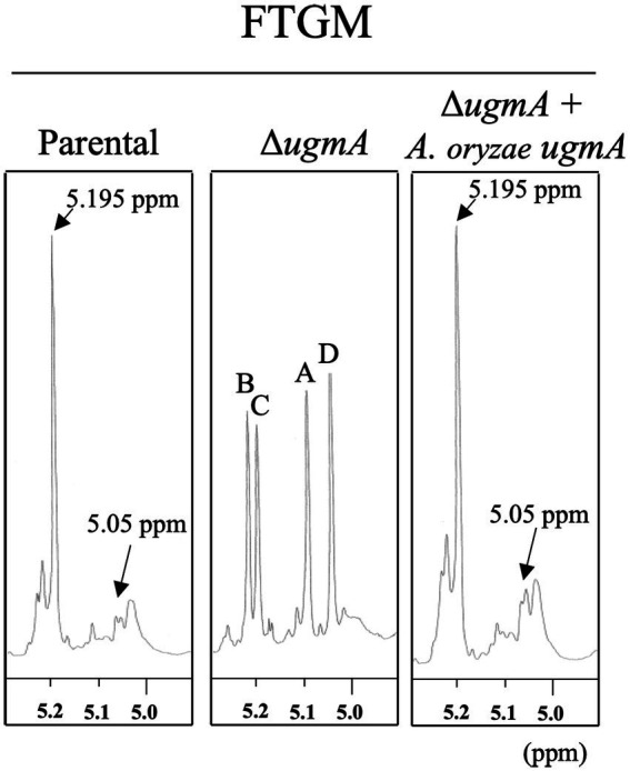
Filamentous fungi belonging to the genus Aspergillus are known to possess galactomannan in their cell walls. Galactomannan is highly antigenic to humans and has been reported to be involved in the pathogenicity of pathogenic filamentous fungi, such as A. fumigatus, and in immune responses. In this study, we aimed to confirm the presence of D-galactofuranose-containing glycans and to clarify the biosynthesis of D-galactofuranose-containing glycans in Aspergillus oryzae, a yellow koji fungus. We found that the galactofuranose antigen is also present in A. oryzae. Deletion of ugmA, which encodes UDP-galactopyranose mutase in A. oryzae, suppressed mycelial elongation, suggesting that D-galactofuranose-containing glycans play an important role in cell wall integrity in A. oryzae. Proton nuclear magnetic resonance spectrometry revealed that the galactofuranose-containing sugar chain was deficient and that core mannan backbone structures were present in ΔugmA A. oryzae, indicating the presence of fungal-type galactomannan in the cell wall fraction of A. oryzae. The findings of this study provide new insights into the cell wall structure of A. oryzae, which is essential for the production of fermented foods in Japan.
Keywords: Aspergillus oryzae; UDP-galactopyranose mutase (UGM); cell wall; fungal-type galactomannan; galactofuranose.
Copyright © 2023 Kadooka, Tanaka, Hira, Maruyama, Goto and Oka.
Conflict of interest statement
The authors declare that the research was conducted in the absence of any commercial or financial relationships that could be construed as a potential conflict of interest.
Figures


Similar articles
-
Identification of an α-(1→6)-Mannosyltransferase Contributing To Biosynthesis of the Fungal-Type Galactomannan α-Core-Mannan Structure in Aspergillus fumigatus.mSphere. 2022 Dec 21;7(6):e0048422. doi: 10.1128/msphere.00484-22. Epub 2022 Nov 29. mSphere. 2022. PMID: 36445154 Free PMC article.
-
UDP-Galactopyranose Mutase Mediates Cell Wall Integrity, Polarity Growth, and Virulence in Fusarium graminearum.Appl Environ Microbiol. 2023 Feb 28;89(2):e0123522. doi: 10.1128/aem.01235-22. Epub 2023 Jan 19. Appl Environ Microbiol. 2023. PMID: 36656025 Free PMC article.
-
Transcriptomic and molecular genetic analysis of the cell wall salvage response of Aspergillus niger to the absence of galactofuranose synthesis.Cell Microbiol. 2016 Sep;18(9):1268-84. doi: 10.1111/cmi.12624. Epub 2016 Jul 29. Cell Microbiol. 2016. PMID: 27264789 Free PMC article.
-
Biosynthesis of galactomannans found in filamentous fungi belonging to Pezizomycotina.Biosci Biotechnol Biochem. 2018 Feb;82(2):183-191. doi: 10.1080/09168451.2017.1422383. Epub 2018 Jan 15. Biosci Biotechnol Biochem. 2018. PMID: 29334321 Review.
-
Structure, mechanism, and dynamics of UDP-galactopyranose mutase.Arch Biochem Biophys. 2014 Feb 15;544:128-41. doi: 10.1016/j.abb.2013.09.017. Epub 2013 Oct 3. Arch Biochem Biophys. 2014. PMID: 24096172 Free PMC article. Review.
Cited by
-
Substrate binding and catalytic mechanism of UDP-α-D-galactofuranose: β-galactofuranoside β-(1→5)-galactofuranosyltransferase GfsA.PNAS Nexus. 2024 Oct 25;3(11):pgae482. doi: 10.1093/pnasnexus/pgae482. eCollection 2024 Nov. PNAS Nexus. 2024. PMID: 39507050 Free PMC article.
-
Functional study of two ER localized sterol C-14 reductases in Aspergillus oryzae.3 Biotech. 2024 May;14(5):136. doi: 10.1007/s13205-024-03988-7. Epub 2024 Apr 25. 3 Biotech. 2024. PMID: 38682096 Free PMC article.
-
The postbiotic potential of Aspergillus oryzae - a narrative review.Front Microbiol. 2024 Oct 23;15:1452725. doi: 10.3389/fmicb.2024.1452725. eCollection 2024. Front Microbiol. 2024. PMID: 39507340 Free PMC article. Review.
-
Research advances in fungal polysaccharides: production, extraction, characterization, properties, and their multifaceted applications.Front Cell Infect Microbiol. 2025 Jun 9;15:1604184. doi: 10.3389/fcimb.2025.1604184. eCollection 2025. Front Cell Infect Microbiol. 2025. PMID: 40552121 Free PMC article. Review.
References
-
- Afroz S., El-Ganiny A. M., Sanders D. A., Kaminskyj S. G. (2011). Roles of the Aspergillus nidulans UDP-galactofuranose transporter, UgtA in hyphal morphogenesis, cell wall architecture, conidiation, and drug sensitivity. Fungal Genet. Biol. 48, 896–903. doi: 10.1016/j.fgb.2011.06.001, PMID: - DOI - PubMed
-
- Alam M. K., van Straaten K. E., Sanders D. A., Kaminskyj S. G. (2014). Aspergillus nidulans cell wall composition and function change in response to hosting several Aspergillus fumigatus UDP-galactopyranose mutase activity mutants. PLoS One 9:e85735. doi: 10.1371/journal.pone.0085735, PMID: - DOI - PMC - PubMed
-
- Arentshorst M., de Lange D., Park J., Lagendijk E. L., Alazi E., van den Hondel C. A. M. J. J., et al. . (2020). Functional analysis of three putative galactofuranosyltransferases with redundant functions in galactofuranosylation in Aspergillus Niger. Arch. Microbiol. 202, 197–203. doi: 10.1007/s00203-019-01709-w, PMID: - DOI - PMC - PubMed
-
- Chihara Y., Tanaka Y., Izumi M., Hagiwara D., Watanabe A., Takegawa K., et al. . (2020). Biosynthesis of β-(1→5)-galactofuranosyl chains of fungal-type and O-mannose-type galactomannans within the invasive pathogen Aspergillus fumigatus. mSphere 5, e00770–e00719. doi: 10.1128/mSphere.00770-19, PMID: - DOI - PMC - PubMed
LinkOut - more resources
Full Text Sources