Case Reports
. 2023 Feb 19;11(2):e6918.
doi: 10.1002/ccr3.6918. eCollection 2023 Feb.

Radius-Maumenee syndrome: A case series with a long-term follow-up

Affiliations
Case Reports

Radius-Maumenee syndrome: A case series with a long-term follow-up

Eva Elksne et al. Clin Case Rep. .

Abstract

The aim of the case series is to highlight the surgical challenges experienced like failed intervention, choroidal effusion, a postoperative cystoid macular oedema, and describe treatment options for Radius-Maumenee syndrome. Authors reported on 3 bilateral cases of Radius-Maumenee syndrome which underwent medical treatment, trabeculectomy with Mitomycin C, implantation with XEN45, Ahmed glaucoma valve, Baerveldt glaucoma implant, and cyclophotocoagulation.

Keywords: Radius‐Maumenee syndrome; ab‐interno subconjunctival gel stent; glaucoma drainage device; idiopathic elevated episcleral venous pressure; secondary open‐angle glaucoma; trabeculectomy with mitomycin C.


Conflict of interest statement

The authors declare no potential conflicts of interest with respect to the research, authorship, and/or publication of this article.

Figures

FIGURE 1
FIGURE 1
Slit‐lamp appearance of dilated episcleral vessels in both eyes. No signs of active inflammation of ocular surface were observed.
FIGURE 2
FIGURE 2
Patient's optic nerve head and surrounded retinal vasculature changes in the right (OD) and left eye (OS). A cup‐to‐disc ratio was 0.7 in both eyes.
FIGURE 3
FIGURE 3
Visual field of both eyes. In the right eye (OD), a paracentral scotoma was noticed mostly due to changes of post‐cystoid macular oedema and epiretinal membrane. In the left eye (OS), scotoma affected the nasal part. The mean deviation was −9.31 dB (OD) and −6.75 dB (OS).
FIGURE 4
FIGURE 4
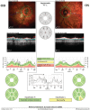
Peripapillary retinal nerve fiber layer for both eyes. Significant artifacts were seen in the scan for the right eye (OD). The temporal inferior segment showed borderline value in the left eye (OS).
FIGURE 5
FIGURE 5
Location of subconjunctival gel stent on the first post‐surgical day. The anterior segment optical coherence tomography revealed an appropriate position of the gel implant. A bleb was rather flat.
FIGURE 6
FIGURE 6
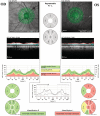
Visual field and peripapillary retinal nerve fiber layer (pRNFL) in the left eye (OS). pRNFL from the baseline examination revealed changes in inferior quadrants. The pRNFL for the right eye (OD) was not available. The visual field of the OS showed a mean deviation of −8.92 decibels (dB). The visual field for the OD was not possible to be performed.
FIGURE 7
FIGURE 7
Visual field for both eyes. The right eye (OD) was within normal limits. However, the left eye (OS) displayed significant glaucomatous damage (MD −14.14 dB).
FIGURE 8
FIGURE 8
Optical coherence tomography for optic nerve head measuring peripapillary retinal nerve fiber layer (pRNFL). The right eye (OD) had pRNFL within normal limits. The left eye (OS) demonstrated advance glaucomatous changes.
FIGURE 9
FIGURE 9
Visual fields at the last follow‐up for both eyes. The right eye (OD) did not indicate significant changes. The left eye (OS) showed progression (MD −17.10 dB).
FIGURE 10
FIGURE 10
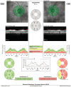
Peripapillary retinal nerve fiber layer for both eyes. No significant changes at the last follow‐up were observed in the right eye (OD). The left eye (OS) reflected slight changes.
FIGURE 11
FIGURE 11
Visual field at the last follow‐up. The right eye (OD) was without glaucomatous damages. The left eye (OS) reflected the inferior arcuate scotoma (MD −6.89 dB).

References

    1. Minas TF, Podos SM. Familial glaucoma associated with elevated episcleral venous pressure. Arch Ophthalmol. 1968;80(2):202‐208. doi:10.1001/ARCHOPHT.1968.00980050204010 - DOI - PubMed
    1. Radius RL, Maumenee AE. Dilated episcleral vessels and open‐angle glaucoma. Am J Ophthalmol. 1978;86(1):31‐35. doi:10.1016/0002-9394(78)90010-7 - DOI - PubMed
    1. Goldmann H. Out‐flow pressure, minute volume and resistance of the anterior chamber flow in man. Doc Ophthalmol. 1951;5‐6(1):278‐356. doi:10.1007/BF00143664 - DOI - PubMed
    1. Kass MA, Heuer DK, Higginbotham EJ, et al. The ocular hypertension treatment study: a randomized trial determines that topical ocular hypotensive medication delays or prevents the onset of primary open‐angle glaucoma. Arch Ophthalmol. 2002;120:701. doi:10.1001/archopht.120.6.701 - DOI - PubMed
    1. Jorgensen JS, Guthoff R. The role of episcleral venous pressure in the development of secondary glaucomas. Klin Monbl Augenheilkd. 1988;193(5):471‐475. doi:10.1055/S-2008-1050284 - DOI - PubMed

Publication types

LinkOut - more resources