

AFM evaluation of a humanized recombinant antibody affecting C. auris cell wall and stability
- PMID: 36814881
- PMCID: PMC9940460
- DOI: 10.1039/d2ra07217c
AFM evaluation of a humanized recombinant antibody affecting C. auris cell wall and stability
Abstract
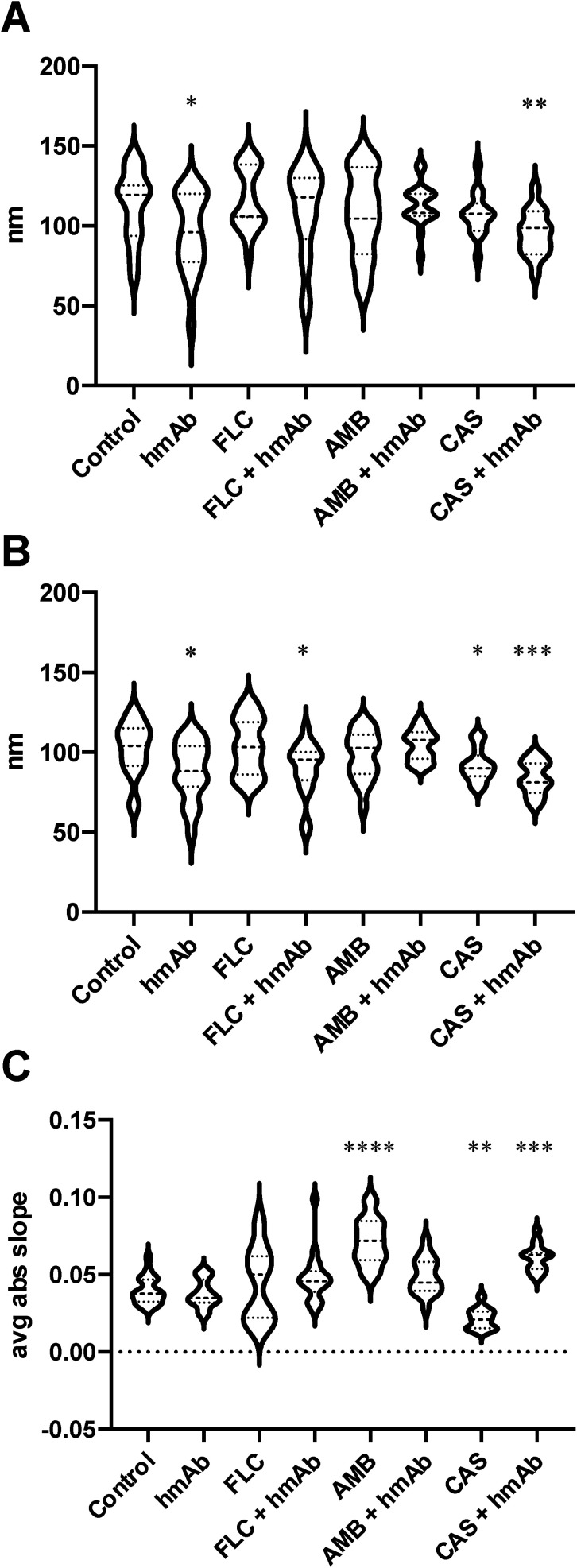
Fungal infections are increasingly impacting on the health of the population and particularly on subjects with a compromised immune system. The resistance phenomenon and the rise of new species carrying sometimes intrinsic and multi-drug resistance to the most commonly used antifungal drugs are greatly concerning healthcare organizations. As a result of this situation, there is growing interest in the development of therapeutic agents against pathogenic fungi. In particular, the Candida genus is responsible for severe life-threatening infections and among its species, C. auris is considered an urgent threat by the Center for Disease Control and Prevention, and is one of the three leading causes of morbidity and mortality worldwide. H5K1 is a humanized monoclonal antibody (hmAb) that selectively binds to β-1,3-glucans, vital components of the fungal cell wall. It has been previously demonstrated that it is active against Candida species, especially against C. auris, reaching its greatest potential when combined with commercially available antifungal drugs. Here we used atomic force microscopy (AFM) to assess the effects of H5K1, alone and in combination with fluconazole, caspofungin and amphotericin B, on C. auris cells. Through an extensive exploration we found that H5K1 has a significant role in the perturbation and remodeling of the fungal cell wall that is reflected in the loss of whole cell integrity. Moreover, it contributes substantially to the alterations in terms of chemical composition, stiffness and roughness induced specifically by caspofungin and amphotericin B. In addition to this, we demonstrated that AFM is a valuable technique to evaluate drug-microorganism interaction.
This journal is © The Royal Society of Chemistry.
Conflict of interest statement
Diatheva s.r.l. supported the study but was not involved in carrying out or managing the investigation, nor was it involved in the analysis and interpretation of data, and in the preparation of the manuscript. T. D. M. is an employee of Diatheva s.r.l., M. Magnani holds shares in Diatheva s.r.l. This does not alter our adherence to RSC policies on sharing data and materials. All the other authors do not have any conflict of interest.
Figures


Similar articles
-
Caspofungin-resistance in Candida auris is cell wall-dependent phenotype and potential prevention by zinc oxide nanoparticles.Med Mycol. 2021 Dec 3;59(12):1243-1256. doi: 10.1093/mmy/myab059. Med Mycol. 2021. PMID: 34612496
-
A new humanized antibody is effective against pathogenic fungi in vitro.Sci Rep. 2021 Sep 30;11(1):19500. doi: 10.1038/s41598-021-98659-5. Sci Rep. 2021. PMID: 34593880 Free PMC article.
-
In Vitro Interaction and Killing-Kinetics of Amphotericin B Combined with Anidulafungin or Caspofungin against Candida auris.Pharmaceutics. 2021 Aug 25;13(9):1333. doi: 10.3390/pharmaceutics13091333. Pharmaceutics. 2021. PMID: 34575409 Free PMC article.
-
Candida and candidaemia. Susceptibility and epidemiology.Dan Med J. 2013 Nov;60(11):B4698. Dan Med J. 2013. PMID: 24192246 Review.
-
Caspofungin: a major breakthrough in treatment of systemic fungal infections.J Assoc Physicians India. 2006 Dec;54:943-8. J Assoc Physicians India. 2006. PMID: 17334012 Review.
Cited by
-
Nanotechnology meets medicine: applications of atomic force microscopy in disease.Biophys Rev. 2025 Apr 3;17(2):359-384. doi: 10.1007/s12551-025-01306-w. eCollection 2025 Apr. Biophys Rev. 2025. PMID: 40376402 Free PMC article. Review.
-
Next-generation antifungal drugs: Mechanisms, efficacy, and clinical prospects.Acta Pharm Sin B. 2025 Aug;15(8):3852-3887. doi: 10.1016/j.apsb.2025.06.013. Epub 2025 Jun 23. Acta Pharm Sin B. 2025. PMID: 40893690 Free PMC article. Review.
-
Old and new strategies in therapy and diagnosis against fungal infections.Appl Microbiol Biotechnol. 2024 Jan 19;108(1):147. doi: 10.1007/s00253-023-12884-8. Appl Microbiol Biotechnol. 2024. PMID: 38240822 Free PMC article. Review.
-
Antifungal immunity: advances in PRR recognition, adaptive responses, and immune-based therapies.Sci China Life Sci. 2025 Aug;68(8):2206-2224. doi: 10.1007/s11427-024-2835-y. Epub 2025 Mar 5. Sci China Life Sci. 2025. PMID: 40055278 Review.
-
Current Perspectives of Antifungal Therapy: A Special Focus on Candida auris.J Fungi (Basel). 2024 Jun 6;10(6):408. doi: 10.3390/jof10060408. J Fungi (Basel). 2024. PMID: 38921394 Free PMC article. Review.
References
-
- Koehler P. Stecher M. Cornely O. A. Koehler D. Vehreschild M. J. G. T. Bohlius J. Wisplinghoff H. Vehreschild J. J. Clin. Microbiol. Infect. 2019;25:1200–1212. - PubMed
-
- Pappas P. G. Lionakis M. S. Arendrup M. C. Ostrosky-Zeichner L. Kullberg B. J. Nat. Rev. Dis. Primer. 2018;4:18026. - PubMed
-
- Candida auris|Candida auris|Fungal Diseases|CDC, https://www.cdc.gov/fungal/candida-auris/index.html, accessed October 31, 2021
LinkOut - more resources
Full Text Sources
Research Materials
Miscellaneous