. 2023 Feb 6:14:1100499.
doi: 10.3389/fimmu.2023.1100499. eCollection 2023.

FcγRI FG-loop functions as a pH sensitive switch for IgG binding and release

Affiliations

FcγRI FG-loop functions as a pH sensitive switch for IgG binding and release

Jinghua Lu et al. Front Immunol. .

Abstract

Understanding the molecular mechanism underlying the hierarchic binding between FcγRs and IgG antibodies is critical for therapeutic antibody engineering and FcγR functions. The recent determination of crystal structures of FcγRI-Fc complexes, however, resulted in two controversial mechanisms for the high affinity receptor binding to IgG. Here, we describe high resolution structures of a bovine FG-loop variant of FcγRI in complex with the Fc fragment of IgG1 crystallized in three different conditions at neutral pH, confirming the characteristic FG loop-Fc interaction is critical to the high affinity immunoglobulin binding. We showed that the FcγRI D2-domain FG-loop functioned as a pH-sensing switch for IgG binding. Further live cell imaging of FcγRI-mediated internalization of immune complexes showed a pH sensitive temporal-spatial antibody-antigen uptake and release. Taken together, we demonstrate that the structures of FcγRI-Fc crystallized at neutral and acidic pH, respectively, represent the high and low affinity binding states of the receptor for IgG uptake and release. These results support a role for FcγRI in antigen delivery, highlight the importance of Fc glycan in antibody binding to the high affinity receptor and provide new insights to future antibody engineering.

Keywords: Fc-glycan recognition; antibody recognition; human FcγRI; live cell tracking of FcγRI-antibody endocytosis; pH sensitive binding; receptor FG-loop.


Conflict of interest statement

The authors declare that the research was conducted in the absence of any commercial or financial relationships that could be construed as a potential conflict of interest.

Figures

Figure 1
Figure 1
Sequence analysis of FG loop residues in FcγRs D2 domain. (A) Logo display of FG loop residues among all FcγRs. (B) Usage of positively charged residues K,H,R, and other residues on FG loop. (C) Binding affinities of human and bovine H174R variant FcγRI to human IgG1.
Figure 2
Figure 2
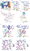
Structures of H174R FcγRI in complex with IgG1 Fc. (A) Structure superposition of three structures of FcγRI H174R variant in complex with Fc (PDB code: 8DIR, 8DIN and 8DJ7). All structural alignments were based on D2 domain of FcγRI. D1, D2 and D3 domain of FcγRI were highlighted as cartoons in green, blue, and magenta, respectively. The Fc chain (A, B) were colored in orange and cyan, respectively, with glycosylation attached to Asn 297 shown in stick model. (B, D) Structures of individual FcγRI-Fc complexes, 8DIR (B), 4X4M (C) and 4W4O (D). The acetate and Zn atom bound to H174 are shown in spheres. E-F) Interface contact region between FcγRI D2 domain and Fc-A chain as observed in 8DIR (E) and 4W4O (F). (G, H) Interface between H174R FcγRI D2 domain and Fc-B chains observed in 8DIR (G) and 4W4O (H). FcγRI residues were shown as magenta sticks and labelled in black. Residues from Fc chains were shown as cyan or green sticks and labelled in red.
Figure 3
Figure 3
Sequence alignment of FcγR D2 domains. The interface residues in FcγRI-Fc complex are shaded in cyan or blue. V132 and Y176 of FcγRI are highlighted in the red box. They correspond to L118 and V163 in FcγRIII.
Figure 4
Figure 4
Interactions between FG loop and Fc. (A) (2Fo-Fc) electron density map contoured at 1σ showing R174 of FcγRI forming salt-bridge and hydrogen bond contacts with D265 and glycan, respectively, on Fc. (B) Stick model depicting R174 contacting D265 and glycan on Fc in the H174R variant FcγRI-Fc complex. (C) H174 in 4W4O is situated too far from D265 and glycan but instead is coordinated by a Zn ion. (D, E) Ligplot and cartoon drawing to illustrate the interactions from R174 of the receptor to D265 and glycans on Fc-B chain. (F, G) interactions between R174 and Fc observed in 8DIR (F) are replaced by water molecules in 4W4O (G). (H, I) Contacts of Fc-B chain L235 with the receptor hydrophobic pocket as observed in 8DIR (H) and 4W4O (I).
Figure 5
Figure 5
Binding of wildtype or mutant recombinant FcγRI and FcγRIII to IgG1. (A, B) Biolayer interferometry (BLI) measurements of FcγRI binding to the wildtype and D265R mutant of VRC01. (C, D) SPR measurements of FcγRI V132L/Y176V (C) and 173KHR175 to 173EEE175 (D) mutants binding to IgG1. (E–H) SPR measurements of FcγRIII WT and mutants binding to IgG1.
Figure 6
Figure 6
pH sensitive binding between FcγRI and IgG. (A) Binding of FcγRI to IgG1 at pH 7.5, 4.6 and 4.0. (B) pH-titration of the FcγRI wildtype, H174R and H174E mutants binding to IgG1. (C) Binding of BSA and FcγRI to rabbit polyclonal anti-BSA antibody at pH7.5, 4.6 and 4.0. (D) Binding of FcγRII and FcγRIII to IgG1 at pH7 and 4.6.
Figure 7
Figure 7
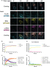
Temporal-spatial interaction between FcγRI and IgG during internalization of immune complex (IC). (A) Time lapse of fluorescent images showing FcγRI-mediated phagocytosis of IC by live monocyte-derived macrophages. The boxed area (10μm x 10 μm) was further enlarged to show the distribution of IgG(Alexa633), BSA(pHrodo) and FcγRI(Alexa488) with a scale bar of 5μm. (B, D) Time-dependent fluorescent intensities of labeled FcγRI, BSA and anti-BSA during FcγRI-mediated internalization of BSA-anti-BSA immune complex (B) or its control without anti-BSA (D). The fluorescence of pHrodo increased as the pH declined. More BSA are internalized in FcγRI-mediated process (B) than spontaneous internalization (D). (C–E) Manders coefficient analyses of live cell images for colocalization between pairs of fluorescent probes during FcγRI-mediated internalization of BSA-anti-BSA immune complex (C) or its control without anti-BSA (E). Manders coefficients were calculated for pairwise colocalization between FcγRI, BSApHrodo, and anti-BSA antibody with respect to either fluorescent probe. For example, FcγRI-α-BSA refers to the percentage of FcγRI (cyan) that colocalizes with anti-BSA (magenta), while α-BSA-FcγRI is magenta-cyan and is the percentage of α-BSA that colocalizes with FcγRI. While BSA and anti-BSA remain colocalized throughout the duration of the live cell imaging, FcγRI showed optimal colocalization with anti-BSA at the beginning and dissociated at a later time. FcγRI was stained by Alexa488 antibody(cyan), BSA protein was labeled with pHrodo(yellow), and anti-BSA antibody was labeled with Alexa633 (magenta).
Figure 8
Figure 8
A cartoon showing the pH dependent IgG binding and release by FcγRI.

Similar articles

Cited by

References

    1. Chenoweth AM, Trist HM, Tan PS, Wines BD, Hogarth PM. The high-affinity receptor for IgG, FcgammaRI, of humans and non-human primates. Immunol Rev (2015) 268:175–91. doi: 10.1111/imr.12366 - DOI - PubMed
    1. Bruhns P, Iannascoli B, England P, Mancardi DA, Fernandez N, Jorieux S, et al. . Specificity and affinity of human fcgamma receptors and their polymorphic variants for human IgG subclasses. Blood (2009) 113:3716–25. doi: 10.1182/blood-2008-09-179754 - DOI - PubMed
    1. Nimmerjahn F, Ravetch JV. Fcgamma receptors as regulators of immune responses. Nat Rev Immunol (2008) 8:34–47. doi: 10.1038/nri2206 - DOI - PubMed
    1. Lux A, Yu X, Scanlan CN, Nimmerjahn F. Impact of immune complex size and glycosylation on IgG binding to human FcgammaRs. J Immunol (2013) 190:4315–23. doi: 10.4049/jimmunol.1200501 - DOI - PubMed
    1. Hogarth PM. Fc receptors: Introduction. Immunol Rev (2015) 268:1–5. doi: 10.1111/imr.12372 - DOI - PubMed

Publication types

LinkOut - more resources