

doi: 10.1016/j.eats.2022.09.001.
eCollection 2023 Jan.
Arthroscopic Saucerization and Repair of a Torn Medial Discoid Meniscus
Affiliations
- PMID: 36814971
- PMCID: PMC9939740
- DOI: 10.1016/j.eats.2022.09.001
Item in Clipboard
Arthroscopic Saucerization and Repair of a Torn Medial Discoid Meniscus
Arthrosc Tech.
.
Abstract
A discoid meniscus is a congenital abnormality that usually affects the lateral meniscus, leading to instability and increased risk of tearing. A discoid medial meniscus is an extremely rare pathology that is seldom described in literature. In this report, we present the technique of operative treatment of a symptomatic, torn discoid medial meniscus. The meniscus is saucerized to 6-8 mm of stable rim, and the inside-out technique is used as the modality of meniscal fixation. Although a discoid medial meniscus is an uncommon finding, all treating surgeons should be aware of the possibility during surgical intervention.
© 2022 The Authors.
Figures
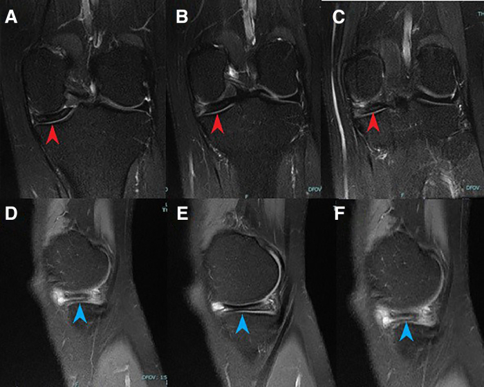
Left knee, preoperative T2 MRI coronal cuts (A-C) demonstrate increased meniscal body width (red arrowheads) and sagittal cuts (D-F) show increased central thickness consistent with discoid meniscus (blue arrowheads).
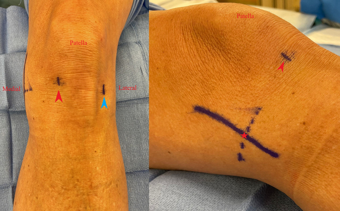
Left knee, outside view of arthroscopic incision sites marked prior to fluid insufflation to prevent distortion. The red arrowhead marks the inferomedial portal site, the blue arrowhead marks the inferolateral portal site, and the red star marks the medial incision site.

Left knee, arthroscopic view through the inferomedial (A) and inferolateral (B) portals visualizing a left discoid medial meniscus. Stability is tested by placing a hook probe through the tear and pulling the posterior horn anteriorly. Anterior subluxation of the posterior horn is indicative of a meniscocapsular injury.
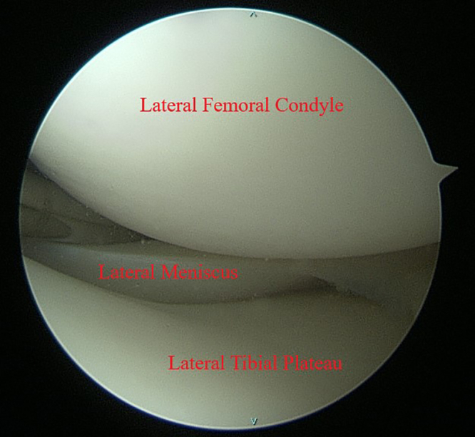
Left knee, arthroscopic view through the inferolateral portal. The lateral meniscus is visualized during the diagnostic arthroscopy to assess for any additional pathologies that need to be addressed.
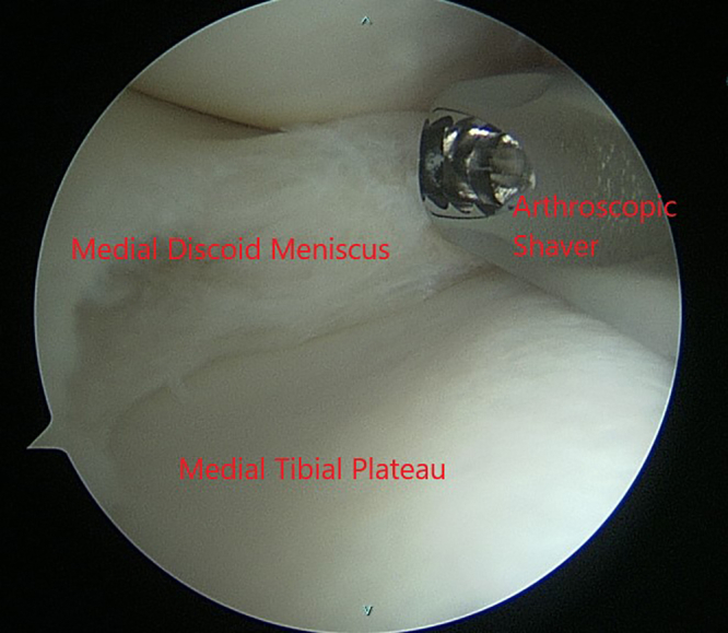
Left knee, arthroscopic view through the inferomedial portal. The discoid medial meniscus is saucerized to 6-8 mm of stable rim using an arthroscopic punch and shaver.

Left knee, arthroscopic view of the saucerized medial discoid meniscus through the inferolateral portal. A hook probe (blue arrowhead) is used to assess for meniscal stability after saucerization is complete.

Left knee, arthroscopic view through the inferolateral portal using a 70° scope. The Guillquist maneuver can be used to better visualize the posterior horn of the medial discoid meniscus through the notch to assess meniscocapsular injury.

Left knee, arthroscopic view through the inferolateral portal. Vertical mattress sutures are placed posteriorly to secure the meniscus to the posterior capsule and carried on through the meniscal body as needed (blue arrowheads). Absence of subluxation of the meniscus should be noted upon completion of the repair.

Left knee, arthroscopic view through the inferolateral portal. An anomalous insertion of the discoid medial meniscus anterior horn on the anterior cruciate ligament.
Similar articles
-
Arthroscopic Saucerization of a Symptomatic Posterior Horn Tear in a Discoid Medial Meniscus.Ochsner J. 2021 Spring;21(1):115-118. doi: 10.31486/toj.19.0125. Ochsner J. 2021. PMID: 33828437 Free PMC article.
-
Symptomatic Bilateral Torn Discoid Medial Meniscus Treated with Saucerization and Suture.Case Rep Orthop. 2016;2016:8487194. doi: 10.1155/2016/8487194. Epub 2016 Aug 30. Case Rep Orthop. 2016. PMID: 27656305 Free PMC article.
-
Saucerization and suture of symptomatic bilateral medial discoid meniscus in a 13 years old male football player: a case report and literature review.Orthop Rev (Pavia). 2022 Apr 25;14(2):33699. doi: 10.52965/001c.33699. eCollection 2022. Orthop Rev (Pavia). 2022. PMID: 35774929 Free PMC article.
-
Symptomatic Complete Discoid Medial Meniscus Completely Coalesced with the Anterior Cruciate Ligament: A Case Report and Literature Review.Orthop Surg. 2022 Sep;14(9):2391-2395. doi: 10.1111/os.13377. Epub 2022 Aug 1. Orthop Surg. 2022. PMID: 35913195 Free PMC article. Review.
-
The discoid lateral meniscus in children: a narrative review of pathology, diagnosis and treatment.Ann Jt. 2022 Oct 15;7:38. doi: 10.21037/aoj-21-31. eCollection 2022. Ann Jt. 2022. PMID: 38529145 Free PMC article. Review.
Cited by
-
Medial Discoid Meniscus: A Rare Case Report.Cureus. 2023 Jun 5;15(6):e39971. doi: 10.7759/cureus.39971. eCollection 2023 Jun. Cureus. 2023. PMID: 37416007 Free PMC article.
References
-
- Cave E.F., Staples O.S. Congenital discoid meniscus. A cause of internal derangement of the knee. Am J Surg. 1941;54:371–376.
-
- Woodmass J.M., LaPrade R.F., Sgaglione N.A., Nakamura N., Krych A.J. Meniscal repair: Reconsidering indications, techniques, and biologic augmentation. J Bone Joint Surg Am. 2017;99:1222–1231. - PubMed
-
- Kocher M.S., Logan C.A., Kramer D.E. Discoid lateral meniscus in children: Diagnosis, management, and outcomes. J Am Acad Orthop Surg. 2017;25:736–743. - PubMed
-
- Tachibana Y., Yamazaki Y., Ninomiya S. Discoid medial meniscus. Arthroscopy. 2003;19:E12–E18. - PubMed
-
- Feroe A.G., Hussain Z.B., Stupay K.L., et al. Surgical management of medial discoid meniscus in pediatric and adolescent patients. J Pediatr Orthop. 2021;41:e804–e809. - PubMed
LinkOut - more resources
Full Text Sources
