

doi: 10.1016/j.eats.2022.08.052.
eCollection 2023 Jan.
Proximal Tibiofibular Joint Reconstruction With a Semitendinosus Allograft for Chronic Instability
Affiliations
- PMID: 36814978
- PMCID: PMC9939421
- DOI: 10.1016/j.eats.2022.08.052
Item in Clipboard
Proximal Tibiofibular Joint Reconstruction With a Semitendinosus Allograft for Chronic Instability
Arthrosc Tech.
.
Abstract
Whereas acute proximal tibiofibular joint (PTFJ) dislocation may require urgent reduction, chronic or recurrent instability may initially be approached with conservative treatment. Indications for PTFJ reconstruction include persistent lateral knee pain and/or tibiofibular instability for which conservative treatment has failed. Owing to the low incidence of diagnosed isolated PTFJ instability, there is still no consensus regarding the optimal surgical treatment, with an array of options having been previously described. We describe the treatment of isolated PTFJ instability using an anatomic reconstruction with semitendinosus allograft for chronic instability.
© 2022 The Authors.
Figures
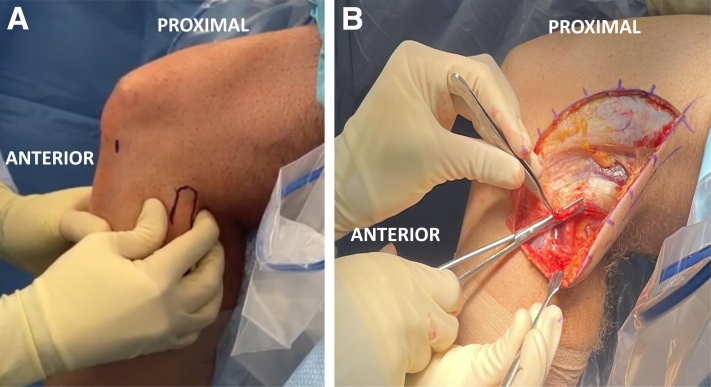
Intraoperative lateral view of left knee. (A) Examination of proximal tibiofibular joint stability is performed with the patient under anesthesia. The proximal fibula is outlined with a marking pen, and a comparative bilateral examination is performed. (B) Initial exposure via a 15-cm hockey-stick incision is performed in line with the iliotibial band fibers.
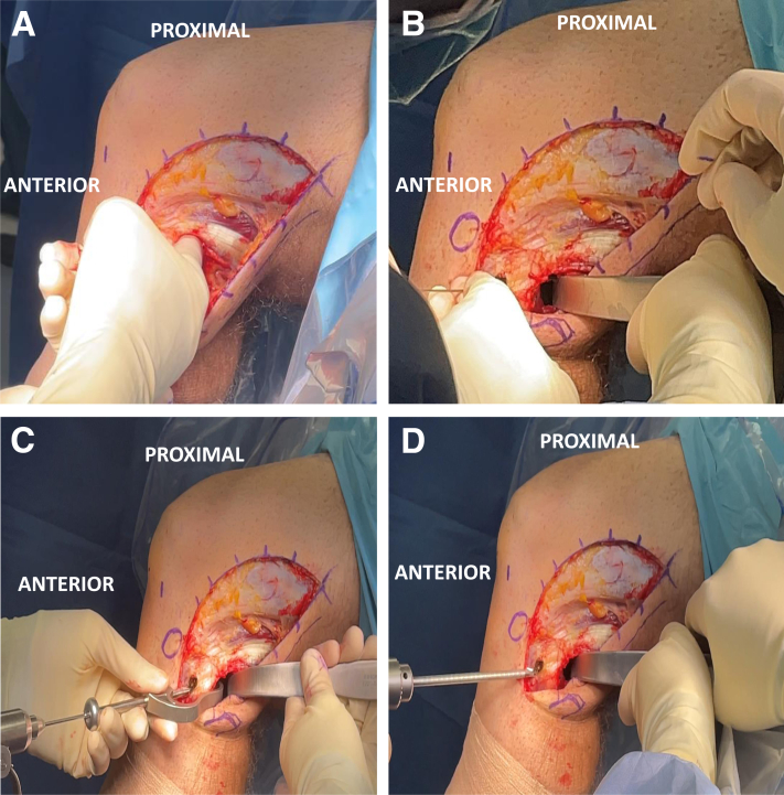
Fibular tunnel formation in left knee (lateral view). (A) Blunt elevation of soleus muscle and fingertip palpation of posterior aspect of proximal tibiofibular joint. (B) Chandler retractor placement protects the common peroneal nerve and vascular bundle during tunnel formation. (C) Fibular aiming guide and 2.4-mm guide pin in place on fibular head with entry point on anterolateral aspect and aimed posteromedially, exiting just distal to fibular insertion of popliteofibular ligament. (D) Final fibular tunnel formation with 6-mm cannulated reamer over guide pin while Chandler retractor is kept in place to protect neurovascular bundle.
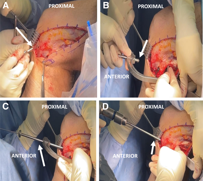
Tibial tunnel formation in left knee (lateral view). (A) Exposure of anterolateral tibial flat spot (arrow) between Gerdy tubercle and tibial tubercle. (B) Positioning of tibial aiming guide (arrow). The posterior tunnel emergence should be 1 cm medial and 1 cm proximal to the posterior fibular tunnel opening. (C) With manual reduction of the proximal tibiofibular joint, by use of a posterior retractor and tibial aiming guide, a 2.4-mm guide pin (arrow) is placed through the posterior aspect of the tibia. (D) Final tibial tunnel formation with 6-mm cannulated reamer (arrow) over guide pin. It should be noted that the Chandler retractor remains in place throughout tunnel formation and proximal tibiofibular joint reduction is maintained.
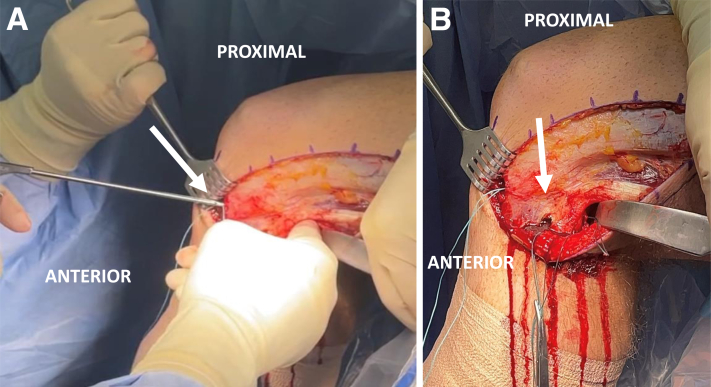
Passing stitch placement for future passage of semitendinosus allograft in left knee (lateral view). (A) Placement of tibial passing stitch (arrow) from anterior to posterior. (B) Tibial and fibular passing stitches in place (arrow). These stitches will allow for graft shuttling in both tunnels.
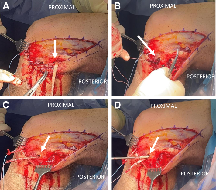
Graft passage and fixation of semitendinosus allograft in left knee (lateral view). (A) The graft is initially shuttled in the tibial tunnel from posterior (arrow) to anterior. (B) The proximal limb of the graft is then shuttled through the fibular tunnel from posterior to anterior (arrow). (C) Graft in place through tibial and fibular tunnels (arrow). (D) Initial fixation of graft in fibular tunnel (arrow) with 7 × 25 -mm interference screw. The semitendinosus allograft will subsequently be tensioned and fixed into the tibial tunnel at 70° of knee flexion with an additional 7 × 25 -mm interference screw.
References
-
- Anavian J., Marchetti D.C., Moatshe G., et al. The forgotten joint: Quantifying the anatomy of the proximal tibiofibular joint. Knee Surg Sports Traumatol Arthrosc. 2018;26:1096–1103. - PubMed
-
- See A., Bear R.R., Owens B.D. Anatomic mapping for surgical reconstruction of the proximal tibiofibular ligaments. Orthopedics. 2013;36:e58–e63. - PubMed
-
- Ogden J.A. Subluxation and dislocation of the proximal tibiofibular joint. J Bone Joint Surg Am. 1974;56:145–154. - PubMed
LinkOut - more resources
Full Text Sources
