. 2022 Nov 29;8(2):341-356.
doi: 10.1016/j.ekir.2022.11.015. eCollection 2023 Feb.

High Macrophage Densities in Native Kidney Biopsies Correlate With Renal Dysfunction and Promote ESRD

Affiliations

High Macrophage Densities in Native Kidney Biopsies Correlate With Renal Dysfunction and Promote ESRD

Maren B Pfenning et al. Kidney Int Rep. .

Abstract

Introduction: Macrophages and monocytes are main players in innate immunity. The relevance of mononuclear phagocyte infiltrates on clinical outcomes remains to be determined in native kidney diseases.

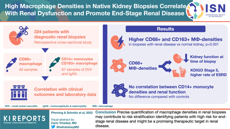
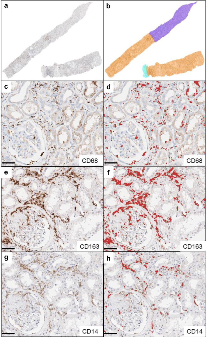
Methods: Our cross-sectional study included 324 patients with diagnostic renal biopsies comprising 17 disease entities and normal renal tissues for comparison. All samples were stained for CD68+ macrophages. Selected groups were further subtyped for CD14+ monocytes and CD163+ alternatively activated macrophages. Using precise pixel-based digital measurements, we quantified cell densities as positively stained areas in renal cortex and medulla as well as whole renal tissue. Laboratory and clinical data of all cases at the time of biopsy and additional follow-up data in 158 cases were accessible.

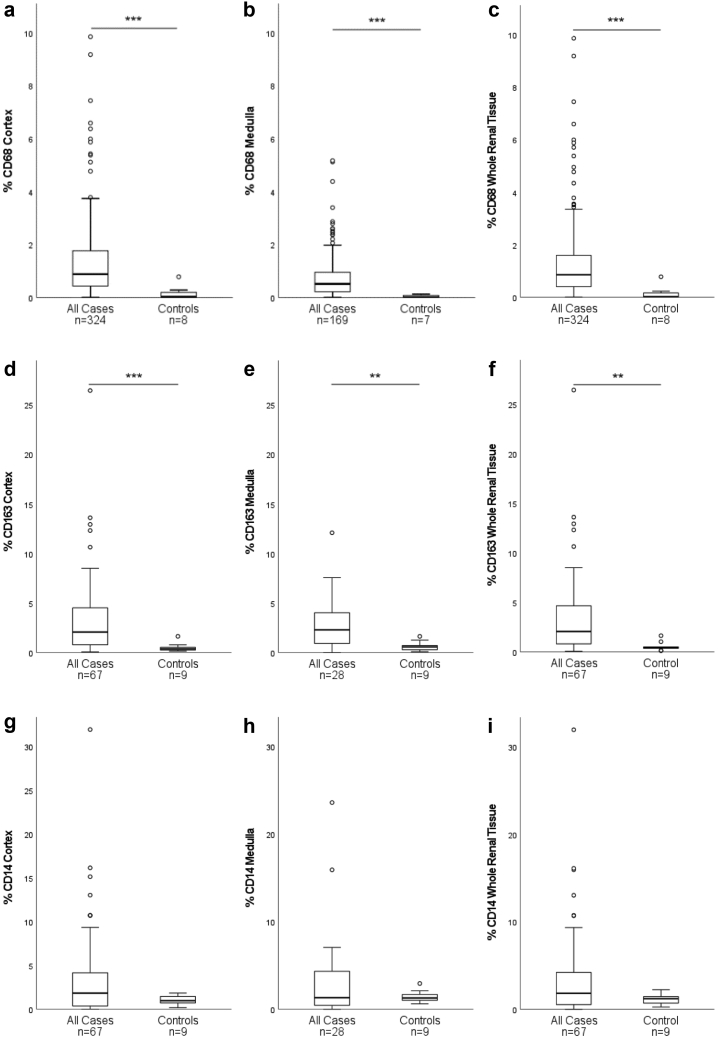
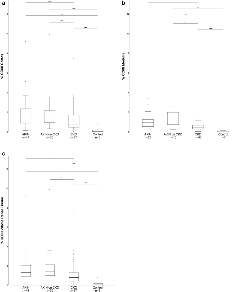
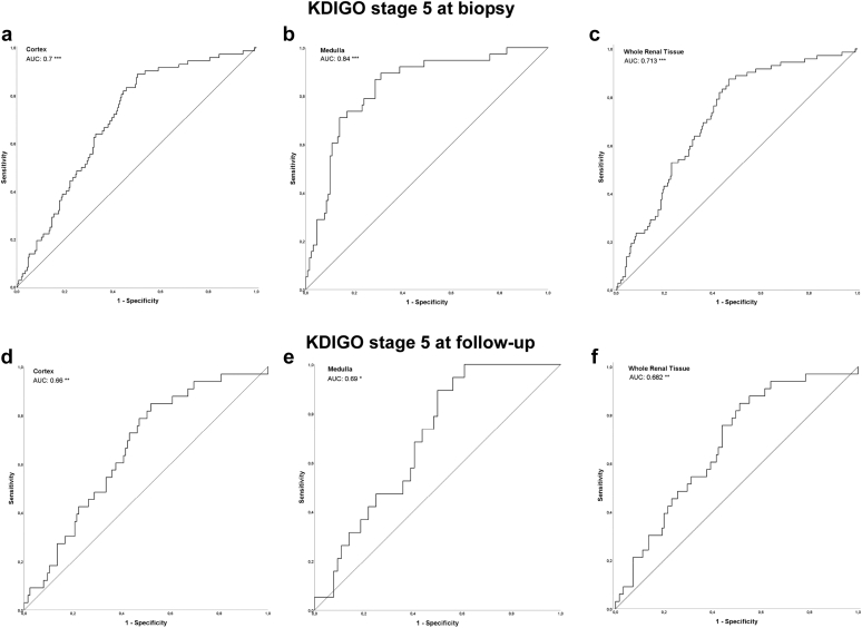
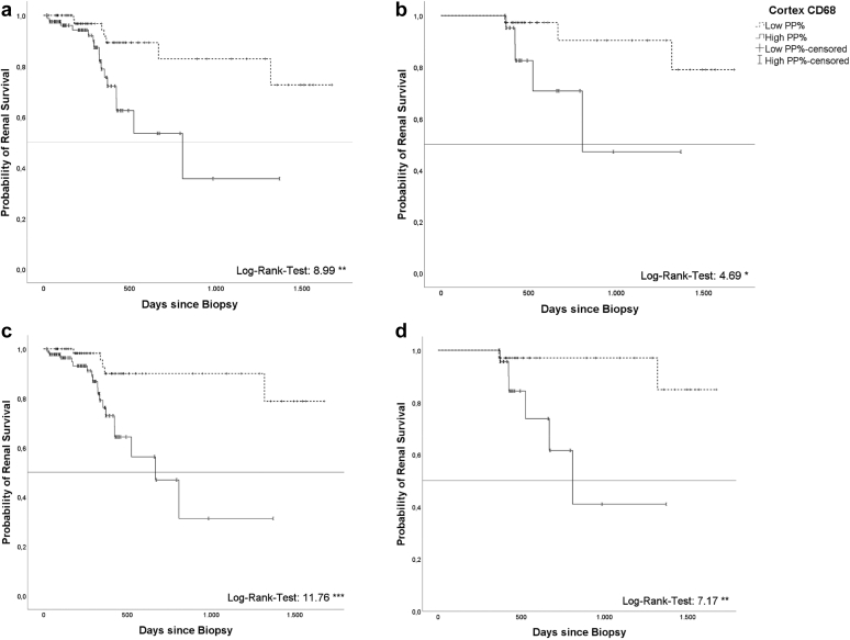
Results: Biopsies with renal disease consistently revealed higher CD68+-macrophage densities and CD163+-macrophage densities in cortex and medulla compared to controls. High macrophage densities correlated with impaired renal function at biopsy and at follow-up in all diseases and in diseases analyzed separately. High cortical CD68+-macrophage densities preceded shorter renal survival, defined as requirement of permanent dialysis. CD14+ monocyte densities showed no difference compared to controls and did not correlate with renal function.

Conclusion: Precise quantification of macrophage densities in renal biopsies may contribute to risk stratification to identify patients with high risk for end-stage renal disease (ESRD) and might be a promising therapeutic target in renal disease.

Keywords: Dialysis risk; Digital pathology; Disease progression; Macrophage; Native kidney disease; Renal pathology.


Figures

None
Graphical abstract
Figure 1
Figure 1
Visualization of histologic and digital methods in a renal biopsy with vasculitis: (a) Whole-slide image of section stained for CD68 (brown). (b) Annotated regions of interests: orange: cortical tissue, dark blue: medullary tissue, light blue: extrarenal tissue. Bar represents 500 μm. (c) Detail of cortical tissue from vasculitis case with CD68-stain for macrophages, (e) CD163-stain for M2-macrophages and (g) CD14-stain for monocytes. Corresponding positive pixel detections shown red; (d): CD68, (f): CD163, (h): CD14. Bars represent 50 μm.
Figure 2
Figure 2
Macrophage and monocyte densities (% area) in all diseased cases compared to controls. CD68+-densities in cortex (a), medulla (b), whole renal tissue (c) and CD163+-densities in cortex (d), medulla (e) and whole renal tissue (f) are higher in native kidney diseases (cases) compared to normal kidneys (controls). CD14+-densities in cortex (g), medulla (h) and whole renal tissue (i) do not differ between cases and controls. (∗P < 0.05, ∗∗P < 0.01, ∗∗∗P < 0.001; box: 1. -3. quartile, low whisker: 1. quartile – 1.5 × interquartile range, high whisker: 3. quartile + 1.5 × interquartile range; °: outlier).
Figure 3
Figure 3
Macrophage densities (% CD68-immunostained area) in renal cortex (a), medulla (b) and whole renal tissue (c) in acute kidney injury of all stages according to RIFLE-criteria (AKIN), AKIN with an underlying chronic kidney disease (AKIN on CKD), and chronic kidney disease of all stages according to KDIGO (CKD), compared to controls with normal renal tissue. Boxes show the medians with quartile ranges, whiskers display 1.5 × interquartile range of the 1. and 3. quartile. (∗∗∗ P < 0.001; °outlier).
Figure 4
Figure 4
Receiver operating characteristics (bold curve) with AUC-values of severe impairment of renal function (KDIGO stage 5) and CD68+-macrophage densities at biopsy in cortex (a; n = 324), medulla (b; n = 167) and whole renal tissue (c; n = 324), as well as at follow-up (complete cohort) in cortex (d; n = 158), medulla (e; n = 83) and whole renal tissue (f; n = 158). The fair curve is the reference AUC = 0.5 in a–f. (∗ P < 0.05, ∗∗ P < 0.01, ∗∗∗ P < 0.001). AUC, area under the curve.
Figure 5
Figure 5
Kaplan-Meyer curve showing cumulative kidney survival at follow-up for entire follow-up cohort (a; n = 158) comparing low (< median; n = 76) and high (> median; n = 82) cortical CD68+densities (log rank 8.989 ∗∗) and for subcohort with outcome data >365 days (b; n = 60, < median n = 37; > median n = 23), respectively. c and d depict the cumulative kidney survival using a ROC-derived (receiver operating characteristics derived) separation value (c: entire cohort, n = 158, < ROC-derived separation value n = 68, > ROC-derived separation value n = 90; d: subcohort with outcome data >365 days, n = 60, < ROC-derived separation value n = 34, > ROC-derived separation value n = 26). (∗ P < 0.05, ∗∗ P < 0.01, ∗∗∗ P < 0.001). Median of 0.875% and ROC-derived separation value of 0.7998% were calculated in whole study cohort (n = 324). ROC, receiver operating characteristics.

References

    1. Duffield J.S. Cellular and molecular mechanisms in kidney fibrosis. J Clin Invest. 2014;124:2299–2306. doi: 10.1172/JCI72267. - DOI - PMC - PubMed
    1. Netea M.G., Balkwill F., Chonchol M., et al. A guiding map for inflammation. Nat Immunol. 2017;18:826–831. doi: 10.1038/ni.3790. - DOI - PMC - PubMed
    1. Gurtner G.C., Werner S., Barrandon Y., Longaker M.T. Wound repair and regeneration. Nature. 2008;453:314–321. doi: 10.1038/nature07039. - DOI - PubMed
    1. Chawla L.S., Eggers P.W., Star R.A., Kimmel P.L. Acute kidney injury and chronic kidney disease as interconnected syndromes. N Engl J Med. 2014;371:58–66. doi: 10.1056/NEJMra1214243. - DOI - PMC - PubMed
    1. Murray P.J., Wynn T.A. Protective and pathogenic functions of macrophage subsets. Nat Rev Immunol. 2011;11:723–737. doi: 10.1038/nri3073. - DOI - PMC - PubMed