. 2022 Oct 31;8(2):317-329.
doi: 10.1016/j.ekir.2022.10.024. eCollection 2023 Feb.

Modeling of ACTN4-Based Podocytopathy Using Drosophila Nephrocytes

Affiliations

Modeling of ACTN4-Based Podocytopathy Using Drosophila Nephrocytes

Johanna Odenthal et al. Kidney Int Rep. .

Abstract

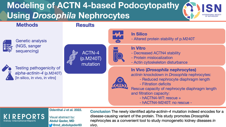
Introduction: Genetic disorders are among the most prevalent causes leading to progressive glomerular disease and, ultimately, end-stage renal disease (ESRD) in children and adolescents. Identification of underlying genetic causes is indispensable for targeted treatment strategies and counseling of affected patients and their families.

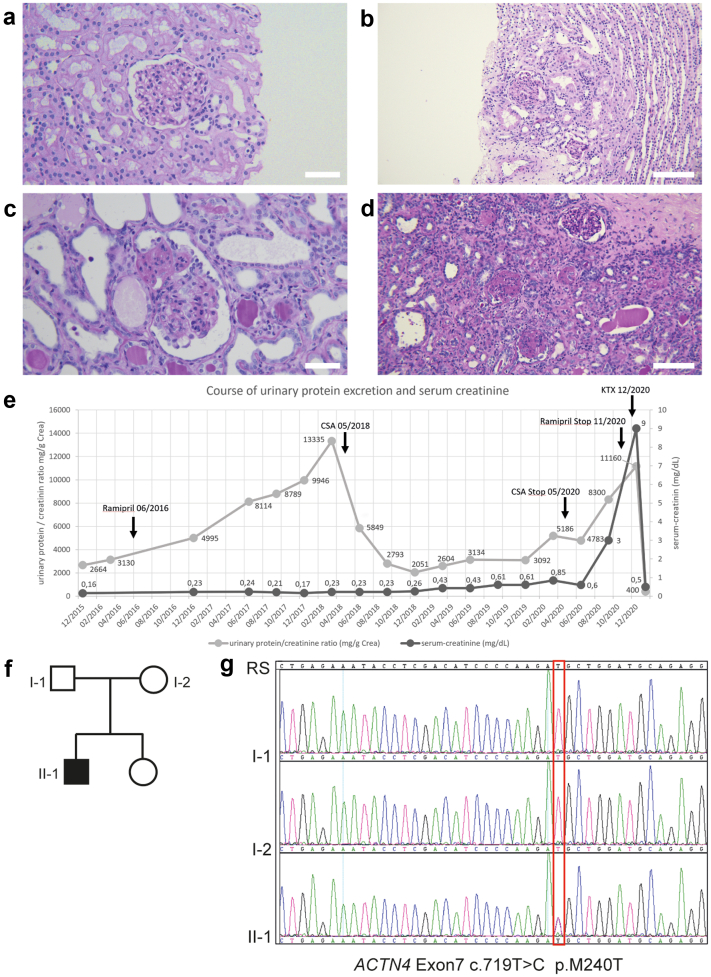
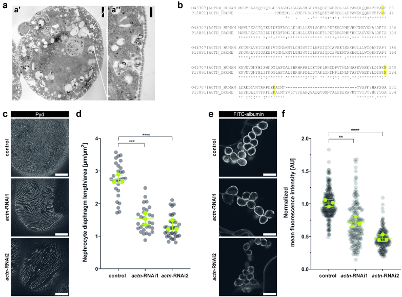
Methods: Here, we report on a boy who presented at 4 years of age with proteinuria and biopsy-proven focal segmental glomerulosclerosis (FSGS) that was temporarily responsive to treatment with ciclosporin A. Molecular genetic testing identified a novel mutation in alpha-actinin-4 (p.M240T). We describe a feasible and efficient experimental approach to test its pathogenicity by combining in silico, in vitro, and in vivo analyses.

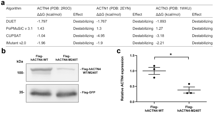
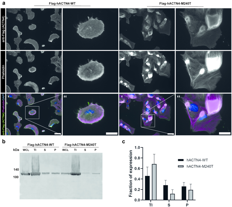
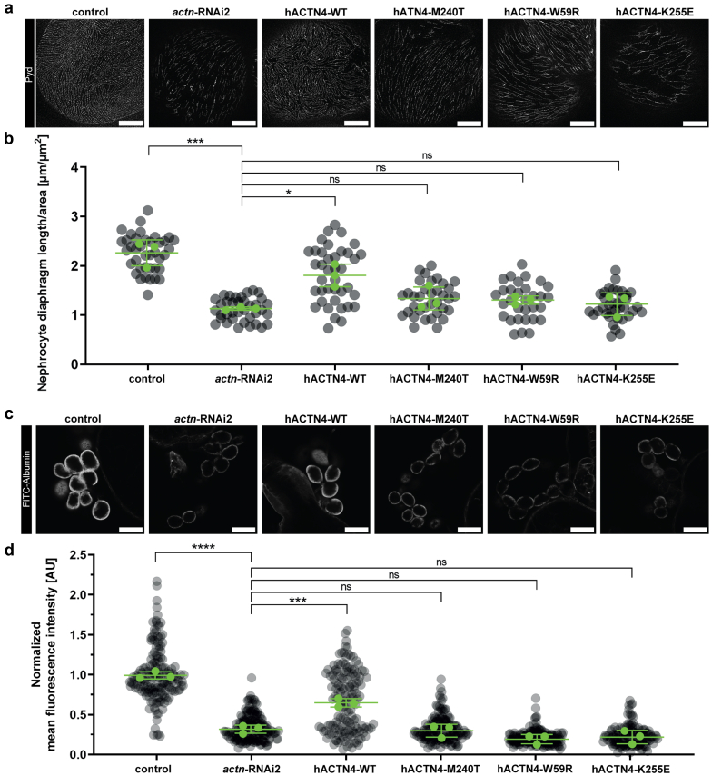
Results: The de novo p.M240T mutation led to decreased alpha-actinin-4 stability as well as protein mislocalization and actin cytoskeleton rearrangements. Transgenic expression of wild-type human alpha-actinin-4 in Drosophila melanogaster nephrocytes was able to ameliorate phenotypes associated with the knockdown of endogenous actinin. In contrast, p.M240T, as well as other established disease variants p.W59R and p.K255E, failed to rescue these phenotypes, underlining the pathogenicity of the novel alpha-actinin-4 variant.

Conclusion: Our data highlight that the newly identified alpha-actinin-4 mutation indeed encodes for a disease-causing variant of the protein and promote the Drosophila model as a simple and convenient tool to study monogenic kidney disease in vivo.

Keywords: ACTN4; Drosophila; FSGS; nephrocyte; nephrotic syndrome; podocyte.


Figures

None
Graphical abstract
Figure 1
Figure 1
Identification of a novel ACTN4 mutation in a patient presenting with steroid-resistant nephrotic syndrome. (a–d) Periodic acid–Schiff stainings of kidney biopsies of the index patient. (a) Initial kidney biopsy in 2015 showing glomeruli with normal appearing capillary tuft. Electron microscopy revealed foot process effacement of the podocytes leading to the diagnosis of minimal change glomerulopathy. (b) Kidney biopsy in 2017 with 1 glomerulum showing a perihilar focal and segmental glomerulosclerosis. (c,d) Nephrectomy specimen of (c) right and (d) left native kidney with increased globally sclerosed glomeruli and increased interstitial fibrosis and tubular atrophy. (e) Overview of the clinical course of the patient. Gray: urinary protein/creatinine ratio in mg/g. Black: serum-creatinine level in mg/dl. The patient first presented with proteinuria in December 2015. After a first biopsy (2015) showing minimal change glomerulopathy and, in the absence of clinical nephrotic syndrome, rampiril was initiated in June 2016. A second biopsy in June 2017 confirmed focal segmental glomerulosclerosis. Therapy with CSA resulted in a prompt but transient decrease of proteinuria and was stopped in May 2020 when renal function declined and proteinuria increased despite treatment. The patient was then prepared for kidney transplantation and received a living-donor graft in December 2020. (f,g) Pedigree of the index patient and targeted Sanger sequencing of ACTN4 exon 7. Electropherograms depict the relevant sequence section around c.719T>C (p.M240T) in the index patient (II-1) and both parents (I-1 and I-2). Scale bars: 50 μm in (a) and (c), 100 μm in (b) and (d). CSA, ciclosporin A; KTx, kidney transplantation; RS, reference sequence.
Figure 2
Figure 2
In silico and in vitro characterization of hACTN4-M240T stability. (a) In silico prediction of M240T mutation on alpha-actinin protein stability (delta-delta-G) using 4 different computational algorithms. M>T amino acid exchange in the conserved domain has a destabilizing effect in ACTN4 as well as ACTN1 and ACTN3. (b) Representative western blot analysis of whole-cell lysates cotransfected with Flag-hACTN4-WT or Flag-hACTN4-M240T and Flag-GFP serving as expression control. (c) Densitometric quantification of 3 independent experiments as shown in (b). Compared with the WT variant, hACTN4-M240T is significantly lower expressed (n = 3, error bars indicate SD, ∗P < 0.05, two-tailed t test). GFP, green fluorescent protein; WT, wild-type.
Figure 3
Figure 3
ACTN4-M240T mutation leads to perturbed cellular localization and aggregate formation. (a) Immunofluorescence analysis of podocyte cell lines stably expressing Flag-tagged hACTN4-WT or hACTN4-M240T. Cells were stained with anti-FLAG antibody as well as Phalloidin and 4′,6-diamidino-2-phenylindole to visualize filamentous actin and the nucleus, respectively. In contrast to cells expressing the WT variant, where the protein is located in the cortical region together with F-actin, localization of ACTN4-M240T can be detected more toward the center of the cells, partially associated with large F-actin positive aggregates. Single channels are shown in gray, and scale bar indicates 50 μm in ‘ and 25 μm in ‘‘. (b) Representative western blot analysis of fractionation assays performed in cells transiently expressing either hACTN4-WT or hACTN4-M240T. Whole-cell lysates were subjected to differential centrifugation and the Triton X-100 insoluble as well as the supernatant and pellet derived from the Triton X-100 soluble fraction were analyzed by western blot stained with anti-FLAG antibodies. (c) Densitometric analysis of ACTN4 distribution in TI, S, and P fractions for ACTN4-WT and ACTN4-M240T (n = 4). P, pellet; S, supernatant; TI, Triton X-100 insoluble; WCL, whole-cell lysates; WT, wild-type.
Figure 4
Figure 4
Knockdown of Drosophila actinin in nephrocytes results in decreased ND length and reduced filtration function. (a) Electron micrograph depicting a wild-type nephrocyte. Scale bars indicate 5 μm in A’ and 500 nm in A’’ (b) Protein alignment of the N-terminal actin-binding domain of human ACTN4 and Drosophila actinin. Highlighted are amino acids known to be causative of monogenic nephrotic syndrome. Actinin shares 68.83% overall identity with ACTN4, the actin-binding domain of the 2 proteins shares 78.8% identity. (c,d) Representative micrographs of nephrocytes stained with (c) anti-Pyd and (d) quantification of the ND length. Nephrocytes derived from either control larvae, or larvae with nephrocyte-specific knockdown of actinin (ACTN), by using 2 different UAS-RNAi-lines (actn-RNAi1 and actn-RNAi2). Compared with control nephrocytes, the ND length of actn-depleted nephrocytes is significantly reduced (gray dots show all nephrocytes measured, green dots represent means of n = 3 independent experiments performed in 3 experimental crossings, error bars indicate SD, ∗∗P < 0.01, ∗∗∗P < 0.001, one-way ANOVA with Tukey’s post hoc test). (e,f) Representative micrographs of nephrocytes subjected to (e) FITC-albumin tracer and (f) quantification of fluorescence intensity as a measure of uptake capacity. Control and ACTN knockdown nephrocytes were incubated in 0.2 mg/ml FITC-albumin solution for 30 seconds, and fluorescence intensity was quantified using Fiji. The data are presented as normalized to control levels. Both, actn-RNAi1 and actn-RNAi2 nephrocytes show a significantly reduced capacity of FITC-albumin uptake with respect to control nephrocytes, indicating severe filtration defects (gray dots show all nephrocytes measured, green dots indicate means of n = 3 independent experiments performed in 3 experimental crossings, error bars indicate SD, ∗∗P < 0.01, ∗∗∗∗P < 0.0001, one-way ANOVA with Tukey’s post hoc test). Scale bars indicate 5 μm in (c) and 25 μm in (f). FITC, fluorescein isothiocyanate; ND, nephrocyte diaphragm; Pyd, polychaetoid; RNAi, RNA interference.
Figure 5
Figure 5
hACTN4-M240T reexpression does not ameliorate actinin knockdown–associated phenotypes. (a,b) Representative micrographs of nephrocytes stained with (a) anti-Pyd and (b) quantification of the ND length. Nephrocytes derived from either control larvae, larvae with nephrocyte-specific knockdown of actinin (actn-RNAi2) as well as larvae reexpressing the indicated hACTN4-variant in the knockdown background. hACTN4-WT is able to partially rescue the actinin knockdown–associated reduction in ND length, whereas reexpression of hACTN-M240T does not lead to increased ND length. This is also true for FSGS-associated mutations FSGS-W59R and FSGS-K255E (gray dots indicate all nephrocytes measured, green dots show means of n = 3 independent experiments performed in 3 experimental crossings, error bars indicate SD, ∗∗∗P < 0.001, ∗P < 0.05, one-way analysis of variance with Tukey’s post hoc test). (c,d) Representative micrographs of nephrocytes subjected to (c) FITC-albumin tracer and (d) quantification of fluorescence intensity as a measure of uptake capacity. Nephrocytes were incubated in 0.2 mg/ml FITC-albumin solution for 30 seconds, and fluorescence intensity was quantified using Fiji. The data are presented as normalized to control levels. Reexpression of hACTN4-WT also leads to a significant increase in tracer uptake capacity, compared with hACTN4-M240T, hACTN4-W59R, and hACTN4-K255E, where a rescue of actinin knockdown–associated reduction of tracer uptake cannot be observed (gray dots indicate all nephrocytes measured, green dots show means of n = 3 independent experiments performed in 3 experimental crossings, error bars indicate SD, ∗∗∗P < 0.001, ∗∗∗∗P < 0.0001, one-way analysis of variance with Tukey’s post hoc test). Scale bars indicate 5 μm in (a) and 25 μm in (c). FITC, fluorescein isothiocyanate; ND, nephrocyte diaphragm; Pyd, polychaetoid; WT, wild-type.

References

    1. Rosenberg A.Z., Kopp J.B. Focal segmental glomerulosclerosis. Clin J Am Soc Nephrol. 2017;12:502–517. doi: 10.2215/CJN.05960616. - DOI - PMC - PubMed
    1. McGrogan A., Franssen C.F., de Vries C.S. The incidence of primary glomerulonephritis worldwide: a systematic review of the literature. Nephrol Dial Transplant. 2011;26:414–430. doi: 10.1093/ndt/gfq665. - DOI - PubMed
    1. Shabaka A., Ribera A.T., Fernández-Juárez G. Focal segmental glomerulosclerosis: state-of-the-art and clinical perspective. Nephron. 2020;144:413–427. doi: 10.1159/000508099. - DOI - PubMed
    1. Sadowski C.E., Lovric S., Ashraf S., et al. A single-gene cause in 29.5% of cases of steroid-resistant nephrotic syndrome. J Am Soc Nephrol. 2015;26:1279–1289. doi: 10.1681/ASN.2014050489. - DOI - PMC - PubMed
    1. Liu J., Wang W. Genetic basis of adult-onset nephrotic syndrome and focal segmental glomerulosclerosis. Front Med. 2017;11:333–339. doi: 10.1007/s11684-017-0564-1. - DOI - PubMed

LinkOut - more resources