

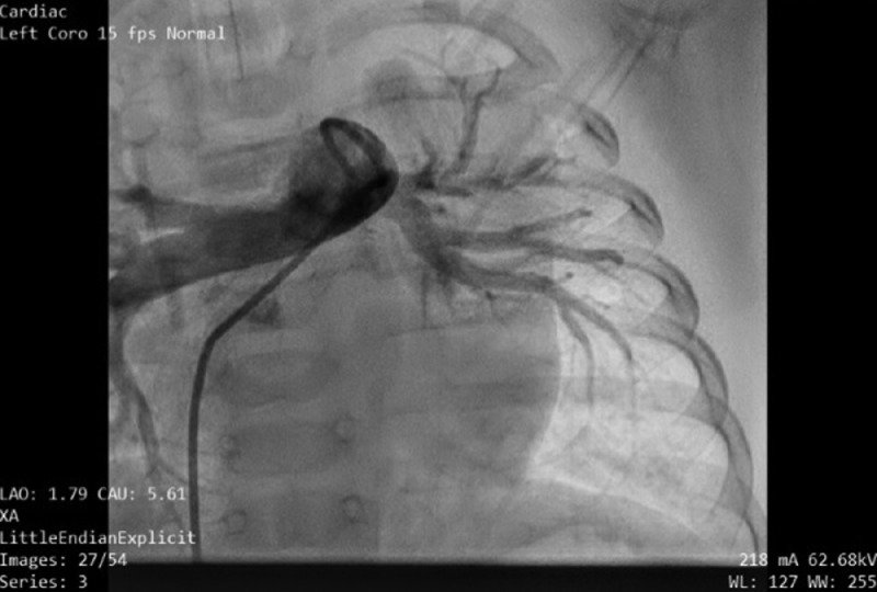
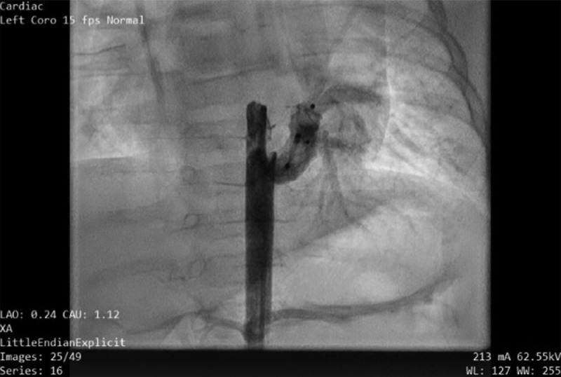
Large isolated major aortopulmonary collateral artery causing dilated left ventricle
- PMID: 36815146
- PMCID: PMC9939544
- DOI: 10.1016/j.radcr.2023.01.063
Large isolated major aortopulmonary collateral artery causing dilated left ventricle
Abstract
Isolated major aortopulmonary collateral artery (MAPCA), in the absence of evidence of structural heart disease, is a very rare observation. This anomaly usually appears in preterm newborns. In the majority of babies, isolated MAPCAs cause no symptoms and regress spontaneously after birth and their conservative management is usually sufficient. We report a case of an asymptomatic full-term 5-month-old infant presenting with heart murmur as the only sign during clinical evaluation. Echocardiography revealed a dilated left ventricle, with no pulmonary hypertension. Computed tomography angiogram showed a large MAPCA arising from the descending thoracic aorta and supplying blood to the left lower lobe. The condition was managed successfully by percutaneous obliteration with Amplatzer vascular plugs. Isolated MAPCA is usually a benign anomaly, presenting no clinical finding and requiring no specific treatment. However, in a small minority of infants, this congenital disorder may progress, with detrimental impacts on cardiac structure before clinical symptoms appear. Early intervention may be required to prevent irreversible sequelae.
Keywords: Amplatzer vascular plugs; Asymptomatic; Closure; Isolated major aortopulmonary collateral artery.
© 2023 The Authors. Published by Elsevier Inc. on behalf of University of Washington.
Figures


References
-
- Wernovsky G, Anderson R., Kumar K, Redington A, Tweddell J, Tweddell J, editors. Anderson's pediatric cardiology. 4th ed. Elsevier; Philadelphia, PA: 2019. editors.
-
- Tinmaswala MA, Saple PP, Gupta A, Prachi N, Nitinkumar A, Amin K. Isolated major aortopulmonary collateral artery causing CCF in a newborn: a case report. Int J Med Res Health Sci. 2015;4:471–473.
Publication types
LinkOut - more resources
Full Text Sources
