. 2023 Jun;601(12):2513-2532.
doi: 10.1113/JP284004. Epub 2023 Mar 7.

Novel regenerative drug, SPG302 promotes functional recovery of diaphragm muscle activity after cervical spinal cord injury

Affiliations

Novel regenerative drug, SPG302 promotes functional recovery of diaphragm muscle activity after cervical spinal cord injury

Matthew J Fogarty et al. J Physiol. 2023 Jun.

Abstract

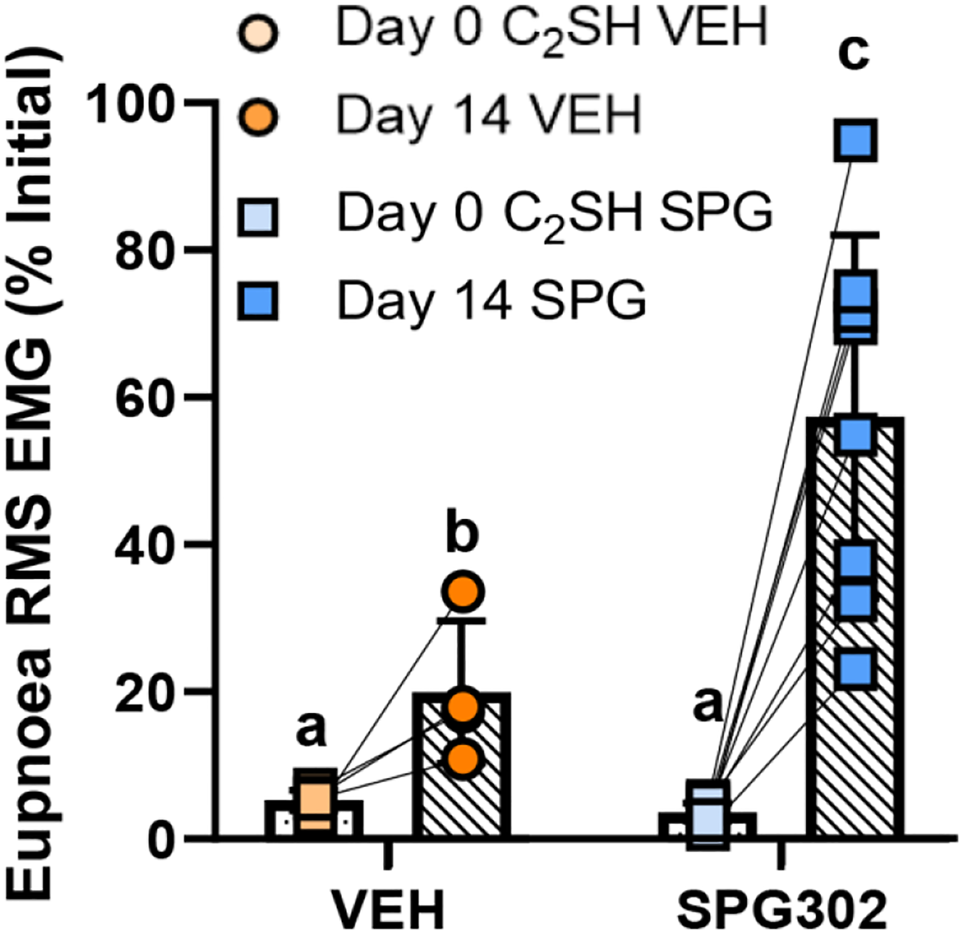
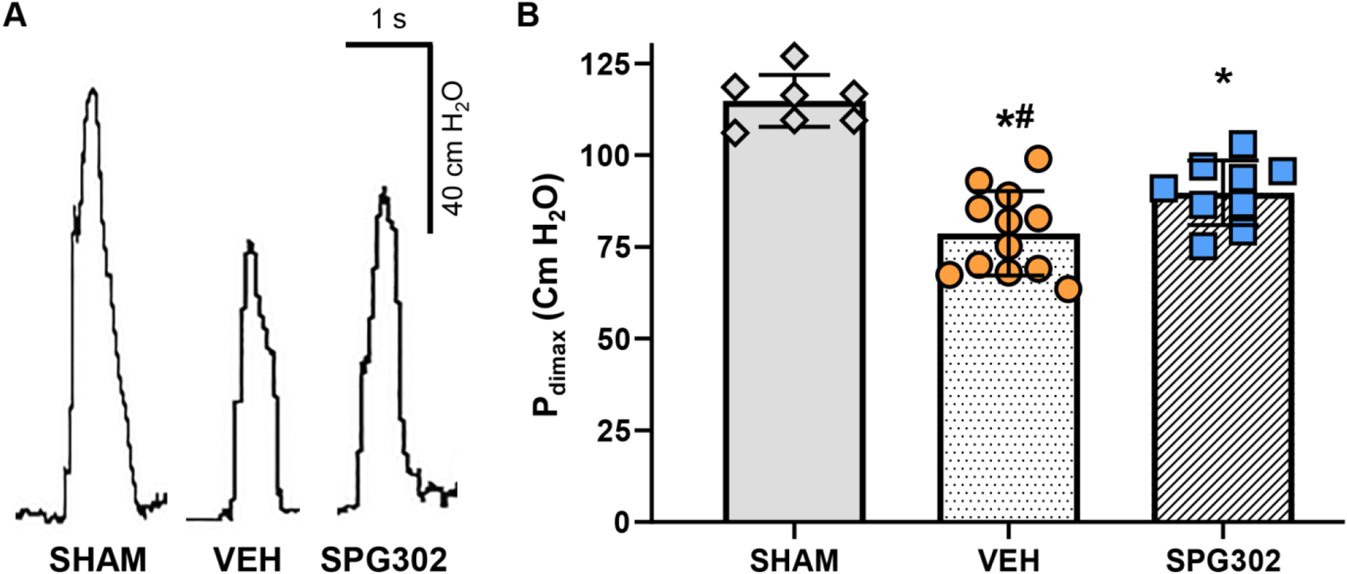
Spinal cord hemisection at C2 (C2 SH), sparing the dorsal column is widely used to investigate the effects of reduced phrenic motor neuron (PhMN) activation on diaphragm muscle (DIAm) function, with reduced DIAm activity on the injured side during eupnoea. Following C2 SH, recovery of DIAm EMG activity may occur spontaneously over subsequent days/weeks. Various strategies have been effective at improving the incidence and magnitude of DIAm recovery during eupnoea, but little is known about the effects of C2 SH on transdiaphragmatic pressure (Pdi ) during other ventilatory and non-ventilatory behaviours. We employ SPG302, a novel type of pegylated benzothiazole derivative, to assess whether enhancing synaptogenesis (i.e., enhancing spared local connections) will improve the incidence and the magnitude of recovery of DIAm EMG activity and Pdi function 14 days post-C2 SH. In anaesthetised Sprague-Dawley rats, DIAm EMG and Pdi were assessed during eupnoea, hypoxia/hypercapnia and airway occlusion prior to surgery (C2 SH or sham), immediately post-surgery and at 14 days post-surgery. In C2 SH rats, 14 days of DMSO (vehicle) or SPG302 treatments (i.p. injection) occurred. At the terminal experiment, maximum Pdi was evoked by bilateral phrenic nerve stimulation. We show that significant EMG and Pdi deficits are apparent in C2 SH compared with sham rats immediately after surgery. In C2 SH rats treated with SPG302, recovery of eupneic, hypoxia/hypercapnia and occlusion DIAm EMG was enhanced compared with vehicle rats after 14 days. Treatment with SPG302 also ameliorated Pdi deficits following C2 SH. In summary, SPG302 is an exciting new therapy to explore for use in spinal cord injuries. KEY POINTS: Despite advances in our understanding of the effects of cervical hemisection (C2 SH) on diaphragm muscle (DIAm) EMG activity, very little is understood about the impact of C2 SH on the gamut of ventilatory and non-ventilatory transdiaphragmatic pressures (Pdi ). Recovery of DIAm activity following C2 SH is improved using a variety of approaches, but very few pharmaceuticals have been shown to be effective. One way of improving DIAm recovery is to enhance the amount of latent local spared connections onto phrenic motor neurons. A novel pegylated benzothiazole derivative enhances synaptogenesis in a variety of neurodegenerative conditions. Here, using a novel therapeutic SPG302, we show that 14 days of treatment with SPG302 ameliorated DIAm EMG and Pdi deficits compared with vehicle controls. Our results show that SPG302 is a compound with very promising potential for use in improving functional outcomes post-spinal cord injury.

Keywords: diaphragm muscle; electromyography; rehabilitation; spinal cord injury; transdiaphragmatic pressure.


Conflict of interest statement

Competing Interests: VFS, PV and STS declare that they are members of the executive team and shareholders of Spinogenix. All other authors declare no competing interests.

Figures

Figure 1.
Figure 1.
Representative recordings of right (red) and left (blue) DIAm EMG activity from a rats that underwent sham surgery (SHAM) or C2SH surgery (right side, dotted rectangles) and were subsequently treated with DMSO vehicle (VEH) or SPG302. Recordings across all behaviours were obtained immediately before laminectomy (pre-surgery) on day 0, immediately after C2SH on day 0 or laminectomy in sham controls (post-surgery) and 14 days after surgery.
Figure 2:
Figure 2:
A: Schematic of the C2SH lesion employed in the present study, note the severing of the lateral and ventral funiculi, and the preservation of the dorsal funiculus and some sparing adjacent to the central canal (CC). Descending motor tracts (magenta) comprising the lateral and ventral coticospinal tracts (LCT and VCT) along with ascending sensory (green) lateral and ventral spinothalamic tracts (L&VST) are depicted as lesioned (grey semitransparency) on the right hand side. B: The % (mean ± SD) of rats that recovered (> 10% initial RMS DIAm EMG during eupnoea) was greater in SPG302 treated rats (blue squares) compared to VEH rats (orange circles) at 7- (P=0.032) and 14-days post-C2SH (P=0.036; n=14 VEH, n=10 SPG302; Fisher’s exact test). C: Epifluorescence images of spinal cord histology of horizontal sections (70 μm) at the level of the central canal (yellow dashed line) in non-revovered (Non-Rec.) and recovered (Rec.) vehicle and SPG302 treated rats. The quantification of the spared region following right sided C2SH is illustrated by the red line. D: Scatterplot shows the spared portion (from the central canal to the cut lesion edge) of the spinal cord at the middle “equatorial” latitude of the C2 spinal cord being unchanged between non-recovered (n=7) and recovered rats (n=7; P=0.687, Student’s unpaired t-test), regardless of treatment with VEH (orange circles) or SPG302 (blue squares). E: Scatterplot shows the spared portion expressed as a % of the radius at the middle “equatorial” latitude of C2 being unchanged between non-recovered (n=7) and recovered rats (n=7; P=0.855, Student’s unpaired t-test), regardless of treatment with VEH (orange circles) or SPG302 (blue squares).
Figure 3.
Figure 3.
DIAm RMS EMG activity was calculated for each animal and behaviour and normalized to the corresponding day 0 pre-surgery mean DIAm RMS EMG (i.e., 100 is the starting point for eupnoea, HH and occlusion within each rat). The scatterplot shows mean DIAm RMS EMG for the right injured side for all behaviours with mean (± SD) values of each VEH (orange circle symbols) or SPG302 treated (blue square symbols) rats represented by each dotpoint. Within each behaviour, different levels of DIAm RMS EMG activity are indicated by different letters (ie., a ≠ b ≠ c, with P<0.05). Immediately following C2SH, DIAm RMS EMG activity was reduced to <10% of initial during eupnoea and HH and <20% of initial during occlusion in both VEH and SPG302 treated rats. In VEH rats, 14-days post-C2SH (darkened symbols) did not improve DIAm RMS EMG in eupnoea or HH (P>0.05) compared to immediate post-C2SH values. However, there was an increase at 14-days following C2SH in the occlusion DIAm RMS EMG compared to immediately post-C2SH (P<0.05). Treatment with SPG302 improved the magnitude of DIAm RMS EMG at 14 days following C2SH (darkened symbols) in all behaviours. DIAm RMS EMG activity was returned to ~50% of initial values in SPG302 rats during eupnoea, HH and occlusion (P<0.05 in all comparisons). In addition, compared immediately post C2SH, 14-day treatment with SPG302 led to greater DIAm RMS EMG compared to 14-day VEH rats during eupnoea and HH (P<0.05). Three-way ANOVA with Bonferroni post-test, n=14 VEH, n=10 SPG302.
Figure 4.
Figure 4.
In exclusively recovered rats (ie., those that exhibited >10% of initial DIAm RMS EMG druring eupnoea) at 14 days following C2SH, DIAm RMS EMG (mean ± SD) was greater in SPG302 treated (blue squares), compared to VEH rats (orange circles). Different letters indicate significant differences (P<0.05). Two-way ANOVA with Bonferroni post-test, n=4 VEH, n=8 SPG302.
Figure 5.
Figure 5.
A: Example Pdi traces during eupnoea immediately prior to surgery (day 0), immediately following C2SH (day 0) and 14-days after C2SH in VEH and SPG302 rats. B: Scatterplot of Pdi (mean ± SD), (with SHAM values represented by a dashed line) illustrate a marked reduction in Pdi during eupnoea immediately following C2SH in VEH (light orange circles) and SPG302 (light blue squares) treated rats compard to pre-surgery (grey symbols; *, P<0.05). By 14-days following C2SH, VEH treated rats (dark orange circles) continue to have reduced Pdi during eupnoea compared to SHAM (and pre-injury) (*, P<0.05) and to SPG302 treated rats (dark blue squares; #, P<0.05). Furthermore, Pdi during eupnoea in SPG302 treated rats has returned to SHAM (and pre-injury) levels by day 14. Two-way ANOVA with Bonferroni post-test, n=7 SHAM, n=13 VEH, n=9 SPG302.
Figure 6.
Figure 6.
A: Example Pdi traces during HH immediately prior to surgery (day 0), immediately following C2SH (day 0) and 14-days after C2SH in VEH and SPG302 rats. B: Scatterplot of Pdi (mean ± SD), (with SHAM values represented by a dashed line) illustrate a marked reduction in Pdi during HH immediately following C2SH in VEH (light orange circles) and SPG302 treated rats (light blue squares) compard to pre-surgery (grey symbols; *, P<0.05). By 14-days following C2SH, VEH treated rats (dark orange circles) continue to have reduced Pdi during HH compared to SHAM (and pre-injury) (*, P<0.05) and to SPG302 treated rats (dark blue squares; #, P<0.05). Furthermore, Pdi during HH in SPG302 treated rats has returned to SHAM (and pre-injury) levels by day 14. Two-way ANOVA with Bonferroni post-test, n=7 SHAM, n=13 VEH, n=9 SPG302.
Figure 7.
Figure 7.
A: Example Pdi traces during occlusion immediately prior to surgery (day 0), immediately following C2SH (day 0) and 14-days after C2SH in VEH and SPG302 rats. B: Scatterplot of Pdi (mean ± SD), (with SHAM values represented by a dashed line) illustrate a marked reduction in Pdi during occlusion immediately following C2SH in VEH (light orange circle) and SPG302 treated rats (light blue squares) compared to presurgery values (grey symbols; *, P<0.05). By 14-days following C2SH, VEH treated rats (dark orange circles) continue to have reduced Pdi during occlusion compared to SHAM (and pre-injury) (*, P<0.05) and to SPG302 treated rats (dark blue squares; #, P<0.05). Furthermore, Pdi during occlusion in SPG302 treated rats has returned to SHAM (and pre-injury) levels by day 14. Two-way ANOVA with Bonferroni post-test, n=7 SHAM, n=13 VEH, n=9 SPG302.
Figure 8.
Figure 8.
A: Example Pdimax traces (achieved via bilaterl phrenic nerve stimulation) at terminal experiments (day 14) in SHAM, VEH and SPG302 rats. B: Scatterplot of Pdimax (mean ± SD), illustrate a marked reduction in Pdimax 14-days after C2SH in VEH (orange circles) and SPG302 treated (blue squares) compared to SHAM rats (grey diamonds; *, P<0.05). However, 14-day treatement with SPG302 improved Pdimax in SPG302 rats compared to VEH (#, P<0.05). One-way ANOVA with Tukey’s post-test, n=7 SHAM, n=12 VEH, n=9 SPG302.
Figure 9.
Figure 9.
Correlation of normalised Pdi values to estmated preinjury Pdimax and nomalised RMS EMG (on the injured side) to RMS EMGmax across the gamut of ventilatory (eupnoea, HH and occlusion) and non-ventilatory behaviours 14-days post C2SH. XY plot shows the linearity of increased Pdi with increased RMS EMG with increased DIAm motor unit recruitment. Cluster analyses shows increased Pdi and RMS EMG of SPG302 treated rats (blue squares) compared to VEH (orange circles)

References

    1. Alvarez-Argote S, Gransee HM, Mora JC, Stowe JM, Jorgenson AJ, Sieck GC, and Mantilla CB (2016). The Impact of Midcervical Contusion Injury on Diaphragm Muscle Function. J Neurotrauma 33, 500–509. - PMC - PubMed
    1. Aubier M, Murciano D, Lecocguic Y, Viires N, and Pariente R (1985). Bilateral phrenic stimulation: a simple technique to assess diaphragmatic fatigue in humans. J Appl Physiol (1985) 58, 58–64. - PubMed
    1. Bandaru SP, Liu S, Waxman SG, and Tan AM (2015). Dendritic spine dysgenesis contributes to hyperreflexia after spinal cord injury. J Neurophysiol 113, 1598–1615. - PMC - PubMed
    1. Bellemare F, and Bigland-Ritchie B (1984). Assessment of human diaphragm strength and activation using phrenic nerve stimulation. Respir Physiol 58, 263–277. - PubMed
    1. Bellemare F, Bigland-Ritchie B, and Woods JJ (1986). Contractile properties of the human diaphragm in vivo. J Appl Physiol 61, 1153–1161. - PubMed

Publication types

Substances