. 2023 Mar 17;132(6):674-689.
doi: 10.1161/CIRCRESAHA.122.321228. Epub 2023 Feb 23.

Smooth Muscle Mineralocorticoid Receptor Promotes Hypertension After Preeclampsia

Affiliations

Smooth Muscle Mineralocorticoid Receptor Promotes Hypertension After Preeclampsia

Lauren A Biwer et al. Circ Res. .

Abstract

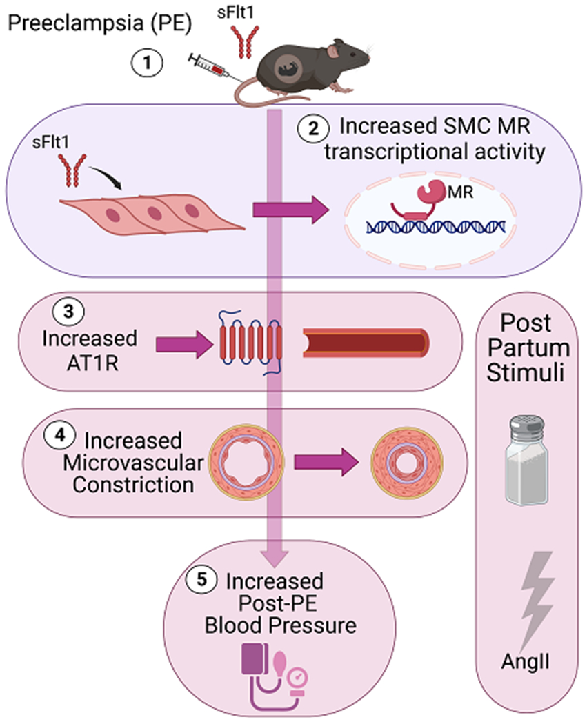
Background: Preeclampsia is a syndrome of high blood pressure (BP) with end organ damage in late pregnancy that is associated with high circulating soluble VEGF receptor (sFlt1 [soluble Fms-like tyrosine kinase 1]). Women exposed to preeclampsia have a substantially increased risk of hypertension after pregnancy, but the mechanism remains unknown, leaving a missed interventional opportunity. After preeclampsia, women have enhanced sensitivity to hypertensive stress. Since smooth muscle cell mineralocorticoid receptors (SMC-MR) are activated by hypertensive stimuli, we hypothesized that high sFlt1 exposure in pregnancy induces a postpartum state of enhanced SMC-MR responsiveness.

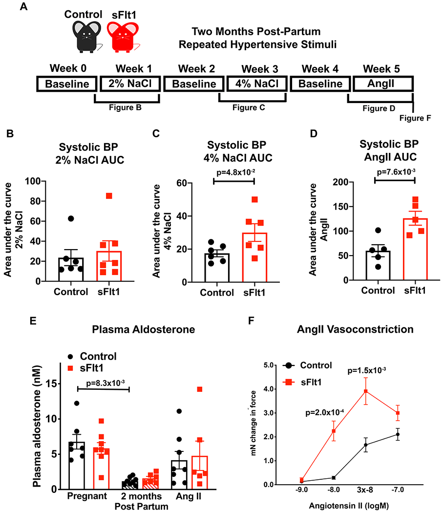
Methods: Postpartum BP response to high salt intake was studied in women with prior preeclampsia. MR transcriptional activity was assessed in vitro in sFlt1-treated SMC by reporter assays and PCR. Preeclampsia was modeled by transient sFlt1 expression in pregnant mice. Two months post-partum, mice were exposed to high salt and then to AngII (angiotensin II) and BP and vasoconstriction were measured.

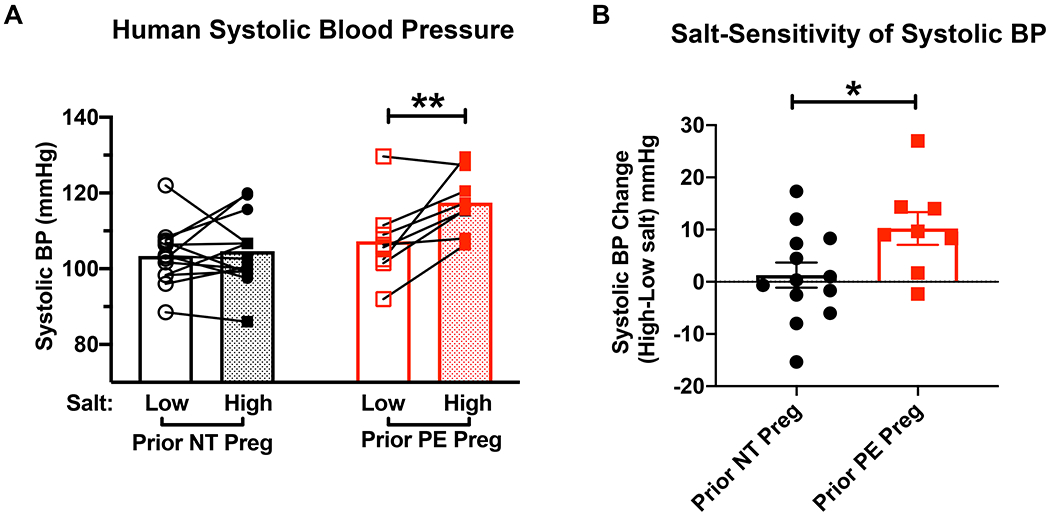
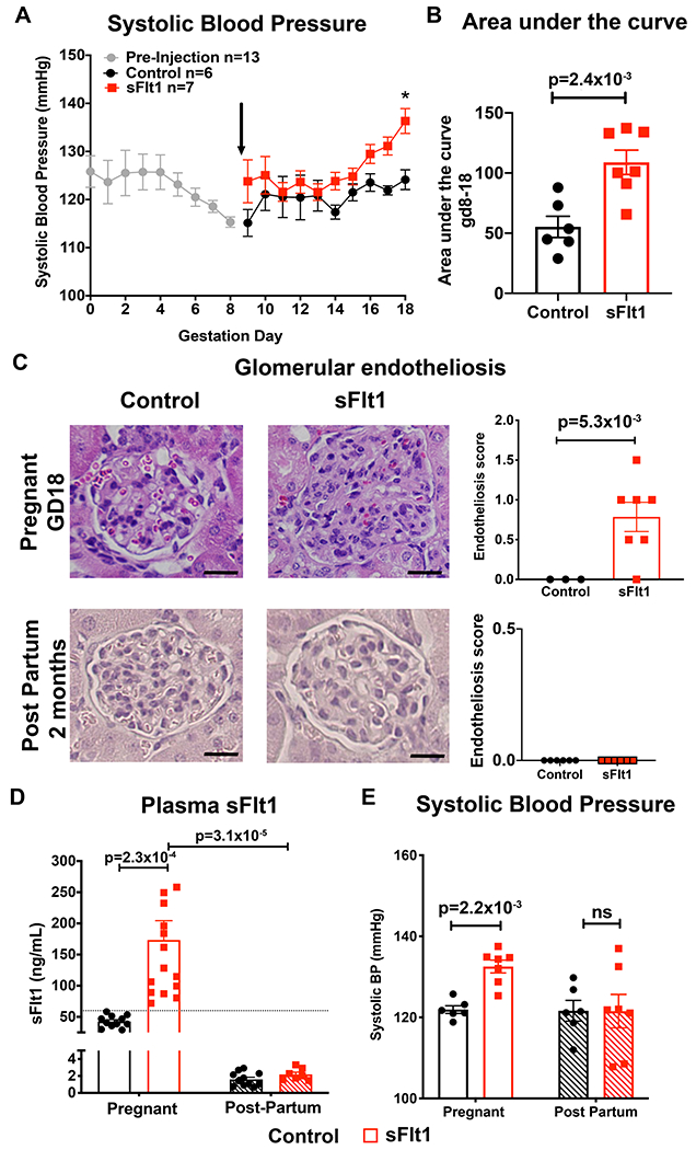
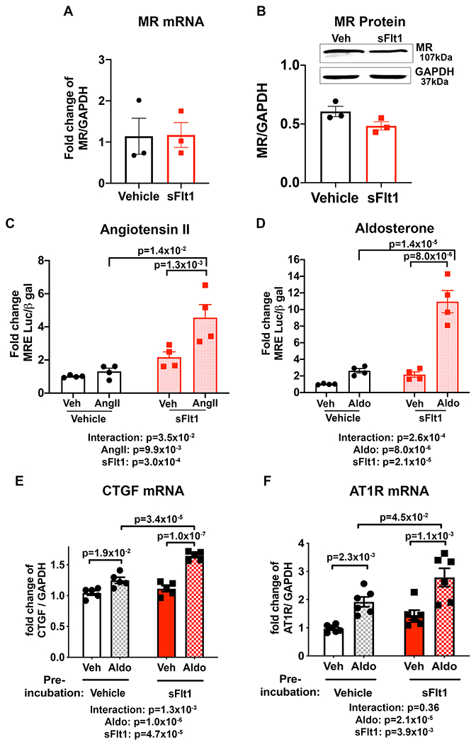
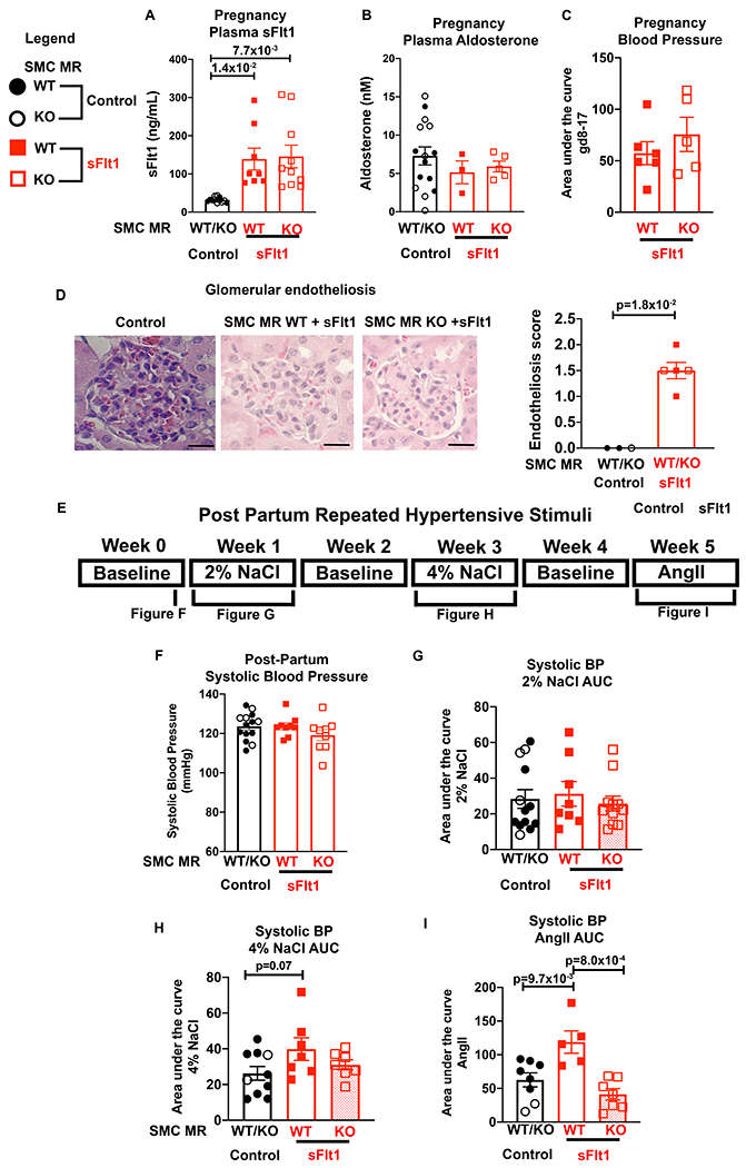
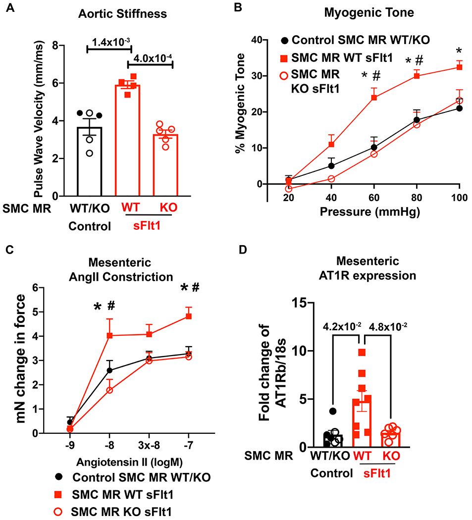
Results: Women exposed to preeclampsia had significantly enhanced salt sensitivity of BP verses those with a normotensive pregnancy. sFlt1 overexpression during pregnancy in mice induced elevated BP and glomerular endotheliosis, which resolved post-partum. The sFlt1 exposed post-partum mice had significantly increased BP response to 4% salt diet and to AngII infusion. In vitro, SMC-MR transcriptional activity in response to aldosterone or AngII was significantly increased after transient exposure to sFlt1 as was aldosterone-induced expression of AngII type 1 receptor. Post-partum, SMC-MR-KO mice were protected from the enhanced response to hypertensive stimuli after preeclampsia. Mechanistically, preeclampsia mice exposed to postpartum hypertensive stimuli develop enhanced aortic stiffness, microvascular myogenic tone, AngII constriction, and AngII type 1 receptor expression, all of which were prevented in SMC-MR-KO littermates.

Conclusions: These data support that sFlt1-induced vascular injury during preeclampsia produces a persistent state of enhanced sensitivity of SMC-MR to activation. This contributes to postpartum hypertension in response to common stresses and supports testing of MR antagonism to mitigate the increased cardiovascular risk in women after PE.

Keywords: angiotensin II; hypertension; mineralocorticoid receptor; myocytes, smooth muscle; pre-eclampsia.


Figures

Figure 1.
Figure 1.. Prior preeclampsia is associated with salt sensitive blood pressure in women.
(A) Women with prior normotensive (NT, n=13) or preeclamptic (preeclampsia, n=8) pregnancies were subjected to one week of low salt diet and one week of high salt diet in a crossover design. Systolic blood pressure was measured at the end of each condition. **P=7.6x10⁻³ via repeated measures 2 way ANOVA with Sidak post hoc. (B)The change in systolic blood pressure from low to high salt measurements in mmHg. *P=3.5x10⁻² via unpaired t-test
Figure 2.
Figure 2.. Exposure to elevated plasma sFlt1 in pregnant C57Bl/6 females is sufficient to phenocopy hypertension and renal damage seen in human preeclampsia.
(A) Systolic blood pressure (SBP) measured during pregnancy with radiotelemetry. Black arrow denotes randomization to injection with either “Control” (CMV-Null transgene) or “sFlt1” adenovirus at gestation day (GD) 9. All mouse BP data were averaged together prior to randomization (GD0-8, n=13). Control n=6, sFlt1 n=7. *p=1.6x10⁻² via two way repeated measures ANOVA with Sidak post hoc. (B) Area under the curve for SBP between GD8 before injection and the end of pregnancy (GD18) for each mouse. Control n=6, sFlt1 n=7. p value determined by unpaired student t test. (C) Representative images of glomeruli from kidneys at GD18 and 2 months post-partum, stained with hematoxylin and eosin. Average glomerular endotheliosis severity scored based on blinded analysis. Scale bar=20μm. Control n=3, sFlt1 n=7. p value determined by unpaired t-test with Welch correction. (D) Plasma was taken on GD17 and two months post-partum and sFlt1 measured using ELISA. Dashed line indicates exclusion value for sFlt1 levels of 60ng/mL. Control pregnant n=11, sFlt1 pregnant n=15, Control post-partum n=11 , sFlt1 post-partum n=7. p values determined via two way ANOVA with Sidak post hoc. (E) SBP at the end of pregnancy (GD17) and two months post-partum. Control pregnant n=6, sFlt1 pregnant n=7, Control post-partum n=6, sFlt1 post-partum n=7. p value determined by two way ANOVA with Sidak post hoc.
Figure 3.
Figure 3.. Prior preeclampsia results in sensitivity to post-partum hypertensive stimuli in mice.
(A) Timeline of post-partum hypertensive stimuli protocol with baseline measurements on standard chow (0.5% NaCl) taken at two months post-partum. Mice were then subjected to 2%NaCl diet for one week, a week of washout, 4% NaCl for one week, a week of washout, and then one week of Angiotensin II (AngII) infusion (600ng/kg/day). The area under the SBP curve quantifies the change in BP response to 1 week exposure to: (B) 2% NaCl (Control n=6, sFlt1 n=7, p=9.5x10⁻¹ via Mann-Whitney test), (C) 4% NaCl (Control n=6, sFlt1 n=6, p value determined by unpaired t-test), and (D) AngII infusion compared to each individual mouse’s baseline SBP (Control n=5, sFlt1 n=5, p value determined by unpaired student t test.) (E) Plasma Aldo at the end of pregnancy, two months post-partum and after repeated hypertensive stimuli. Control pregnant n=7, sFlt1 pregnant n=8, Control post-partum n=8, sFlt1 post-partum n=6, Control AngII n=8, sFlt1 AngII n=6, p value determined by Kruskal-Wallis with Dunn post hoc. (F) Ex vivo microvascular mesenteric artery vasoconstriction to AngII. Control n=7, sFlt n=9, p values determined by 2 way repeated measures mixed effects model, Sidak post hoc test.
Figure 4.
Figure 4.. Transient exposure of smooth muscle cells (SMC) to sFlt1 in vitro enhances mineralocorticoid receptor (MR) transcriptional activity and target gene expression upon receptor stimulation.
Pac1 SMC biological replicates were exposed to 50 ng/mL sFlt1 for 24 hours and expression of MR; (A) mRNA and (B) protein was measured. Control n=3, sFlt1 n=3. Representative western blots of MR and GAPDH in SMC and quantification are shown. 24 hours after sFlt1 was removed, MR transcriptional reporter activity was measured in response to MR stimulation with (C) angiotensin II (AngII, all groups n=4 independent experiments, p values determined by 2-way ANOVA with Sidak post hoc or (D) aldosterone (Aldo, all groups n=4 independent experiments, p values determined by 2-way ANOVA with Sidak post hoc compared to vehicle treated controls. Aldo-stimulated mRNA expression of MR target genes; (E) connective tissue growth factor (CTGF, all groups n=5 independent experiments, p values determined by 2-way ANOVA with Sidak post hoc and, (F) AngII type 1 receptor (AT1R), all groups n=6 independent experiment, p values determined by 2-way ANOVA with Sidak post hoc.
Figure 5.
Figure 5.. SMC-MR-KO mice exposed to high sFlt1 in pregnancy are protected from exacerbated response to post-partum hypertensive stress.
Pregnant SMC-MR-WT or SMC-MR-KO females were injected with Control or sFlt1 on GD9, allowed to deliver, and then exposed to hypertensive stimuli 2 months post-partum. Control injected females were combined for analyses as there were no statistically significant differences between WT and KO. (A) Plasma sFlt1 levels on GD17. Control n=12, WT sFlt n=8, KO sFlt n=10, p values determined via Welch’s one way ANOVA and Dunnett’s multiple comparisons. (B) Plasma aldosterone level on GD17. Control n=15, SMC-MR-WT sFlt n=3, SMC-MR-KO sFlt n=5. (C) Area under the curve for SBP between GD8 before injection and the end of pregnancy (GD17) for each mouse. SMC-MR-WT sFlt1 n=6, SMC-MR-KO sFlt1 n=5. (D) Representative images of glomeruli from mice on GD18, stained with hematoxylin and eosin. Scale bar=20μm. Average glomerular endotheliosis severity scored based on blinded analysis. Control n=3, WT sFlt n=3, KO sFlt n=2, p values determined by Mann-Whitney test. E) Timeline of post-partum hypertensive stimuli protocol. (F) SBP was measured via radiotelemetry two months post-partum. Control n=10, WT sFlt n=5, KO sFlt n=5. (G) The SBP area under the curve (AUC) for 2% NaCl. Control n=13, WT sFlt n=8, KO sFlt n=11. P=8.3x10⁻¹ via Kruskal-Wallis test. (H) The SBP AUC during 4% NaCl. Control n=10, WT sFlt n=7, KO sFlt n=7, p values determined by one way ANOVA and Dunnett’s post hoc test. (I) The SBP AUC during AngII. Control n=8, WT sFlt n=5, KO sFlt n=7, p values determined by one way ANOVA and Dunnett’s post hoc test. All SBP was compared to each individual mouse’s baseline.
Figure 6.
Figure 6.. SMC-MR is necessary for post- preeclampsia aortic stiffness, microvascular dysfunction and AT1R upregulation in response to hypertensive stimuli.
In vivo aortic stiffness and ex vivo mesenteric microvessels were compared between Control mice (SMC-MR-WT and SMC-MR-KO exposed to control pregnancy, SMC-MR-WT exposed to sFlt1-induced preeclampsia, and SMC-MR-KO exposed to sFlt1-induced preeclampsia, all after hypertensive stimuli. (A) Aortic stiffness was measured using pulse wave velocity of the abdominal aorta. Control n=5, WT sFlt n=4, KO sFlt n=5, p values determined by one way ANOVA with Tukey post hoc test. (B) Myogenic tone was measured in control mice (n=15), SMC-MR-WT sFlt (n=11) and SMC-MR-KO mice (n=9). At 60mmHg: *p=7.0x10⁻⁴ Control versus SMC-MR-WT sFlt, #p=7.0x10⁻⁴ SMC-MR-WT sFlt vs SMC-MR-KO sFlt. At 80mmHg: *p=4.7x10⁻³ Control versus SMC-MR-WT sFlt, #p=6.0x10⁻³ SMC-MR-WT sFlt vs SMC-MR-KO sFlt. At 100mmHg: *p=1.1x10⁻² Control versus SMC-MR-WT sFlt. p values determined by 2 way repeated measures ANOVA with Sidak post hoc. (C) Ex vivo microvascular mesenteric constriction to AngII was measured in controls (n=14), SMC-MR-WT sFlt (n=5), and SMC-MR-KO mice (n=9). At 10−8 concentration: *p=3.0x10⁻² Control vs SMC-MR-WT sFlt, #p=7.0x10⁻⁴ SMC-MR-WT sFlt vs SMC-MR-KO sFlt. At 10−7 concentration: *p=1.8x10⁻² Control vs SMC-MR-WT sFlt, #p=1.6x10⁻² SMC-MR-WT sFlt vs SMC-MR-KO sFlt via two-way repeated measures ANOVA with Sidak post hoc. (D) Mesenteric vessels were isolated and mRNA expression of AT1Rb was quantified. Control n=7, WT sFlt n=8, KO sFlt n=6, p values determined by Brown-Forsyth/Welch ANOVA and Dunnett’s post hoc test.
Figure 7.
Figure 7.. Summary Figure.
(1) Adenoviral sFlt1 injection during mid-gestation induces preeclampsia -like signs in mice that resolve post-partum. (2) sFlt1 exposure enhances SMC-MR transcriptional activity and target gene expression, including increased AT1R expression. When formerly preeclamptic mice are challenged post-partum with hypertensive stimuli (high dietary salt, AngII) they exhibit; (3) increased microvascular AT1R and (4) increased microvascular constriction that contributes to (5) exacerbated BP response to hypertensive stress. SMC-MR gene deletion in mice protects against the increased microvascular AT1R expression, enhanced myogenic tone and vasoconstriction and mitigates the exacerbated BP responsiveness after preeclampsia. Thus, the sFlt1-induced preeclampsia model in mice reproduces the post-partum hypertensive sensitivity seen in post- preeclampsia women in a manner that is independent of preexisting risk factors and depends on the presence of SMC-MR.

References

    1. Chappell LC, Cluver CA, Kingdom J,Tong S. Pre-eclampsia. Lancet. 2021;398:341–354. - PubMed
    1. Bellamy L, Casas JP, Hingorani AD,Williams DJ. Pre-eclampsia and risk of cardiovascular disease and cancer in later life: systematic review and meta-analysis. BMJ. 2007;335:974. - PMC - PubMed
    1. Ray JG, Vermeulen MJ, Schull MJ,Redelmeier DA. Cardiovascular health after maternal placental syndromes (CHAMPS): population-based retrospective cohort study. Lancet. 2005;366:1797–1803. - PubMed
    1. Tooher J, Thornton C, Makris A, Ogle R, Korda A,Hennessy A. All Hypertensive Disorders of Pregnancy Increase the Risk of Future Cardiovascular Disease. Hypertension. 2017;70:798–803. - PubMed
    1. Mongraw-Chaffin ML, Cirillo PM,Cohn BA. Preeclampsia and cardiovascular disease death: prospective evidence from the child health and development studies cohort. Hypertension. 2010;56:166–171. - PMC - PubMed

Publication types

Substances